
Radiographic study of proximal ulna dorsal angulation in south Indian population
Abstract
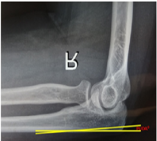
Background: The normal structure of proximal ulna is unique among long bones as described in the literature. Proximal dorsal angulation of ulna (PUDA) was not detailed in standard anatomical textbooks. The identification of PUDA is an important anatomic landmark for surgeons treating proximal ulna fractures, nonunions, malunions or osteotomies of proximal forearm in results of fractures. The study was conducted to identify the PUDA and to measure the distance from tip-to-apex of PUDA in bilateral elbow radiographs.
Materials & Methods: The present study was conducted on 56 bilateral elbow radiographs (26 Male, 30 Female), which are obtained from department of radiology. The radiographs were analyzed and measurements were recorded on each radiograph to identify the PUDA, later olecranon tip-to-apex distance of the PUDA was also measured.
Results: No significant differences were observed in mean age of male and female patients. The measurements were recorded and entered in standard excel data sheet for analyzing PUDA in various patients. It was identified that 84% of the radiographs presents PUDA.
Conclusion: Determination of the PUDA may be helpful in anatomic plating of the ulna for fractures, nonunions or malunions. Side to side correlation of PUDA with gender difference is reliable for recommendation to medical manufacturers for modelling ulna plates and components of artificial elbow joints.
Downloads
References
2. Hewins EA, Gofton WT, Dubberly J, MacDermid JC, Faber KJ, King GJ. Plate fixation of olecranon osteotomies. J Orthop Trauma 2007;21(2):58-62.[pubmed]
3. Gray H. The ulna. Gray’s Anatomy. 37th ed., Ch. 3, London, Churchill Livingstone Publishers, 1989. p. 413.
4. Kulkarni N. Elbow joint. In: Clinical Anatomy for Students;Problem Solving Approach. New Delhi: Jaypee BrothersPublishers; 2007. p. 63.
5. Warwick D. The elbow and forearm. In: Solomon L, Warwick D, Nayagam S, editors. Apley’s System ofOrthopaedics and Fractures. 8th ed. Hodder Arnold Publishers; 2001. p. 304.
6. Das S. A Manual of Clinical Surgery. 4th ed. S. Das’sPublications; 1996. p. 139.
7. Laino DK, Petchprapa CN, Lee SK. Ulnar variance: correlation of plain radiographs, computed tomography, and magnetic resonance imaging with anatomic dissection. J Hand Surg Am. 2012;37:90–97.[pubmed]
8. Dhillon MS, Gopinathan NR, Kumar V. Misconceptions about the three point bony relationship of the elbow. Indian J Orthop2014;48:453-7.[pubmed]
9. Grechenig W, Clement H, Pichler W, Tesch NP, Windisch G. The influence oflateral and anterior angulation of the proximal ulna on the treatment of aMonteggia fracture: an anatomical cadaver study. J Bone Joint Surg Br.2007 Jun;89(6):836–8.[pubmed]
10. Windisch G, Clement H, Grechenig W, Tesch NP, Pichler W. Amorphometrical study of the medullary cavity of the ulna referred tointramedullary nailing. Surg Radiol Anat. 2007 Feb;29(1):47–53.[pubmed]
11. Puchwein P, Schildhauer TA, Schöffmann S, Heidari N, Windisch G, PichlerW. Three-dimensional morphometry of the proximal ulna: a comparison tocurrently used anatomically preshaped ulna plates. J Shoulder Elb Surg.2012;21:1018–23.
12. Duggal N, Dunning CE, Johnson JA, King GJ. The flat spotof the proximal ulna: a useful anatomic landmark in totalelbow arthroplasty. J Shoulder Elbow Surg 2004;13:206–7.[pubmed]
13. Dumont CE, Pfirrmann CW, Ziegler D, Nagy L. Assessment of radial and ulnar torsion profiles with cross-sectional magnetic resonance imaging. A study of volunteers. J Bone Joint Surg Am 2006;88: 1582-8.[pubmed]
14. Akpinar F, Aydinlioglu A, Tosun N, Tuncay I. Morphologic evaluation of the ulna. Acta OrthopScand2003;74:415-9.[pubmed]
15. Wang AA, Mara M, Hutchinson DT. The proximal ulna: An anatomic study with relevance to olecranon osteotomy and fracture fixation. J Shoulder Elbow Surg 2003;12:293-6.[pubmed]
16. Matzon JL, Widmer BJ, Draganich LF, Mass DP, Phillips CS. Anatomy of the coronoid process. J Hand Surg [Am] 2006; 31:1272-8.[pubmed]
17. Mall G, Hubig M, Buttner A, Kuznik J, Penning R, Graw M. Sex determination and estimation of stature from the long bones of the arm. Forensic Sci Int 2001;117:23-30.[pubmed]
18. Wadia F, Kamineni S, Dhotare S, Amis A. Radiographic measurements of normal elbows: clinical relevance to olecranon fractures. Clin Anat2007;20:407-10.[pubmed]
19. Goldberg SH, Omid R, Nassr AN, Beck R, Cohen MS. Osseous anatomy of the distal humerus and proximal ulna: implications for total elbow arthroplasty. J Shoulder Elbow Surg 2007;16(3 suppl): S39-46.[pubmed]

 OAI - Open Archives Initiative


