A study on uterine and vaginal arteries and their clinical significance
Abstract
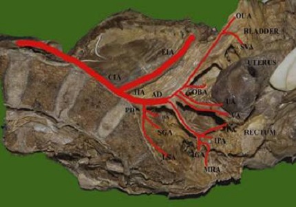
Background: The anterior division of the internal iliac artery usually gives origin to the uterine artery and vaginal artery in common. Uterine artery travels in cardinal ligament to reach uterus by crossing the ureter anteriorly & parametrium of the inferior broad ligament of the uterus gives way for the uterine artery. Uterus is mainly supplying by uterine artery and it is anastomosing usually with ovarian artery. Vaginal artery is source of blood supply to vagina and lower part of urinary bladder.
Objectives: The present study conducted to study the morphological features of uterine and vaginal arteries, and their variations, in origin and branching pattern. Materials and
Methods: Dissection of 50 adult human pelvic halves was procured from embalmed cadavers of J.J.M. Medical College and S.S.I.M.S & R.C, Davangere for the study.
Results: Out of 50 specimens uterine and vaginal arteries we had traced only in 17 specimens. Out of these 17 specimens, uterine artery took origin from anterior division directly in 15 specimens and double uterine artery was observed in 2 specimens. Vaginal artery took origin from anterior division directly in 15 specimens and with internal pudendal artery in 2 specimens.
Conclusion: Uterine and vaginal arteries in majority of cases taken origin from anterior division of internal iliac artery and in few cases vaginal artery taken origin from internal pudendal artery. The knowledge of uterine and vaginal arteries is very important during obstetrics and pelvic surgeries and in treating of diseases related female pelvic organs.
Downloads
References
2. Lipschutz B. A composite study of the hypogastric artery and its branches. Ann Surg 1918; 67(5):584-608.
3. Havaldar PP, Taz S, Angadi AV, Saheb SH. Morphological study of obturator artery. Int J Anat Res. 2014;2(2):354-7.
4. Gomez-Jorge J, Keyoung A, Levy EB, Spies JB. Uterine artery anatomy relevant to uterine leiomyomata embolization. Cardiovasc Intervent Radiol. 2003 Nov-Dec;26(6):522-7. [PubMed]
5. Pelage JP, Le Dref O, Soyer P, et al. Arterial anatomy of the female genital tract: variations and relevance to transcatheter embolization of the uterus. AJR Am J Roentgenol. 1999;172: 989-994. 10.2214/ajr.172.4.10587133.
6. Havaldar PP, Taz S, Angadi AV, Saheb SH. Study of posterior division of internal iliac artery. Int J Anat Res. 2014;2(2):375-9.
7. Pavan P Havaldar et al. Study of medial circumflex artery Int J Anat Res.2014;Vol 2(2):380-82.
8. Pelage JP, Le Dref O, Soyer P, Jacob D, Kardache M, Dahan H, Lassau JP, Rymer R. Arterial anatomy of the female genital tract: variations and relevance to transcatheter embolization of the uterus. AJR Am J Roentgenol. 1999 Apr;172(4):989-94.
9. Saraiya PV, Chang TC, Pelage JP, Spies JB. Uterine artery replacement by the round ligament artery: an anatomic variant discovered during uterine artery embolization for leiomyomata. J Vasc Interv Radiol. 2002;13(9):939–41. [PubMed]
10. Worthington-Kirsch L, Walker WJ, Adler L, Hutchins FL. Anatomic variation in the uterine arteries: a cause of failure of uterine artery embolisation for the management of symptomatic myomata. Min Invas Ther Allied Technol. 1999;8(6):397–402.
11. Spies JB. The pitfalls of uterine embolization: avoiding the failed procedure. In: Spies JB, Pelage JP, editors. Uterine artery embolization and gynecologic embolotherapy. Philadelphia: Lippincott Williams & Wilkins; 2005. p. 95–105.
12. Lee KH, Park TC, Park JS. Duplicated uterine arteries in laparoscopic hysterectomy. J Minim Invasive Gynecol. 2008 Jan-Feb;15(1):3. doi: 10.1016/j.jmig.2007.05.006. [PubMed]
13. Bergman RA, Thompson SA, Afifi AK and Saadeh FA. Compendium of human anatomic variation. Baltimore and Munich: Urban and Schwazenberg; 1988 .p.84-85.

 OAI - Open Archives Initiative


