E-ISSN:2455-5436
P-ISSN:2456-9518
RNI:MPENG/2017/70870

Case Report

septic Nonunion

Surgical Review: International Journal of Surgery Trauma and Orthopedics

2023 Volume 9 Number 2 April-June
Publisherwww.medresearch.in

Masquelet technique to treat a septic nonunion after plating of a femoral open fracture

Patel M.1*, Patel G.2, Chaudhary J.3, Patel A.4, Joshi M.5
DOI: https://doi.org/10.17511/ijoso.2023.i02.01

1* Maulik Patel, DNB Ortho Resident, Diploma in Orthopaedics, GMERS medical college and Hospital, Dharpur, Patan, Gujrat, India.

2 Gaurang Patel, Associate Professor, MS Orthopaedics, SSG Hospital and Medical College, Baroda, Vadodara, Gujrat, India.

3 Jaydeep Chaudhary, Senior Resident, MS Orthopaedics, GMERS medical college and hospital, Dharpur, Patan, Gujrat, India.

4 Abhay Patel, Junior Resident, Diploma in Orthopaedics, GMERS medical college and hospital, Dharpur, Patan, Gujrat, India.

5 Mayur Joshi, DNB Ortho Resident, Diploma in Orthopaedics, GMERS medical college and hospital, Dharpur, Patan, Gujrat, India.

Septic nonunion is one of the most common complications after an open fracture. The risk of infection and bony defects is high following an open fracture , and it is essential to address both issues to improve patient outcomes. Treatment can be complex and costly , and outcome is often uncertain. In the 1980s, Masquelet first described the induced membrane technique and autologous bone grafting to manage critical-size bone defects. In cases of Septic non-union, two different surgical steps are required for management. In the former step, radical management to control infection and in the latter step, biological stimulus to promote bone healing. In this case, we present a 28 years old female patient with an open grade 3A Femoral shaft Fracture. Initially , the patient received wound debridement and Femur plating which unfortunately resulted in septic non-union. Subsequently, the patient was managed with the induced membrane technique and a double-plate osteosynthesis to protect the biological chamber.

Keywords: Septic Nonunion, Masquelet Technique, Femoral Open Fracture

Corresponding Author How to Cite this Article To Browse
Maulik Patel, DNB Ortho Resident, Diploma in Orthopaedics, GMERS medical college and Hospital, Dharpur, Patan, Gujrat, India.
Email:
Maulik Patel, Gaurang Patel, Jaydeep Chaudhary, Abhay Patel, Mayur Joshi, Masquelet technique to treat a septic nonunion after plating of a femoral open fracture. Surgical Rev Int J Surg Trauma Orthoped. 2023;9(2):6-12.
Available From
https://surgical.medresearch.in/index.php/ijoso/article/view/270

Manuscript Received Review Round 1 Review Round 2 Review Round 3 Accepted
2023-03-25 2023-03-27 2023-04-03 2023-04-10 2023-04-16
Conflict of Interest Funding Ethical Approval Plagiarism X-checker Note
Nil Nil Yes 17%

© 2023by Maulik Patel, Gaurang Patel, Jaydeep Chaudhary, Abhay Patel, Mayur Joshiand Published by Siddharth Health Research and Social Welfare Society. This is an Open Access article licensed under a Creative Commons Attribution 4.0 International License https://creativecommons.org/licenses/by/4.0/ unported [CC BY 4.0].

Introduction

Despite significant advancements in open fracture care, nonunion remains a challenging issue, especially in young patients with high-energy trauma, with reported prevalence ranging from 0% to 14%, and a median incidence of 2.4% for diaphyseal femur fractures [1,2].

Septic non-union is a devastating complication of an open femur fracture, requiring a prolonged, expensive, and uncertain treatment course, as it requires addressing both infection and bone defects [3].

The treatment of infection necessitates surgical radical debridement of infected tissue and antibiotics, followed by bone healing promotion. The bone transport technique and vascularized bone transfer are treatments for long bone defects, but they are characterized by prolonged healing times and serious complications, with an incidence exceeding 20% [4].

The induced membrane technique, introduced by Masquelet et al. 30 years ago, is an alternative for managing large bone defects secondary to chronic infections or nonunion [5].

This paper presents a case of septic non-union following plating of an open femur fracture treated with the induced membrane technique (Masquelet) and plate osteosynthesis. The double-plating construct provides an effective option for protecting the biological chamber while waiting for graft integration.

Case Report

A healthy 28-year-old female presented to the orthopedics outpatient department with a primary complaint of pain over the right lower third thigh and a pus-discharging sinus from a previous operated suture line for the past 3 months.

Eight (8) months ago, the patient sustained a road traffic accident and was diagnosed with an open grade 3A lower third femur shaft fracture and patella fracture. She underwent wound debridement followed by distal femur plating and patella encirclage at another hospital. Postoperatively, the patient was afebrile, and dressings were clean. Suture removal was done on postoperative day 15. Three months ago, pus discharge was coming out

from the suture line, and antibiotics were started for the same. There was no improvement even after starting antibiotics.

The patient was conscious and well-oriented to time, place, and person. Her temperature was 37.6°C, heart rate was 86 beats/minute, respiratory rate was 12 breaths/minute, and blood pressure was 120/80 mmHg.

On local examination, by palpation, the right lower limb temperature was mildly increased compared to the left lower limb. Seropurulent pus discharge was coming out from the suture line. Limb length discrepancy was approximately 4 cm shortening compared to the left lower limb. The range of movement of the knee was 0-30 degrees, and the range of movement of the hip and ankle was normal.

X-ray of the right lower limb showed distal femur plating with encirclage at the femur fracture site with no callus formation at the fracture site with sclerotic fracture ends. Patella encirclage with bony union was seen on digital radiographs.

surgery_270_01.JPG
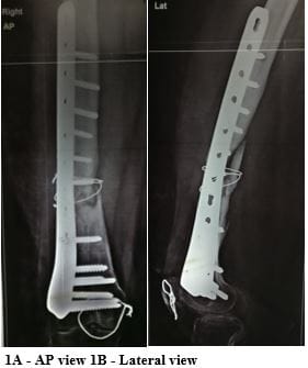
Figure 1 : X ray of right femur at the time of presentation .No callus formation at femur fracture end with visible sclerotic femur fracture end.

In the first stage of the Masquelet surgical technique, the implant was removed


followed by removal of necrosed fracture ends and infected and non-viable tissue. The fracture was stabilized with a mono-axial external fixator, and an intra-operative microbiological pus culture was sent. Initially, empirical antibiotics were started followed by culture-specific antibiotics. On regular follow-up, dressing was clean, and suture removal was done at postoperative 15 days (Figure 2). After three weeks, the external fixator was removed. After one week of external fixator removal, the pin track site was healthy. Antibiotic-loaded cement space was positioned together with distal femur plating in the first stage (Figure 3).

There was gradual improvement in the local condition of the lower limb after the first stage.

surgery_270_02.JPG
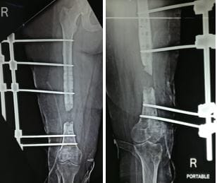
Figure 2 : 2A & 2B- Fracture temporary stabilise with mon-axial external fixator.

surgery_270_03.JPG
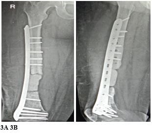
Figure 3 ( 3A & 3B ) : Antibiotic loaded cement space positioned together with distal femur plating.

Blood infective biomarkers were monitored regularly and came within the normal range after 1.5 months of the first surgical intervention. After 2 months, antibiotic therapy was stopped.

After 8 weeks of the first surgical stage, the second stage of the Masquelet technique was planned. The previous incision was used for exposure. The induced membrane over the spacer was opened longitudinally and loaded on suture threads to allow subsequent closure. Once the cement spacer was removed, the bone defect was filled with bone graft taken from the posterior superior iliac spine (PSIS) and fibula strut graft. After putting the bone graft, the non-union site was additionally stabilized with an anterior reconstruction plate.

An intra-operative microbiological sample was sent for culture, and the sample demonstrated the absence of infection.

surgery_270_04.JPG

surgery_270_05.JPG

Figure 4 : Second stage of Masquelet Technique …4A - Intra-operative apperance of induced membrane (black arrow ) , 4B - Post-operative X ray.


Post-operative broad-spectrum antibiotic treatment was given for 3 weeks. During follow-up, the dressing was clean and suture removal was done on postoperative day 21.

surgery_270_06.JPG
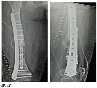
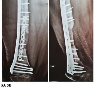
Figure 5 ( 5A & 5B ) : X rays at one month follow up.

surgery_270_07.JPG
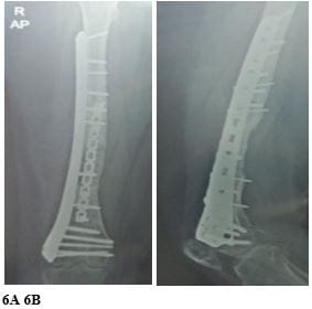
Figure 6 ( 6A & 6B ) : X rays at six months follow up showed complete graft integration as well as bony union.

The postoperative patient was advised non-weight bearing walking for the initial 3 weeks. After 3 weeks, the patient was advised for active and passive mobilization of the knee. The patient was advised regular follow-up. Post-op 4 weeks, digital X-rays were taken, and then after the patient was advised toe touch walking.

On further follow-up, X-ray showed significant progression in graft integration and bone healing. During her post-op 6 months, the patient no longer complained of pain or any sign of infection. X-rays showed complete remodelling of the graft together with complete healing at the non-union site.

In local examination, the range of movement of the knee was 0-100 degrees with a limb length discrepancy of 0.5 cm shortening present on the right side compared to the left.

Discussion

Nowadays, nonunions are continue to be a technically demanding obstacle to bone healing: in femoral shaft fractures, this complication can occur with an incidence traditionally reported of 2.4% [2].

Recent data published by Zura et al. has showed a significant increase in its prevalence, up to 13.9%, due to the increase of complex and open fractures resulting from high-energy trauma [6].

In femur fracture, nonunion is more frequently seen in the transition area between the middle third and distal third and in the distal third of the diaphysis, due to poor vascularisation in this area[7].

Intramedullary nailing of femur provides excellent functional outcomes and union rates, being the treatment of choice for femoral shaft fractures; despite this, femur nonunion after nailing is not a rare occurrence, with reported rates ranging from 1 to 20% [8–10].

Nandra et al. reported nonunion rates of 1.1% for antegrade nailing and of 5.8% for retrograde nailing: this confirms that the fracture of the distal diaphysis and of the distal third, in which a retrograde approach is most suitable, are those most vulnerable to consolidation issues [11].

Infection is a well-documented problem after open femur fractures, with incidence rising with the grade of bone exposure, according to the Gustilo and Anderson classification: early surgical and medical management must be undertaken to minimise the risk of this serious issue [12,13].

Elniel et al. emphasise the importance of an accurate debridement with enlargement of skin injury and excision of all non-viable tissues, even in low-grade open fractures [14].


Radical debridement would have prevented the septic complication, and therefore significantly reduced the overall duration of treatment.Bone defect is the other main challenging issue in the management of a nonunion after an open fracture.

In this scenario, bone defects can be divided into acute and chronic: acute defects are the consequence of bone loss after the initial injury or after early surgical debridement; chronic defects resuting from the removal of infected and necrotic bone [15,16]. bone defects more than six centimetres are the most difficult to treat [16].

Historically, distraction osteogenesis, massive autograft or allograft and free vascularised bone transfer have been proposed to deal with this challenging condition. Replacement of the defect by the implant of megaprosthesis or cages has also been advocated [17,18].

Vascularised bone transfer use is limited by the anatomical conformation of the pedicle, a difficulty in microsurgical anastomosis technique, risk of donor site morbidity and stress fracture [19,20]. The main issues for distraction osteogenesis are the long healing time and several complications such as pin tract infection, nonunion at the docking site and, especially in the femur, the potential risk of articular stiffness [4].

In 1986, Masquelet described the technique of the induced membrane and autologous bone grafting as an alternative method for managing critical bone defects [5]. This procedure is based on two surgical phases [21]. The first is characterised by the debridement of soft tissues and bone with application of a cement spacer, application of an external fixator to stabilise the spacer, and soft tissue coverage if necessary. The second phase takes place involves the removal of the cement spacer and the application of cancellous bone graft obtained from the iliac crest.

Azi et al. [22] reported that the Masquelet technique is a kind of foreign body reaction. This process begins with the presence of the cement spacer [23,24]. Pelissier et al., Liu et al. have discovered that this pseudosynovial membrane is highly vascu-larised and rich in growth factors [25,26]. Moreover, it can form bone and cartilage and it has osteogenic and osteoinductive properties related to the presence of various key genes connected to angiogenesis and osteogenesis

[25,27]. The cement spacer in the bone defect prevents the formation of fibrotic tissue and keeps the volume free for the subsequent bone graft [5]. Lastly, given the presence of local antibiotics, the cement can also operate as a local antibiotic delivery system, which may be important in infected bone defects.

The technique initially proposed by Masquelet for treating septic nonunions involved the use of external fixation. However, more recent studies have demonstrated similar success rates using both plates and nails [23].

Intramedullary nails have the advantage of reducing the amount of graft required while not interfering with bone healing [28]. Plates, on the other hand, have been found to be more effective in cases involving joint proximities and complex patterns [28].

Jiang et al. have recently suggested the use of a double-plate fixation in conjunction with an autogenous fibular bone graft for treating nonunion of the femur shaft. They noted that a single plate on the lateral side of the femur could increase bending forces, potentially leading to mechanical failure [29]. Consequently, double-plate fixation is considered a better option compared to a single plate.

To increase the mechanical stability of our construct, we adopted the double-plating technique. This technique allowed us to position a greater quantity of autologous bone graft, creating a mechanical chamber to support the biological chamber. In our opinion, the double-plating technique improved the mechanical stability while waiting for integration of the graft.

Conclusion

In summary, the Masquelat technique has proven to be a valid alternative method for treating septic nonunions by eradicating the infection and promoting good union rates.

Reference

01. Mills LA, Aitken SA, Simpson AH. The risk of non-union per fracture: current myths and revised figures from a population of over 4 million adults. Acta orthopaedica. 2017 Jul 4;88(4):434-9. [Crossref][PubMed][Google Scholar]


02. Simpson AH, Tsang JS. Current treatment of infected non-union after intramedullary nailing. Injury. 2017 Jun 1;48:S82-90. [Crossref][PubMed][Google Scholar]

03. Hak DJ, Fitzpatrick D, Bishop JA, Marsh JL, Tilp S, Schnettler R, Simpson H, Alt V. Delayed union and nonunions: epidemiology, clinical issues, and financial aspects. Injury. 2014 Jun 1;45:S3-7. [Crossref][PubMed][Google Scholar]

04. El‐Gammal TA, Shiha AE, El‐Deen MA, El‐Sayed A, Kotb MM, Addosooki AI, Ragheb YF, Saleh WR. Management of traumatic tibial defects using free vascularized fibula or Ilizarov bone transport: a comparative study. Microsurgery: Official Journal of the International Microsurgical Society and the European Federation of Societies for Microsurgery. 2008;28(5):339-46. [Crossref][PubMed][Google Scholar]

05. Masquelet AC, Fitoussi F, Begue T, Muller GP. Reconstruction of the long bones by the induced membrane and spongy autograft. InAnnales de chirurgie plastique et esthetique 2000 Jun 1 (Vol. 45, No. 3, pp. 346-353) [Crossref][PubMed][Google Scholar]

06. Zura R, Xiong Z, Einhorn T, Watson JT, Ostrum RF, Prayson MJ, Della Rocca GJ, Mehta S, McKinley T, Wang Z, Steen RG. Epidemiology of fracture nonunion in 18 human bones. JAMA surgery. 2016 Nov 1;151(11):e162775-. [Crossref][PubMed][Google Scholar]

07. Santolini E, Goumenos SD, Giannoudi M, Sanguineti F, Stella M, Giannoudis PV. Femoral and tibial blood supply: A trigger for non-union?. Injury. 2014 Nov 1;45(11):1665-73. [Crossref][PubMed][Google Scholar]

08. Gelalis ID, Politis AN, Arnaoutoglou CM, Korompilias AV, Pakos EE, Vekris MD, Karageorgos A, Xenakis TA. Diagnostic and treatment modalities in nonunions of the femoral shaft. A review. Injury. 2012 Jul 1;43(7):980-8 [Crossref][PubMed][Google Scholar]

09. Winquist RA, Hansen Jr ST, Clawson DK. Closed intramedullary nailing of femoral fractures. A report of five hundred and twenty cases. JBJS. 1984 Apr 1;66(4):529-39 [Crossref][PubMed][Google Scholar]

10. Wolinsky PR, McCarty E, Shyr Y, Johnson K.

Reamed intramedullary nailing of the femur: 551 cases. Journal of Trauma and Acute Care Surgery. 1999 Mar 1;46(3):392-9. [Crossref][PubMed][Google Scholar]

11. Nandra R, Grover L, Porter K. Fracture non-union epidemiology and treatment. Trauma. 2016 Jan;18(1):3-11. [Crossref][PubMed][Google Scholar]

12. Papakostidis C, Kanakaris NK, Pretel J, Faour O, Morell DJ, Giannoudis PV. Prevalence of complications of open tibial shaft fractures stratified as per the Gustilo–Anderson classification. Injury. 2011 Dec 1;42(12):1408-15. [Crossref][PubMed][Google Scholar]

13. Thakore RV, Francois EL, Nwosu SK, Attum B, Whiting PS, Siuta MA, Benvenuti MA, Smith AK, Shen MS, Mousavi I, Obremskey WT. The Gustilo–Anderson classification system as predictor of nonunion and infection in open tibia fractures. European Journal of Trauma and Emergency Surgery. 2017 Oct;43:651-6. [Crossref][PubMed][Google Scholar]

14. Elniel AR, Giannoudis PV. Open fractures of the lower extremity: Current management and clinical outcomes. EFORT open reviews. 2018 May;3(5):316. [Crossref][PubMed][Google Scholar]

15. Kanakaris NK, Tosounidis TH, Giannoudis PV. Surgical management of infected non-unions: an update. Injury. 2015 Nov 1;46:S25-32. [Crossref][PubMed][Google Scholar]

16. Zhang Q, Zhang W, Zhang Z, Zhang L, Chen H, Hao M, Deng J, Tang P. Femoral nonunion with segmental bone defect treated by distraction osteogenesis with monolateral external fixation. Journal of Orthopaedic Surgery and Research. 2017 Dec;12(1):1-7. [Crossref][PubMed][Google Scholar]

17. Calori GM, Colombo M, Malagoli E, Mazzola S, Bucci M, Mazza E. Megaprosthesis in post-traumatic and periprosthetic large bone defects: issues to consider. Injury. 2014 Dec 1;45:S105-10. [Crossref][PubMed][Google Scholar]

18. Lindsey RW, Gugala Z, Milne E, Sun M, Gannon FH, Latta LL. The efficacy of cylindrical titanium mesh cage for the reconstruction of a critical‐size canine segmental femoral diaphyseal defect. Journal of orthopaedic research. 2006 Jul;24(7):1438-53. [Crossref][PubMed][Google Scholar]


19. Ronga M, Ferraro S, Fagetti A, Cherubino M, Valdatta L, Cherubino P. Masquelet technique for the treatment of a severe acute tibial bone loss. Injury. 2014 Dec 1;45:S111-5. [Crossref][PubMed][Google Scholar]

20. Woon CY, Chong KW, Wong MK. Induced membranes—a staged technique of bone-grafting for segmental bone loss: a report of two cases and a literature review. JBJS. 2010 Jan 1;92(1):196-201. [Crossref][PubMed][Google Scholar]

21. Masquelet AC. Induced membrane technique: pearls and pitfalls. Journal of orthopaedic trauma. 2017 Oct 1;31:S36-8. [Crossref][PubMed][Google Scholar]

22. Azi ML, Teixeira AA, Cotias RB, Joeris A, Kfuri Jr M. Membrane induced osteogenesis in the management of posttraumatic bone defects. Journal of orthopaedic trauma. 2016 Oct 1;30(10):545-50. [Crossref][PubMed][Google Scholar]

23. Giannodus PV, Faour O, Goff T, Kanakaris N, Dimitrou R. Masquelet technique for treatment of bone defects: tips-trick and future direction. Injury. 2011;42(6):591-8. [Crossref][PubMed][Google Scholar]

24. Giannoudis PV, Harwood PJ, Tosounidis T, Kanakaris NK. Restoration of long bone defects treated with the induced membrane technique: protocol and outcomes. Injury. 2016 Dec 1;47:S53-61. [Crossref][PubMed][Google Scholar]

25. Liu H, Hu G, Shang P, Shen Y, Nie P, Peng L, Xu H. Histological characteristics of induced membranes in subcutaneous, intramuscular sites and bone defect. Orthopaedics & Traumatology: Surgery & Research. 2013 Dec 1;99(8):959-64. [Crossref][PubMed][Google Scholar]

26. Pelissier PH, Masquelet AC, Bareille R, Pelissier SM, Amedee J. Induced membranes secrete growth factors including vascular and osteoinductive factors and could stimulate bone regeneration. Journal of orthopaedic research. 2004 Jan;22(1):73-9. [Crossref][PubMed][Google Scholar]

27. Gruber HE, Ode G, Hoelscher G, Ingram J, Bethea S, Bosse MJ. Osteogenic, stem cell and molecular characterisation of the human induced membrane from extremity bone defects. Bone & Joint Research. 2016 Apr;5(4):106-15. [Crossref][PubMed][Google Scholar]

28. Olesen UK, Eckardt H, Bosemark P, Paulsen AW, Dahl B, Hede A. The Masquelet technique of induced membrane for healing of bone defects. A review of 8 cases. Injury. 2015 Dec 1;46:S44-7 [Crossref][PubMed][Google Scholar]

29. Jiang Y, Wang X, Huang W, Zhu Y, Zhang K, Feng D. A novel bone graft technique combined with plating for aseptic recalcitrant long bone nonunion. BMC Musculoskeletal Disorders. 2022 Dec;23(1):1-9. [Crossref][PubMed][Google Scholar]