E-ISSN:2455-5436
P-ISSN:2456-9518
RNI:MPENG/2017/70870

Case Report

Osteochondroma

Surgical Review: International Journal of Surgery Trauma and Orthopedics

2023 Volume 9 Number 1 January-March
Publisherwww.medresearch.in

Case Report: An Unusual Location of Osteochondroma Dorsal Scapula

Patel M.1*, Parmar R.2, Modi P.3
DOI: https://doi.org/10.17511/ijoso.2023.i01.01

1* Maulik Patel, Diploma in Orthopaedics, DNB ortho Resident, GMERS medical college and Hospital, Dharpur, Patan, Gujrat, India.

2 Ruchir Parmar, MS Orthopaedics, Senior Resident, GMERS medical college and Hospital, Dharpur, Patan, Gujrat, India.

3 Pulkit Modi, MS Orthopaedics, Head of Department, GMERS medical college and Hospital, Dharpur, Patan, Gujrat, India.

Introduction: Osteochondroma of the scapula is one of the rare tumours of the thorax. It constitutes 14.4% of all tumours of the scapula with the ventral surface being the most common site of presentation. Based on our review of the literature, the dorsal surface of the scapula is a rare site of origin of osteochondroma. Among the osteochondroma of the dorsal surface of the scapula, the sessile variant is more common. Case Report: We reported a rare case of a solitary pedunculated variety of osteochondroma at an unusual site-dorsal surface of the scapula in a 9-year-old male child. The tumours were excised and the diagnosis was confirmed by histopathology. Conclusion: This case is reported for its rarity and unusual site of presentation.

Keywords: Scapula, osteochondroma, dorsal surface

Corresponding Author How to Cite this Article To Browse
Maulik Patel, Diploma in Orthopaedics, DNB ortho Resident, GMERS medical college and Hospital, Dharpur, Patan, Gujrat, India.
Email:
Maulik Patel, Ruchir Parmar, Pulkit Modi, Case Report: An Unusual Location of Osteochondroma Dorsal Scapula. Surgical Rev Int J Surg Trauma Orthoped. 2023;9(1):1-5.
Available From
https://surgical.medresearch.in/index.php/ijoso/article/view/266

Manuscript Received Review Round 1 Review Round 2 Review Round 3 Accepted
2023-02-04 2023-02-07 2023-02-14 2023-02-21 2023-02-28
Conflict of Interest Funding Ethical Approval Plagiarism X-checker Note
Nil Nil Yes 16%

© 2023by Maulik Patel, Ruchir Parmar, Pulkit Modiand Published by Siddharth Health Research and Social Welfare Society. This is an Open Access article licensed under a Creative Commons Attribution 4.0 International License https://creativecommons.org/licenses/by/4.0/ unported [CC BY 4.0].

Introduction

One of the rare tumours of the thorax is osteochondroma of the scapula. It constitutes 14.4% of all tumours of the scapula with the ventral surface being the most common site of presentation[1]. Based on our review of the literature, the dorsal surface of the scapula is a rare site of origin of osteochondroma. Among the osteochondroma of the dorsal surface of the scapula, the sessile variant is more common. We report rare cases of a pedunculated variety of osteochondroma at an unusual site-dorsal surface of the scapula in a 9-year-old child. The tumours were excised and the diagnosis was confirmed by histopathological studies. This case was reported for its rarity and its unusual site of presentation.

Case Report

A 9-year-old child presented to us with a complaint of swelling over the right scapular region for 1 year. He had complained of discomfort while sleeping on his back for 8 months and he had difficulty moving his right shoulder for 6 months. The swelling was spontaneous in onset and gradually increasing in nature. It was not associated with any history of trauma, fever, difficulty in breathing, weight loss, or a history of similar complaints in the past. On examination, a hard, oval-shaped bony swelling of size 4 cm* 3 cm was palpable on the dorsum of the right scapula along the medial border, non-tender, and fixed to the scapula and with normal free overlying skin(Fig. 1).

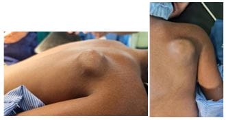
Figure 1: Clinical Pre-operative Pictures (A hard, oval solid palpable mass on the dorsal scapula.)

Right upper limb neurology was normal. There has been no other visible swelling over the body. X-ray of the right shoulder showed a bony swelling arising from the dorsal surface of the right scapula(Fig. 2).

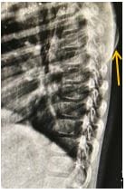
Figure 2: X-ray showing bony mass arising from the dorsal aspect of the scapula.

Magnetic resonance imaging( MRI ) scan showed that a pedunculated mushroom-like mass arising from the dorsal aspect of the medial border of the scapula (Fig. 3).


surgery_266_05.JPG
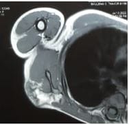
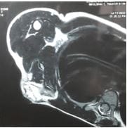
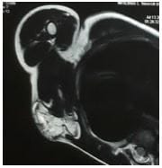
Figure 3: MRI showing a pedunculated mass on the dorsal aspect of the scapula.

There was no evidence of any pathological fracture or ventral extension or chest wall abnormality.Our provisionl diagnosis was pedunculated osteochodroma of right dorsal scapula .An excision biopsy was planned for the patient.

Surgical Technique: General anaesthesia was given. A prone position was given to the patient. Painting and dropping were done over the right scapular region. A 4 cm incision was made parallel to the medial border of the scapula directly over the swelling. Soft tissue was dissected, a bony mass was identified and borders of the mass were delineated ( Fig 4). Peduncle mass was excised in toto. The remnant of the stump was nibbled out and the dorsal surface of the scapula was smoothened

using burr and bone file. A pedunculated mass was sent for histopathology and the diagnosis was confirmed. Histopathological studies confirmed the diagnosis of osteochondroma. Macroscopy showed bony fragments with soft tissue attachments of size 4 cm × 3 cm × 3 cm. Microscopy showed multiple sections of bone fragments were seen with a cartilaginous cap with trabecular bone and fatty tissue. The cartilaginous cap was 5 mm in thickness. The patient was immobilized using an arm pouch for 2 weeks. Suture removal was done on the postoperative 14th day. After suture removal, Pendulum exercise and shoulder muscle strengthening exercises were started. The patient was advised to regular follow up. On postoperative 1-year follow-up, the patient was asymptomatic.

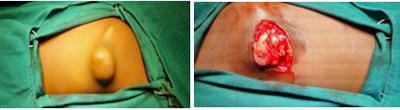
Figure 4: Intra-operative appearance of scapular osteochondroma. 

surgery_266_07.JPG
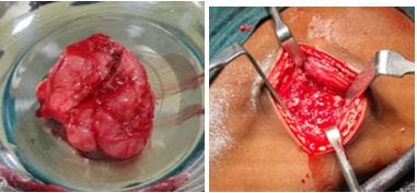
Figure 5: Pedunculated excised mass Figure 6: Post-excision local site.

Discussion

Osteochondroma of the scapula is one of the rare tumours of the thorax. It constitutes 14.4% of all tumours of the scapula with the ventral surface being the most common site of presentation[1]. Based on our review of the literature, the dorsal surface of the scapula is a rare site of origin of osteochondroma. Among the osteochondroma of the dorsal surface of the scapula, the sessile variant is more common Despite its unknown aetiology, a peripheral portion of the physis is herniate from the growth plate [2, 3]. This metaplastic cartilage grows and form exostosis, which is connected to the native bone by a thin stalk. The common age of presentation is <30 years of age, with a male-to-female ratio of 1.5:1 [4].


Our patient was a 9 years old male child. He presented with a complaint of painless bony mass [3]. Pain, if present, is mainly due to the mass effect of the tumour on surrounding tissue. A wide range of other presentations includes a decreased range of motion, underlying bursitis, nerve impingement, fracture of the stalk of the tumour, and “pseudo-winging” of the scapula [2, 4]. Snapping scapula syndrome, which is a syndrome of painful, audible, palpable abnormal scapula thoracic motion, can develop mainly with osteochondroma on the anterior surface of the scapula, especially in adolescence or early adulthood [5]. Osteochondromas of the ventral( anterior ) surface of the scapula lead to potential problems such as bursa formation, snapping syndrome, pseudo-winging of the scapula, and restricted movements of the shoulder, most of the symptoms are relieved by excision of the tumour [6, 7, 8, 9]. Solitary osteochondromas have a 3% chance of converting into osteosarcoma. This was one of the reasons why he was operated on and the tumour excised. This risk may increase up to 10% for patients with hereditary multiple exostoses [5]. Malignant transformation is characterized by a sudden increase in the size of the tumour accompanied by pain. Osteochondromas are usually easy to diagnose clinically, but confirmation is done by histopathological studies of the biopsy. Radiographic studies such as X-ray and MRI scans are essential for isolating the location of the mass and planning surgical approaches [3]. In our patient, an MRI scan of the shoulder was done and it revealed the mass was a pedunculated mushroom-like swelling arising from the dorsal aspect of the scapula along the medial border of the scapula. Magnetic resonance imaging is mainly reserved for cases in which malignancy is suspected. Histopathology of osteochondroma revealed endochondral bone connected to native bone by a thin stalk in continuity with the medullary canal of the native bone. The thickness of the cartilaginous cap seen in the biopsy specimen also is one of the predicting factors for malignant transformation [3]. A cartilaginous cap thickness of <1 cm indicates a benign condition, whereas a cap thicker than 2 cm should raise concern for malignant transformation. the biopsy of our patient revealed the thickness of the cartilaginous cap to be 5 mm, which indicates possibly benign potential. Osteochondromas usually stop growing at the time of closure of the physis and growth into adulthood should

also raise suspicion for possible malignancy [1]. The only definitive treatment of osteochondroma is the en bloc excision of the tumour [3, 4]. After narrowing down the location of the mass with the aid of an MRI scan, we planned a surgical approach parallel to the medial border of the scapula. Endoscopic resection is gaining popularity due to claims of earlier functional recovery, better results in terms of pain relief, post-operative performance, and cosmetic outcomes due to a smaller incision [4, 8]. In our patient, the incision made was only a 4 cm one and we wanted to give importance to resecting the tumour completely. The overall prognosis is good with relapse being very rare, usually occurring when tumour margins are not cleared completely and residual fragments of cartilage cap or periosteum remain following excision [1, 10]. Incomplete excisions lead to a 2% recurrence risk [10]. The decision for surgery was taken because of the increasing size since 1 year and discomfort for the patient since 8 months.

Conclusion

Solitary pedunculated osteochondromas of the scapula are rare tumours and most commonly present on the ventral surface of the scapula which may cause snapping shoulder syndrome. osteochondroma on the dorsal surface of the scapula is a rare and unusual presentation. Excision of the tumour is the treatment for solitary osteochondroma of the scapula which may reduce the risk of recurrence. For symptomatic cases, surgical excision should be done at the earliest to reduce the risk of malignant transformation.

Reference

01. Kwon OS, Kelly JI. Delayed presentation of osteochondroma on the ventral surface of the scapula. International Journal of Shoulder Surgery. 2012 Apr 1;6(2):61. [Crossref][PubMed][Google Scholar]

02. Essadki B, Moujtahid M, Lamine A, Fikry T, Essadki O, Zryouil B. Solitary osteochondroma of the limbs. Clinical review of 76 cases and pathogenic hypothesis. Acta Orthopaedica Belgica. 2000 Apr 1;66(2):146-53 [Crossref][PubMed][Google Scholar]

03. Nekkanti S, Moogali A, Meka A, Nair M. An unusual presentation of osteochondroma on the


dorsal surface of the scapula: a review of two patients. Journal of Orthopaedic Case Reports. 2018 May;8(3. [Crossref][PubMed][Google Scholar]

04. Chillemi C, Franceschini V, Ippolito G, Pasquali R, Diotallevi R, Petrozza V, Rocca CD. Osteochondroma as a cause of scapular winging in an adolescent: a case report and review of the literature. Journal of Medical Case Reports. 2013 Dec;7(1):1-5. [Crossref][PubMed][Google Scholar]

05. Fukunaga S, Futani H, Yoshiya S. Endoscopically assisted resection of a scapular osteochondroma causing snapping scapula syndrome. World Journal of Surgical Oncology. 2007 Dec;5(1):1-7. [Crossref][PubMed][Google Scholar]

06. Danielsson LG, El-haddad I. Winged scapula due to osteochondroma: Report of 3 children. Acta orthopaedica Scandinavica. 1989 Jan 1;60(6):728-9. [Crossref][PubMed][Google Scholar]

07. Okada K, Terada K, Sashi R, Hoshi N. Large bursa formation associated with osteochondroma of the scapula: a case report and review of the literature. Japanese journal of clinical oncology. 1999 Jul 1;29(7):356-60. [Crossref][PubMed][Google Scholar]

08. Mohsen MS, Moosa NK, Kumar P. Osteochondroma of the scapula associated with winging and large bursa formation. Medical Principles and Practice. 2006;15(5):387-90. [Crossref][PubMed][Google Scholar]

09. Roger P, RIET FV. Arthroscopic resection of a symptomatic snapping subscapular osteochondroma. Acta orthopaedica belgica. 2007;73:252-4. [Crossref][PubMed][Google Scholar]

10. Ceberut K, Korkmaz M, Ergin I, Müslehiddinoglu A. Bursa formation with scapular osteochondroma in hereditary multiple exostosis. J Coll Physicians Surg Pak. 2013 Jul 1;23(07):512-4. [Crossref][PubMed][Google Scholar]

11. Unni KK, Inwards CY. Dahlin's bone tumors: general aspects and data on 10,165 cases. Lippincott Williams & Wilkins; 2010. . [Crossref][PubMed][Google Scholar]

12. Rameez R, Ul-Hassan M, Kotwal HA, Kangoo KA, Nazir A. Painful pseudowinging and snapping of scapula due to subscapular osteochondroma: a case report. Journal of Orthopaedic Case Reports. 2016 Nov;6(5):96. [Crossref][PubMed][Google Scholar]

13. Sivananda P, RaO BK, Kumar PV, Ram GS. Osteochondroma of the ventral scapula causing scapular static winging and secondary rib erosion. Journal of Clinical and Diagnostic Research: JCDR. 2014 May;8(5):LD03. [Crossref][PubMed][Google Scholar]

14. Tomo H, Ito Y, Aono M, Takaoka K. Chest wall deformity associated with osteochondroma of the scapula: a case report and review of the literature. Journal of shoulder and elbow surgery. 2005 Jan 1;14(1):103-6. [Crossref][PubMed][Google Scholar]

15. Ermiş MN, Aykut ÜS, Durakbaşa MO, Özel MS, Bozkuş FS, Karakaş ES. Snapping scapula syndrome caused by subscapular osteochondroma. Joint Diseases and Related Surgery. 2012;23(1):040-3. [Crossref][PubMed][Google Scholar]

16. Fukunaga S, Futani H, Yoshiya S. Endoscopically assisted resection of a scapular osteochondroma causing snapping scapula syndrome. World Journal of Surgical Oncology. 2007 Dec;5(1):1-7. [Crossref][PubMed][Google Scholar]

17. Clarke DO, Crichlow A, Christmas M, Vaughan K, Mullings S, Neil I, Whyte N. The unusual osteochondroma: a case of snapping scapula syndrome and review of the literature. Orthopaedics & Traumatology: Surgery & Research. 2017 Dec 1;103(8):1295-8. [Crossref][PubMed][Google Scholar]

18. Jadhav PU, Banshelkikar SN, Seth BA, Goregaonkar AB. Osteochondromas at unusual sites-case series with review of literature. Journal of Orthopaedic Case Reports. 2016 Jan;6(1):52. [Crossref][PubMed][Google Scholar]

19. Frost NL, Parada SA, Manoso MW, Arrington E, Benfanti P. Scapular osteochondromas treated with surgical excision. Orthopedics. 2010 Nov 2;33(11):1-6. [Crossref][PubMed][Google Scholar]

20. Yadkikar SV, Yadkikar VS. International Journal of Medical Research & Health Sciences. . . [Crossref][PubMed][Google Scholar] [Crossref][PubMed][Google Scholar]

21. Salgia A, Biswas SK, Agarwal T, Sanghi S. A rare case presentaion of osteochondroma of scapula. Medical Journal of Dr. DY Patil University. 2013 Jul 1;6(3):338 [Crossref][PubMed][Google Scholar]