E-ISSN:2455-5436
P-ISSN:2456-9518
RNI:MPENG/2017/70870

Case Report

Pseudoaneurysms

Surgical Review: International Journal of Surgery Trauma and Orthopedics

2022 Volume 8 Number 6 November December
Publisherwww.medresearch.in

Incidental Finding of Pseudo Aneurysm of Superficial Femoral Artery and Severe Life-Threatening Bleeding from it During Revision Surgery for Failed Implant: A Case Report

Bisoniya H.1, Varma K.2*, Agarwal S.3, Sharma S.4
DOI: https://doi.org/10.17511/ijoso.2022.i06.01

1 H.S. Bisoniya, Consultant, Plastic and Vascular Surgeon, Bhopal Fracture Hospital, Bhopal, Madhya Pradesh, India.

2* K K Varma, Consultant, Orthopaedics and Director, Bhopal Fracture Hospital, Bhopal, Madhya Pradesh, India.

3 Shashank Agarwal, Consultant, Orthopaedics and Director, Bhopal Fracture Hospital, Bhopal, Madhya Pradesh, India.

4 Sandeep Sharma, Consultant, Orthopaedics and Director, Bhopal Fracture Hospital, Bhopal, Madhya Pradesh, India.

Arterial pseudoaneurysm formation is a rare entity following elective and trauma orthopaedic surgery. Pseudoaneurysms have been reported to occur following total knee arthroplasty, synovectomy and arthroscopic procedures. Here we report a case in which we encountered heavy bleeding while reoperating a case of the broken plate from the operated # shaft femur. On exploration, it was found that there was a pseudoaneurysm in the femoral artery size of rent 3mm, which probably was formed in his first surgery, which incidentally gave way and bled profusely.

Keywords: Pseudoaneurysms, synovectomy, arthroscopic procedures, heavy bleeding, reoperation

Corresponding Author How to Cite this Article To Browse
K K Varma, Consultant, Orthopaedics and Director, Bhopal Fracture Hospital, Bhopal, Madhya Pradesh, India.
Email:
H.S. Bisoniya, K K Varma, Shashank Agarwal, Sandeep Sharma, Incidental Finding of Pseudo Aneurysm of Superficial Femoral Artery and Severe Life-Threatening Bleeding from it During Revision Surgery for Failed Implant: A Case Report. Surgical Rev Int J Surg Trauma Orthoped. 2022;8(6):29-34.
Available From
https://surgical.medresearch.in/index.php/ijoso/article/view/263

Manuscript Received Review Round 1 Review Round 2 Review Round 3 Accepted
2022-09-28 2022-09-30 2022-10-07 2022-10-14 2022-10-21
Conflict of Interest Funding Ethical Approval Plagiarism X-checker Note
Nil Nil Yes 16%

© 2022by H.S. Bisoniya, K K Varma, Shashank Agarwal, Sandeep Sharmaand Published by Siddharth Health Research and Social Welfare Society. This is an Open Access article licensed under a Creative Commons Attribution 4.0 International License https://creativecommons.org/licenses/by/4.0/ unported [CC BY 4.0].

Introduction

Unexpected vascular findings during orthopaedic surgery, do occur and if not addressed may prove fatal. Most vascular complications occur intraoperatively, immediately postoperatively or in the late postoperative period; they most commonly include lacerations, pseudoaneurysms, thrombosis, and arteriovenous fistulas. The surgical approach to repair has varied based on anatomy, the accuracy of diagnosis, infection, and available inventory. [1-3]

A pseudoaneurysm is a pulsatile hematoma that communicates with an artery through a disruption in the arterial wall. Femoral pseudoaneurysm is a rare complication. Pseudoaneurysms are associated with the characteristic findings of a pulsatile mass, a palpable thrill, and an audible to-and-fro murmur. The diagnosis is made by a colour doppler and confirmed by CT angiography. If the pseudoaneurysm is deep-seated and small, the patient does not have any symptoms and it can be missed preoperatively. [2-4]

Case report

A 48-year-old male patient presented to the accident & emergency department on 24th November 21, with pain and deformity over the left thigh for 2 days. He had a history of twisting his leg two days back which resulted in the broken implant. He was an old case of distal femur platting in July 2021. Patient also is a known case of PPRP (Post Polio Residual Paresis) left leg. A clinical examination was done and lab investigations were seen. Both were normal and premedical and pre-anaesthetic check-up was also done. The patient was planned to be taken for surgery on 26/11/21 for exchange platting with bone grafting.

surgery_263_01.JPG
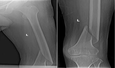
Figure 1: X-Ray of the patient after the accident


surgery_263_02.JPG

Fig 2: X-ray of the patient after the operation


surgery_263_03.JPG

Fig 3: X ray of broken implant


surgery_263_04.JPG

Fig 4: Perforation visible in the superficial formal artery while exploration of pseudoaneurysm, which was the source of bleeding


surgery_263_05.JPG
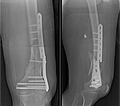
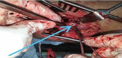
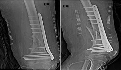
Fig 5: X-ray of the patient after revision surgery

Intraoperatively, while removing the distal part of the broken implant, there was profuse bleeding from the medial side of the bone. As the vascular surgeon was not available in the operation theatre at that time and the bleeding was profuse, we were not able to make out the source of the bleeding, it became a life-threatening condition, so immediately just proximal to the wound tourniquet was applied and the vascular surgeon was called. This proved to be a wise step as the bleeding was minimized and packs were applied tightly till the vascular surgeon arrived.

During the exploration of femoral vessels from the medial aspect of the thigh, it was seen that there was an old perforation in the lateral aspect of the superficial femoral artery and large clot formation. There was a pseudoaneurysm due to a previous injury. The wound was thoroughly explored and the clot was removed. Perforation of the femoral artery was repaired with prolene 6-0 and the wound closed in layers. The femur bone was then stabilised with a plate after the removal of the old implant.

The postoperative course was uneventful and managed with i.v. antibiotics, analgesics and 2 units of blood transfusion. The patient was haemodynamically stable and vitals remained normal throughout. His neurovascular status was normal.

At six months follow-up, there was no sign of recurrence of infection or the pseudoaneurysm.

Discussion

Pseudoaneurysm formation is often secondary to trauma, leading to focal disruption of the arterial vessel wall. As a consequence, blood flow continues to be maintained and both patient and surgeon are initially unaware of vessel injury.

Extravasation of blood through the vessel wall leads to the formation of a pulsating haematoma, the cavity of which is still in direct continuity with the lumen of the artery. The clot liquefies and the pseudoaneurysm is subsequently contained by a fibrous capsule rather than the layers of the vessel wall itself. Unless action is taken to repair the pseudoaneurysm, it will progressively enlarge eroding surrounding structures or will rupture. As a consequence, the presentation can vary from asymptomatic presentation to bruising, swelling, neurological symptoms (due to nerve compression) and even death.

Duplex doppler ultrasonography is the imaging modality of choice if there is clinical doubt regarding the diagnosis between a haematoma or pseudoaneurysm, though a better diagnostic modality is CT angiography. The definitive diagnosis is made by the identification of the connection between the neck of the aneurysm and the injured artery.

In our case, a clinical diagnosis of pseudoaneurysm formation was missed as there were no symptoms and signs of swelling with thrill probably because it was deep-seated and small. Distal pulsation was normal (DP/PT+) so we didn’t suspect any vascular injury but during implant, removal pseudoaneurysm got burst and resulted in severe uncontrolled bleeding.

The overall aim of the article is to make the operating surgeon aware of the potential vascular complications and to be ready to manage them properly if anything happens.

However, it is suggested that the surgeon should be sufficiently acquainted with the exposure of the main vessels above and below the groin to be able to control life-threatening hemorrhage at all times. Surgeons operating both in a hospital and an ambulatory surgical setting should be familiar with techniques to gain proximal control of massive bleeding because the principles can be helpful in primary and revision surgeries.

Kim JW et al presented a case report on pseudoaneurysm of the deep femoral artery caused by a guide wire following a femur intertrochanteric fracture with a hip nail. An 85-year-old woman developed severe swelling and pain in the proximal thigh after internal fixation of an intertrochanteric fracture of the femur with a hip nail.


After reviewing all possible causes, the authors found a mistake in the insertion of a guide wire for the hip nail. Using intraoperative fluoroscopic images, the mal-positioned guide wire was found located posterior to the trochanter on the lateral view of the hip. This case study reminds us that pseudoaneurysm can occur in a guide wire during hip nailing. Surgeons can avoid this complication with confirmation of lateral and anteroposterior views of the hip.

Shaw et al reported two case reports and a review of the literature on geniculate arterial pseudoaneurysm formation following trauma and elective orthopaedic surgery to the knee. Management of pseudoaneurysms around the knee is varied and adjusted according to the size and location of the pseudoaneurysm. Previously, open surgery would have been performed by gaining proximal and distal vascular control of the pseudoaneurysm, evacuating the thrombus and, if the artery was small and deemed non-essential can be ligated, or if essential can be repaired by primary anastomosis or patch repair by insertion of autologous vein graft.[3].

A similar observation was seen by Preuss FR et al in which they observed two cases of delayed vascular complications after orthopaedic surgery. Theoretically, minimally invasive procedures with drilling near vessels increase the chances of vascular injury that goes unnoticed during the index procedure. This report of two cases discusses the diagnosis and repair of pseudoaneurysm and AVF. It is a cautionary warning to consider these vascular injuries as late-presenting, limb-threatening complications of orthopaedic surgery.[4].

Dhal A encountered thirteen pseudoaneurysms of which five were caused as a complication of pin/wire placement of external fixators (one conventional and four Ilizarov frames). The most common symptom was the presence of profuse persistent bleeding from either the wound or the pin/wire site. In nine patients the pseudoaneurysm was directly caused by a fracture or subsequent fracture stabilisation. A high index of suspicion needs to be maintained following penetrating injury in the vicinity of a major vessel, particularly in the presence of persisting symptoms. External fixators are widely used to treat complex orthopaedic problems.

The procedure is technically demanding, requiring a sound knowledge of cross-sectional limb anatomy. Injudicious use may result in potentially life/limb-threatening complications of pseudoaneurysm.[5].

Inamdar D et al report a pseudoaneurysm of the anterior tibial artery caused by a proximal interlocking screw after intramedullary nailing surgery to repair a tibial shaft fracture. The patient experienced complete relief of symptoms following the removal of the nail and the screws, excision of the proximal fibula, resection of the pseudoaneurysm, and ligation of the anterior tibial artery. The authors recommend the oblique placement of the proximal interlocking screws to prevent this rare complication. The management of femoral shaft fractures by retrograde intramedullary nailing is becoming more widespread. Coupe KJ et al presented a case report of injury to a branch of the profunda femoris artery. Toyota T et al presented a similar case of femoral pseudoaneurysm after surgery for an intertrochanteric fracture. In the field of orthopaedic surgery, vascular injury is not rare and not only occurs secondary to fracture but also includes iatrogenic injury. [6-8].

Chan WS et al report the case of an 83-year-old woman who developed a pseudoaneurysm of the profunda femoris artery after dynamic hip screw fixation for an intertrochanteric femoral fracture. 23 days after the fixation, radiological investigations including colour Doppler ultrasonography and computed tomographic angiography identified a pseudoaneurysm surrounded by a large intramuscular haematoma close to the profunda femoris. The patient underwent emergency evacuation and was under intensive care for 3 days and was discharged 6 weeks later, with no complications. At the one-year follow-up, the wound and fracture had healed, but the patient was confined to a wheelchair. A high index of clinical suspicion and radiological imaging is necessary for making the diagnosis.[9].

Pseudoaneurysm of the femoral artery after internal fixation of a trochanteric fracture is caused primarily by overpenetration of the drill bit or screws. Yang KH et al report a case of pseudoaneurysm of the superficial femoral artery after the insertion of a Gamma nail. A probable cause for this injury is adduction and internal rotation of the involved limb.


During the distal interlocking procedure, this maneuver endangers the superficial femoral artery by abutment of the femoral vessels to the femur. It is recommended that the limb be placed in a neutral position during the preparation of the interlocking hole.[10].

Kragh Jr JF studied the practical use of emergency tourniquets to stop bleeding in major limb trauma. The purpose of this study was to measure tourniquet use and complications. They identified potential morbidities from the literature and looked for them prospectively. Morbidity risk was low, and there was a positive risk-benefit ratio in light of the survival benefit. No limbs were lost because of tourniquet use, and tourniquet duration was not associated with increased morbidity. Education for early tourniquet use should continue.[11].

Kronzon I et al did a review on the diagnosis and treatment of iatrogenic femoral artery pseudoaneurysm. Small (less than 2 cm) femoral pseudoaneurysms clot spontaneously and usually require no treatment. Larger femoral pseudoaneurysms may lead to complications including rupture and compression of the adjacent femoral vein (with resulting venous thrombosis) or of the femoral nerve. Treatment may be surgical. However, recently it has been shown that direct, noninvasive compression of the pseudoaneurysm stops the blood flow in the communication and leads to pseudoaneurysm clotting and obliteration.[12].

Conclusion

Traumatic pseudoaneurysm is a typical orthopaedic vascular complication. Pseudoaneurysm has been reported after orthopaedic surgery at various sites, including the foot, tibial diaphysis, knee joint, hip joint, shoulder joint, and lumbar spine. Depending on the site and size of the pseudoaneurysm management consisted of either ligation, resection and end-to-end anastomosis/vein grafting, lateral suture, endoaneurysmorrhaphy or selective embolisation. A high index of suspicion needs to be maintained following penetrating injury in the vicinity of a major vessel, particularly in the presence of persisting symptoms. A high index of clinical suspicion and radiological imaging is necessary for making the diagnosis.


In all the above-mentioned studies, the patient had symptoms and so pseudoaneurysm was diagnosed preoperatively or postoperatively (not intraoperative) as was it in our care. We suggest if there is swelling in the limb, out in absence of thrill and normal distal pulsation a doppler study should be done to rule out any symptomatic and vascular injury.

Reference

01. Rossi G, Mavrogenis A, Angelini A, Rimondi E, Battaglia M, Ruggieri P. Vascular complications in orthopaedic surgery. J Long Term Eff Med Implants. 2011;21(2):127-37. doi: 10.1615/jlongtermeffmedimplants.v21.i2.30 [Crossref][PubMed][Google Scholar]

02. Kim JW, Lee JI, Park KC. Pseudoaneurysm of the deep femoral artery caused by a guide wire following femur intertrochanteric fracture with a hip nail: A case report. Acta Orthop Traumatol Turc. 2017 May;51(3):266-269. doi: 10.1016/j.aott.2017.03.011 [Crossref][PubMed][Google Scholar]

03. Shaw A, Stephen A, Lund J, Bungay P, Denunzio M. Geniculate arterial pseudoaneurysm formation following trauma and elective orthopaedic surgery to the knee: 2 case reports and a review of the literature. J Radiol Case Rep. 2009;3(3):12-6. doi: 10.3941/jrcr.v3i3.42 [Crossref][PubMed][Google Scholar]

04. Preuss, Fletcher R. , Michael M. Hadeed, and David B. Weiss. Delayed vascular complications after orthopaedic surgery: A report of two cases and review of the literature." Journal of Orthopaedics, Trauma and Rehabilitation 26.1 (2019): 48-51 [Crossref][PubMed][Google Scholar]

05. Dhal A, Chadha M, Lal H, Singh T, Tyagi S. Encounters with pseudoaneurysms in orthopaedic practice. Injury. 2001 Dec;32(10):771-8. doi: 10.1016/s0020-1383(01)00094-8 [Crossref][PubMed][Google Scholar]

06. Inamdar D, Alagappan M, Shyam L, Devadoss S, Devadoss A. Pseudoaneurysm of anterior tibial artery following tibial nailing: a case report. J Orthop Surg (Hong Kong). 2005 Aug;13(2):186-9. doi: 10.1177/230949900501300216 [Crossref][PubMed][Google Scholar]


07. Coupe KJ, Beaver RL. Arterial injury during retrograde femoral nailing: a case report of injury to a branch of the profunda femoris artery. J Orthop Trauma. 2001 Feb;15(2):140-3. doi: 10.1097/00005131-200102000-00013 [Crossref][PubMed][Google Scholar]

08. Toyota T, Horiuchi H, Takizawa T, Yamazaki I, Matsunaga D, Nakamura Y, et al. A case of femoral pseudoaneurysm after surgery for intertrochanteric fracture. J Orthop Sci. 2017 Mar;22(2):362-365. doi: 10.1016/j.jos.2015.07.003 [Crossref][PubMed][Google Scholar]

09. Chan WS, Kong SW, Sun KW, Tsang PK, Chow HL. Pseudoaneurysm and intramuscular haematoma after dynamic hip screw fixation for intertrochanteric femoral fracture: a case report. J Orthop Surg (Hong Kong). 2010 Aug;18(2):244-7. doi: 10.1177/230949901001800222 [Crossref][PubMed][Google Scholar]

10. Yang KH, Park HW, Park SJ. Pseudoaneurysm of the superficial femoral artery after closed hip nailing with a Gamma nail: report of a case. J Orthop Trauma. 2002 Feb;16(2):124-7. doi: 10.1097/00005131-200202000-00009 [Crossref][PubMed][Google Scholar]

11. Kragh JF Jr, Walters TJ, Baer DG, Fox CJ, Wade CE, Salinas J, et al. Practical use of emergency tourniquets to stop bleeding in major limb trauma. J Trauma. 2008 Feb;64(2 Suppl):S38-49; discussion S49-50. doi: 10.1097/TA.0b013e31816086b1 [Crossref][PubMed][Google Scholar]

12. Kronzon I. Diagnosis and treatment of iatrogenic femoral artery pseudoaneurysm: a review. J Am Soc Echocardiogr. 1997 Apr;10(3):236-45. doi: 10.1016/s0894-7317(97)70061-0. Erratum in: J Am Soc Echocardiogr 1997 Jun;10(5):30A-31 [Crossref][PubMed][Google Scholar]