E-ISSN:2455-5436
P-ISSN:2456-9518
RNI:MPENG/2017/70870

Research Article

Intestinal Obstruction

Surgical Review: International Journal of Surgery Trauma and Orthopedics

2021 Volume 7 Number 5 Sep-Oct
Publisherwww.medresearch.in

Clinical study of miscellaneous causes of intestinal Obstruction

Chavan A.1, Keniya E.2*, Tamboli A.3, Degaonkar A.4, Kelkar V.5
DOI: https://doi.org/10.17511/ijoso.2021.i05.01

1 Aashish R Chavan, Assistant Professor, Department of General Surgery, Dr Shankarrao Chavan Government Medical College & Hospital, Nanded, Maharashtra, India.

2* Eshita B Keniya, Resident, Department of General Surgery, Dr Shankarrao Chavan Government Medical College & Hospital, Nanded, Maharashtra, India.

3 Abdul R Tamboli, Assistant Professor, Department of General Surgery, Dr Shankarrao Chavan Government Medical College & Hospital, Nanded, Maharashtra, India.

4 Anil Degaonkar, Associate Professor, Department of General Surgery, Dr Shankarrao Chavan Government Medical College & Hospital, Nanded, Maharashtra, India.

5 Vidyadhar Kelkar, Associate Professor, Department of General Surgery, Dr Shankarrao Chavan Government Medical College & Hospital, Nanded, Maharashtra, India.

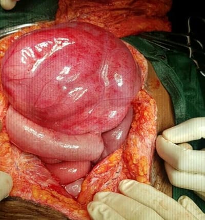
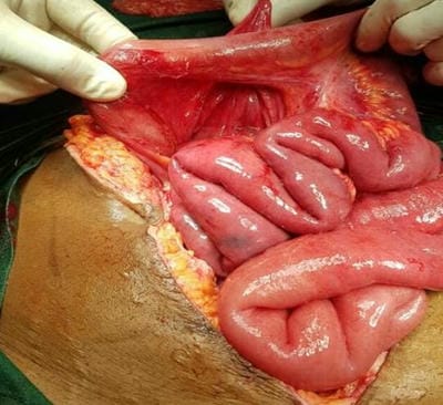
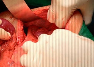
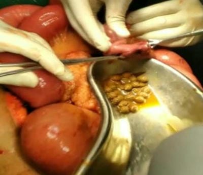
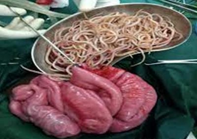
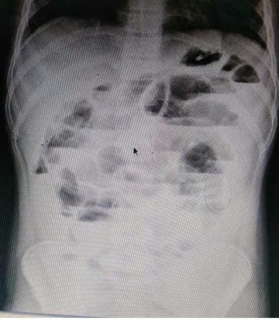
Aims and Objectives: Preoperative diagnosis of intestinal obstruction and timely intervention to reduce postoperative morbidity and mortality. Methods: A retrospective study of 124 cases of intestinal Obstruction from June 2018 to November 2020 by examining previous clinical records in terms of Clinical presentations, Laboratory investigations, Radiological investigations, Operative procedures, and Postoperative outcomes was done. Out of 124 cases, 40 cases were managed conservatively and were excluded from the study. Results: Out of 124 cases, 40 were managed conservatively, 30 with Adhesive Obstruction,20 with malignant growth, 14 with Obstructed External hernias and 20 with miscellaneous causes. There were 20 cases of rare causes of intestinal Obstruction, namely 2 Para duodenal hernia, two foreign body(phytobezoars) obstruction of ileum, 1 - Obstruction by worm infestation, 6 cases of volvulus, one gall stone ileus, 1meconium plug syndrome, and, 1 case of obstructed obturator hernia,1 case of Meckel's Diverticulum with Ileocolic Intussusception, five patients with the varied presentation of abdominal Koch's Out of 20 unusual causes, 17 cases were correctly diagnosed preoperatively by radiological investigations. 4 cases were managed with Resection and Anastomosis, five unusual cases presented with gangrenous changes and 2 cases needed ileostomy. Conclusion: These rare cases can be diagnosed early by a thorough history and clinical evaluation, and appropriate Radiological investigations to prevent hazardous gangrenous changes and prevent morbidity and mortality in patients.

Keywords: Intestinal Obstruction, Mortality, Morbidity, Radiological, Miscellaneous

Corresponding Author How to Cite this Article To Browse
Eshita B Keniya, Resident, Department of General Surgery, Dr Shankarrao Chavan Government Medical College & Hospital, Nanded, Maharashtra, India.
Email:
Aashish R Chavan et al: Clinical study of miscellaneous causes of intestinal Obstruction. Surgical Rev Int J Surg Trauma Orthoped. 2021;7(5):91-99.
Available From
https://surgical.medresearch.in/index.php/ijoso/article/view/247

Manuscript Received Review Round 1 Review Round 2 Review Round 3 Accepted
2021-09-10 2021-09-13 2021-09-20 2021-09-27 2021-10-03
Conflict of Interest Funding Ethical Approval Plagiarism X-checker Note
Nil Nil Yes 17.2%

© 2021by Aashish R Chavan, Eshita B Keniya, Abdul R Tamboli, Anil Degaonkar, Vidyadhar Kelkarand Published by Siddharth Health Research and Social Welfare Society. This is an Open Access article licensed under a Creative Commons Attribution 4.0 International License https://creativecommons.org/licenses/by/4.0/ unported [CC BY 4.0].

Introduction

Acute intestinal obstruction is one of the most common surgical emergencies. Acute intestinal obstruction is one of the most common surgical emergencies. It involves a partial or complete blockage of the bowel, which induces mechanical impairment or complete arrest of content passage through the intestine. Small Bowel Obstruction is mainly of two types, Mechanical and functional obstruction. Mechanical obstruction means that luminal contents cannot pass through the gut tube because the lumen is physically blocked or obstructed. In contrast, functional obstruction means that luminal contents fail to pass because of disturbances in gut motility that prevent coordinated transit from one region of the gut to the next. This form is commonly referred to as as ileus or pseudo-obstruction.[1]. Large Bowel Obstruction can result from either mechanical interruption of the flow of intestinal contents or by the dilation of the colon in the absence of an anatomic lesion.

Miscellaneous causes of bowel obstruction account for 2% to 3% of all cases but should be considered in the differential diagnosis. These include intussusception of the bowel, which in the adult is usually secondary to a pathologic lead point, such as a polyp or tumour; gallstones, which can enter the intestinal lumen by a cholecystoenteric fistula and cause obstruction; enteroliths originating from jejunal diverticula; foreign bodies; and phytobezoars.

Intussusception is a unique type of obstruction that results from the invagination of a segment of the bowel into another. [2]. Volvulus (an axial twist of the gastrointestinal tract around its mesentery),[3]. Gallstone ileus (mechanical bowel obstruction), Adhesions [4,5]. (postoperative or postinflammatory), hernias, worm obstruction due to Ascaris lumbricoides are frequent causes of intestinal obstruction. Sigmoid volvulus, caecal volvulus are also potential causes of this disorder. Indigestible foreign material in the bowel can develop into bezoars. Intestinal obstruction can occur secondary to these bezoars. Phytobezoars specifically refer to those which accumulate as a result of undigested plant or other food material.

surgery_247_01.JPG

Figure-1 Paraduodenal Hernial sac before reduction

surgery_247_02.JPG

Figure-2 Paraduodenal Hernial sac after reduction

surgery_247_03.JPG

Figure-3 Obstructed Obturator Hernia


Symptoms of intestinal Obstruction include abdominal pain (colicky), vomiting, abdominal distension and obstipation (failure to pass flatus and faeces).[6]. Visible peristalsis may be seen in thin patients, while in others, distention may be prominent. Complete obstruction typically is treated with immediate surgery, while partial obstruction seldom requires surgery. Patients with a partial bowel obstruction may be treated conservatively with resuscitation and tube decompression alone.  In some uncommon obstructions, the purpose is to restore normal anatomical conditions post-operatively. Many Indian studies demonstrated that the pattern of intestinal obstruction differs from the Western world.

Therefore, a similar study has been undertaken

  • To identify the various causes of acute intestinal obstruction.
  • To study the clinical profile and appropriate intervention adopted for various causes of intestinal obstruction.
  • To determine morbidity and mortality rate.

Materials and Methods

Setting and Duration: - Patients that attended the outpatient department and the casualty and those who got admitted to the surgical wards of our hospital in 18 months were included in this study.

Type of Study: - A retrospective study of 124 cases diagnosed with intestinal Obstruction from June 2018 to November 2020 was done.

Inclusion Criteria:

  1. Age: Day 1 – 90 years of age
  2. Patients with acute intestinal obstruction who have undergone operative management are included in this study.
  3. The identified cause of intestinal obstruction has an overall incidence of less than 5 percent

Exclusion criteria –

  1. Patients who refused surgical intervention were excluded
  2. Patients who were treated conservatively for subacute intestinal obstruction.
  3. Common causes of intestinal Obstruction like Adhesions, Hernia, Malignancy were excluded

Data Collection Procedure: - included- a detailed record of the patient's history, physical examination, and necessary investigations like baseline, X-Ray abdomen erect and supine in all cases, ultrasound abdomen, CT Abdomen were recorded based on the requirement for each subject. A proforma was recorded of each patient with age, sex, symptom duration, past surgical & medical history, diagnostic workup, etiology of obstruction, the time between arrival and operation, operative information, comorbid factors, morbidity and mortality, length of hospital stay, and the outcome of the patients.

surgery_247_04.JPG

Figure-4 obstruction secondary to Indian Plum Seeds

Analysis: - Postoperative follow up after the discharge of patients was done in the majority of the patients up to 3 months. The results are tabulated mostly stressing on following points - age, etiology, operative management, causes of mortality, postoperative complications adopted. All the information was collected using a structured proforma. Institutional Ethical Committee Approval was taken.

Results

Out of 124 cases, 40 managed conservatively, 30 with Adhesive Obstruction, 20 with malignant growth, 14 with Obstructed External hernias and 20 with miscellaneous causes. Out of 20 unusual reasons, 17 cases were correctly diagnosed by radiological investigations preoperatively. All patients were operated on by laparotomy. All 20 cases were followed up Post Operatively


Table 1. Various Cases of Intestinal Obstruction In Study

Etiology No.Of Patients
Tuberculous Abdomen (Figure-6) 5
Volvulus:Midgut (Ileal ) (Figure 5 a &b ) 2
Volvulus:Transverse Colon (Figure -7) 1
Volvulus:Sigmoid 3
Paraduodenal Hernia        (Figure 1,2) 2
Obstructed Right Obturator Hernia (Figure 3) 1
Gall stone ileus 1
Meconium ileus 1
Meckel”s Diverticulum  with Ileo-ileal intussusceptions 1
Foreign Body Obstruction-Phytobezoars (Figure 4) 2
Foreign Body Obstruction-Worm Infestation (Ascaris L.) (Figure 8) 1

surgery_247_05_a.JPG

Figure -5 (a) Mid gut volvulus

surgery_247_05_b.JPG

Figure -5 (b) Midgut Volvulus with Gangrenous Ileal segment

SEX: Out of the 20 patients, 60% (12) patients were Males, and 40% (08) patients were females.  Male to Female ratio is 1.5: 1

Table 2. Distrubution As Per Sex

Sex No. of patients Percentage
Male 12 60.00%
Female 8 40.00%
Total 20 100.00%

Age: The Majority of patients were in the age group of 21-30 years and 61-70 years.

Table 3. Distrubution As Per Age

Age No. of patients Percentage
0 to 10 1 5.00%
11 to 20 3 15.00%
21 to 30 4 20.00%
31 to 40 3 15.00%
41 to 50 1 5.00%
51 to 60 2 10.00%
61 to 70 4 20.00%
71 to 80 1 5.00%
81 to 90 1 5.00%
Total 20 100.00%

Bowel Involved: Out of 20 cases, majority involved the Small Bowel 75% patients-15 cases, and the Large bowel was affected in 25% patients- 5 cases.

Table 4. Distribution As Per Bowel Involved

  No. of patients Percentage
Large Bowel 5 25.00%
Small Bowel 15 75.00%
Total 20 100.00%

Signs and Symptoms: Out of 20 patients, the majority had pain, tenderness, fever, vomiting. Of the total patients, tenderness was a significant sign.

Table 5. Distribution As Per Presenting Symptoms And Signs

Signs And Symptoms No. of patients Percentage
Pain in abdomen 19 95.00%
Vomiting 18 90.00%
Fever 9 45.00%
Pallor 8 40.00%
Tenderness 1 95.00%
Guarding 6 30.00%
Rigidity 0 0.00%

Duration of Pain: of the total patients, 95%(19) patients complained of pain while 5%(1) patients had no pain. Duration of pain was for up to 2 days in 25% (5) patients, and 70% (14) patients complained of pain for more than two days.


Table 6. Distribution As Per Duration Of Pain

Duration of Pain No. of patients Percentage
<2 Days 5 25.00%
>2 days 14 70.00%
No pain 1 5.00%
Total 20 100.00%

Radiological Findings: Out of 20 patients, x-ray abdomen standing was done in all patients. Multiple air-fluid levels were seen in 70% (14) patients, the Dilated colon was seen in 25%(05)patients. Normal Xray Abdomen was seen in 1 patient.

Table 7. Distribution As Per Radiological Findings

Radiological findings No. of patients Percentage
Multiple Air fluid levels (Figure 9) 14 70.00%
Dilated colon 5 25.00%
Normal 1 5.00%
Total 20 100.00%

Operative Management: All patients were managed with Emergency exploratory Laparotomy. Enterotomy was done in 5 patients (25%). Second most common management done was Resection anastomosis (4 cases-20%) followed by Reduction and Repair of Hernia and Hemicolectomy done in 3 patients each(15%) and ileostomy done in 2 patients(10%). Colostomy done in 1 patient(5%)(16.67%) followed by derotation of volvulus in 1 patient(5%) and Sigmoidectomy in 1 (5%). Gangrenous changes were seen in 5 cases (25%) presenting with obstruction due to various causes.

surgery_247_06.JPG

Figure-6 Coccon Abdomen in A Case of Abdominal Koch's

surgery_247_07.JPG

Figure -7 Transverse Colon Volvulus

surgery_247_08.JPG

Figure 8. Foreign Body Obstruction-Worm Infestation (Ascaris L.)

Post–Operative Complications: Out of patients who had postoperative complications, the most common was fever in 6 patients (30%), followed by Pulmonary Complications in 20% (4)Patients and wound gaping in 15% (3) patients. Prolonged ileus was seen in 10% (2) patients. More severe complications like burst abdomen occurred in 5% (1) patients, faecal fistula in 5% (1) patients. It found that patients with tuberculous obstruction are more prone to complications.

Table 8. Distribution As Per Operative Management

  No. of patients Percentage
Reduction and repair of Hernia 3 15.00%
Enterotomy 5 25.00%
Resection and anastomosis 4 20.00%
Derotation of volvulus 1 5.00%
Hemicolectomy 3 15.00%
Sigmoidectomy 1 5.00%
Illiostomy 2 10.00%
Colostomy 1 5.00%
Total 20 100.00%

surgery_247_09.JPG

Figure 9: Multiple Air Fluid Levels seen on Xray Abdomen Erect in a patient with Small Bowel obstruction

Mortality: Out of 20 patients, death occurred in 4 patients (20%), three patients were of Tuberculous Abdomen (15%), one was of Transverse Colon volvulus (5%). All four patients died due to septicaemia, of which one also developed Pulmonary complications (Pleural effusion).

Table 9. Distribution As Per Postoperative Complications

  No. of patients Percentage
Wound Gaping 3 15.00%
Fever 6 30.00%
Prolonged Illness 2 10.00%
Burst Abdomen 1 5.00%
Septisemia(Death) 4 20.00%
No complications 6 30.00%
Pulmonary Complications 4 20.00%
Faecal fistula 1 5.00%
Total 20 100.00%

Discussion

Acute intestinal obstruction is one of the most common causes that require admission in the hospital emergency surgical departments.  The overall mortality and morbidity of bowel obstruction are substantial. [7,1].

Brewer et al. analysed 1000 consecutive abdominal surgeries and reported an incidence of 2.5%.[8]. The present study showed an increased incidence of intestinal obstruction between 61 – 70 years of age. Similar results are displayed by Playford,9 G. J.Cole,[10]. S.S. Gill.[6]. In the present study, one of the leading causes of intestinal obstruction was tuberculous obstruction in 25 % of patients. The other being volvulus in 30% of patients, Obstructed internal hernia in 15% of patients, foreign body causing intraluminal obstruction in 15% of patients (Phytobezoars in 10%, worm infestation in 5% patients), gall stone ileus in 5% of patients, Meconium ileus in 5% patients, Meckel's diverticulum with ileo-ileal intussusceptions in 5% patients. In volvulus, sigmoid volvulus occurs in 50% of patients, while illeal volvulus occurs in 33% of patients and transverse colon volvulus in 16.67% of patients. Other authors observed similar findings. Arshad M Malik in 2010 studied 229 patients and revealed the exact cause of intestinal tuberculosis as the emerging cause of obstruction identical to our study [12]. Abdominal tuberculosis is a most common type of extrapulmonary tuberculosis, comprising of gastrointestinal tuberculosis, peritoneum, omentum, mesentery and its lymph nodes and other abdominal organs such as liver, spleen and pancreas. Extrapulmonary tuberculosis involves 11-16% of all patients with tuberculosis, of which 3-4% belongs to abdominal tuberculosis [13].

Internal hernias are an uncommon cause of intestinal obstruction and occur when abdominal contents are trapped within a compartment of the abdominal cavity. It is a rare condition with an incidence of < 1% of all cases of bowel obstruction and up to 5.8% of all cases of small bowel obstruction [14,15,16]. The overall male/female sex ratio for internal hernia is approximately three [17]. Para duodenal hernia constitutes 53% of all cases of internal hernias, of which 40% and 13% are of the left and right Para duodenal hernias, respectively [18,19].

In our study, the majority of patients had symptoms of pain (95%), tenderness (95%), vomiting (90%) and fever (45%). However, symptoms of guarding, absent bowel sounds, and palpable mass were seen in fewer subjects. Other authors observed similar findings. In a study by Rehman, the commonest


symptoms were abdominal pain 54 (100%), abdominal distension 49 (90%), vomiting 42 (78%), absolute constipation 37 (68.5%), dehydration 33 (61%), fever 16 (29.6%), mass right iliac fossa 8 (15%), inguinoscrotal swelling 10 (18%).[20]. In a study by Madziga, abdominal pain 88.7%, vomiting 84.8%, and constipation 78.8% were the main symptoms, while tenderness and abdominal masses were common signs.[21]. In a prospective study by Haridimos M et al. in 150 patients, the absence of passage of flatus (90%) and faeces (80.6%) and abdominal distension (65.3%) were the most common symptoms and physical findings, respectively.[22]. The above findings indicate that the clinical profile of Indian patients with intestinal obstruction is the same as that of others.

In a study by Rehman, the commonest etiology were adhesions and bands 23 (42.5%), intestinal tuberculous 13 (24.07%), stricture + ileocecal mass, hernias 10 (18.51%), tumours 03 (5.5%), worm infestations 03 (5.5%), intussusception 02 (3.7%).20 25% underwent enterotomy,20% underwent resection anastomosis, 15% of patients were managed with hemicolectomy, and the same numbers were managed with Reduction and repair of internal hernia, 10% with ileostomy, 5% with derotation of volvulus,5% with colostomy and 5% managed with Sigmoidectomy. The pattern of operative treatment in our study reflects the standard approach to managing patients with intestinal obstruction. In our study, mortality was 20%; three patients had tuberculous obstruction, and 1 had transverse colon volvulus. All of them died due to septicemia. It shows that the mortality in our study is less than that reported by the other studies. In a study by Pal et al., in 150 episodes of mechanical intestinal obstruction, operative mortality was 28%. The highest mortality rate (41.3%) was found in volvulus.[23]. In a retrospective study by Mohamed et al., the mortality rate was 3.5%. In a study by Madziga, the mortality rate was 9.14%.[22]. The study by Chaib E et al. showed operative mortality of 9.09%.[24].

The present study showed that 30% of patients had no complications; however, fever was a postoperative complication in 30% of patients. In a study by Pal et al., the major adverse factors were gangrenous bowel and significant bowel obstruction. [23]. In a survey by Chaib E

et al., complications occurred in 15.7% of patients following operative intervention; wound infection was the most common postoperative complication. [24]. Similar results are shown in the study done by Madziga. [21]. The present study results are in line with published literature, indicating that the sign and symptoms in Indians are more or less as that of the West. The aetiology of acute intestinal obstruction matches that of West. The mortality in our study is less as compared to published literature. For affected patients, high-quality surgical expertise coupled with sound clinical judgment and early surgery when needed will significantly improve survival. Morbidity and mortality associated with intestinal obstruction have declined since the advent of the most sophisticated diagnostic methods, but the condition remains a challenging surgical diagnosis.[25]

Conclusion

It is essential to consider these various causes of intestinal obstruction as treatable causes of obstruction. Intestinal tuberculosis assumes great importance in this part of the country as a cause for obstruction. These multiple causes can be diagnosed early by thorough history taking and clinical evaluation, and appropriate radiological investigations to prevent hazardous gangrenous changes and prevent morbidity and mortality in patients. A high index of suspicion is required in diagnosing patients with these various causes.

Author's Contribution :

  1. Conception or design of the work.: Dr.Anil S Degaonkar4. Dr Aashish R Chavan1. Dr.Keniya Eshita B2 Dr. Vidyadhar Kelkar5
  2. Data collection.: Dr. Keniya Eshita B 2
  3. Data analysis and interpretation: Dr.Keniya Eshita B2
  4. Drafting the article: Dr.Keniya Eshita B2
  5. Critical revision of the article: Dr Aashish R Chavan1.  Dr Abdul R Tamboli3 Dr.Anil S Degaonkar.4
  6. Final approval of the version to be published: Dr Aashish R Chavan1. Dr.Anil S Degaonkar4.  Dr.Vidyadhar Kelkar,5Dr.Keniya Eshita B2


What does the study add to the existing knowledge?

The current study adds to the previous studies and concludes that prompt diagnosis and early management is  key  to  favourable treatment outcomes. Results of our study strongly concur with the existing knowledge  of  intestinal obstruction  in  particular its miscellaneous causes  in  several  of  its  factors.

Reference

01. Sakorafas GH, Poggio JL, Dervenis C, Sarr MG: Small bowel obstruction. Shackelford's surgery of the alimentary tract. W. B. Saunder's Company 2002;5 edition;317-341 [Crossref][PubMed][Google Scholar]

02. Ein SH. Leading points in childhood intussusception. J Pediatr Surg. 1976 Apr;11(2):209-11. doi: 10.1016/0022-3468(76)90289-x [Crossref][PubMed][Google Scholar]

03. Frazee RC, Mucha P Jr, Farnell MB, van Heerden JA. Volvulus of the small intestine. Ann Surg. 1988 Nov;208(5):565-8. doi: 10.1097/00000658-198811000-00004 [Crossref][PubMed][Google Scholar]

04. Akgür FM, Tanyel FC, Büyükpamukçu N, Hiçsönmez A. Anomalous congenital bands causing intestinal obstruction in children. J Pediatr Surg. 1992 Apr;27(4):471-3. doi: 10.1016/0022-3468(92)90340-d [Crossref][PubMed][Google Scholar]

05. Festen C. Postoperative small bowel obstruction in infants and children. Ann Surg. 1982 Nov;196(5):580-3. doi: 10.1097/00000658-198211000-00012 [Crossref][PubMed][Google Scholar]

06. Mann, Charles V. Intestinal obstruction. Bailey and Loves short practice of surgery. 21st edition. Hongkong Chapman and Hall 1175 (1994) [Crossref][PubMed][Google Scholar]

07. Evers BM: Small bowel obstruction. Sabiston's textbook of surgery. Townsend,Beauchamp, Evers, Mattox (Editors). W. B. Saunders Co 2001;16th Ed; 882 -888 [Crossref][PubMed][Google Scholar]

08. Richard, J. B. , et al. Abdominal pain. Am J Surg 131 (1976): 219-223 [Crossref][PubMed][Google Scholar]

09. Playforth RH, Holloway JB, Griffen WO Jr. Mechanical small bowel obstruction: a plea for earlier surgical intervention. Ann Surg. 1970 May;171(5):783-8. doi: 10.1097/00000658-197005000-00018 [Crossref][PubMed][Google Scholar]

10. Cole Gj. A Review Of 436 Cases of Intestinal Obstruction In Ibadan. Gut. 1965 Apr;6(2):151-62. doi: 10.1136/gut.6.2.151 [Crossref][PubMed][Google Scholar]

11. Gill SS, Eggleston FC. Acute intestinal obstruction. Arch Surg. 1965 Oct;91(4):589-91. doi: 10.1001/archsurg.1965.01320160043009 [Crossref][PubMed][Google Scholar]

12. Malik AM, Shah M, Pathan R, Sufi K. Pattern of acute intestinal Obstruction: is there a change in the underlying etiology. Saudi J Gastroenterol. 2010 Oct-Dec;16(4):272-4. doi: 10.4103/1319-3767.70613 [Crossref][PubMed][Google Scholar]

13. Sharma SK, Mohan A. Extrapulmonary tuberculosis. Indian J Med Res. 2004 Oct;120(4):316-53. [Crossref][PubMed][Google Scholar]

14. Davis, Robert. "Surgery of left paraduodenal hernia. " The American Journal of Surgery 129. 5 (1975): 570-573. [Crossref][PubMed][Google Scholar]

15. Gore, Richard M. , and Marc S. Levine. Textbook of gastrointestinal radiology 2 Ed. V. 2. 2000 [Crossref][PubMed][Google Scholar]

16. Newsom, Barry D. , and John S. Kukora. Congenital and acquired internal hernias: unusual causes of small bowel obstruction. The American Journal of Surgery 152.3 (1986): 279-285 [Crossref][PubMed][Google Scholar]

17. Fan, Hsiu-Ping, et al. Clinical spectrum of internal hernia: a surgical emergency. Surgery today 38. 10 (2008): 899-904. [Crossref][PubMed][Google Scholar]

18. Meyers, Morton A. Dynamic radiology of the abdomen: normal and pathologic anatomy. Vol. 179. Springer Science & Business Media, 2000 [Crossref][PubMed][Google Scholar]


19. Dritsas, Eric R. , et al. Para duodenal hernia: a report of two cases. The American Surgeon 67. 8 (2001): 733 [Crossref][PubMed][Google Scholar]

20. Rehman, Aziz Ur, et al. "Pattern of small bowel obstruction in adults. " Journal Of Medical Sciences 18. 2 (2010): 77-78. [Crossref][PubMed][Google Scholar]

21. Madziga AG, Nuhu AI. Causes and treatment outcome of mechanical bowel obstruction in north eastern Nigeria. West Afr J Med. 2008 Apr;27(2):101-5. [Crossref][PubMed][Google Scholar]

22. Markogiannakis H, Messaris E, Dardamanis D, Pararas N, Tzertzemelis D, Giannopoulos P, et al. Acute mechanical bowel obstruction: clinical presentation, etiology, management and outcome. World J Gastroenterol. 2007 Jan 21;13(3):432-7. doi: 10.3748/wjg.v13.i3.432 [Crossref][PubMed][Google Scholar]

23. Pal JC, De SR, Das D. The pattern of acute intestinal obstruction in a peripheral district of eastern India. Int Surg. 1982 Jan-Mar;67(1):41-3. [Crossref][PubMed][Google Scholar]

24. Chaib E, Toniolo CH, Figueira NC, Santana LL, Onófrio PL, de Mello JB. Tratamento cirúrgico da obstrução intestinal [Surgical treatment of intestinal obstruction]. Arq Gastroenterol. 1990 Oct-Dec;27(4):182-6. Portuguese [Crossref][PubMed][Google Scholar]

25. Jackson PG, Raiji MT. Evaluation and management of intestinal Obstruction. Am Fam Physician. 2011 Jan 15;83(2):159-65. [Crossref][PubMed][Google Scholar]