E-ISSN:2455-5436
P-ISSN:2456-9518
RNI:MPENG/2017/70870

Research Article

Distal Humerus Fractures

Surgical Review: International Journal of Surgery Trauma and Orthopedics

2021 Volume 7 Number 5 September October
Publisherwww.medresearch.in

A Clinical Study to Evaluate Functional Outcome of Distal Humerus Fractures in Adult Age Group Treated With Different Modalities

Patel H.1, Patel B.2*, Jetpariya D.3, Patel P.4
DOI: https://doi.org/10.17511/ijoso.2021.i05.04

1 Harsh Girishbhai Patel, Senior Resident, Department Of Orthopedics, Banas Medical College, Palanpur, Gujarat, India.

2* Baiju Mukeshbhai Patel, Senior Resident, Department Of Orthopedics, Smt. NHL Municipal Medical College, Ahmedabad, Gujarat, India.

3 Divyesh Pragajibhai Jetpariya, Senior Resident, Department Of Orthopedics, M.P Shah Medical College, Jamnagar, Gujarat, India.

4 Parth Pravinbhai Patel, Final Year MBBS, Department Of Orthopedics, C U Shah Medical College, Surendranagar, Gujarat, India.

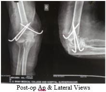
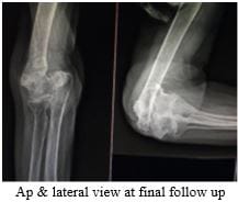
Introduction: We live in a society with a growing elderly population and a young population in which extreme sports and high-speed motor transportation are popular. Therefore the incidence of distal humeral fractures is likely to increase. This study evaluates the functional outcome of distal humeral fractures in adults treated with a different modality. Material and Method: This was a prospective study that includes 29 cases of distal humerus fractures who were treated surgically at C U Shah Medical College and Hospital, Surendranagar, from June 2018 to June 2020, in which 17 were females and 12 were males. Result: According to Mayo Elbow Performance Index (MEPI) scoring system, we had 18(62.07%) patients with excellent results, 8(27.58%) patients with good results, 2(6.9%) patients with the fair result and 1(3.45%) patients with poor outcome. Complications were seen in 15 patients, out of which most common was elbow stiffness seen in 7 patients. Conclusion: Open reduction and internal fixation can be considered as the treatment of choice if there is no contraindication for this because it is essential to maintain length, axial alignment, joint congruity and rotational alignment if a good range of motion is to be restored. This is achieved in the present study.

Keywords: Distal Humerus, Dual Platting, K-wire fixation

Corresponding Author How to Cite this Article To Browse
Baiju Mukeshbhai Patel, Senior Resident, Department Of Orthopedics, Smt. NHL Municipal Medical College, Ahmedabad, Gujarat, India.
Email:
Harsh Girishbhai Patel, Baiju Mukeshbhai Patel, Divyesh Pragajibhai Jetpariya, Parth Pravinbhai Patel, A Clinical Study to Evaluate Functional Outcome of Distal Humerus Fractures in Adult Age Group Treated With Different Modalities. Surgical Rev Int J Surg Trauma Orthoped. 2021;7(5):116-122.
Available From
https://surgical.medresearch.in/index.php/ijoso/article/view/246

Manuscript Received Review Round 1 Review Round 2 Review Round 3 Accepted
2021-10-07 2021-10-09 2021-10-16 2021-10-23 2021-10-30
Conflict of Interest Funding Ethical Approval Plagiarism X-checker Note
Nil Nil Yes 17%

© 2021by Harsh Girishbhai Patel, Baiju Mukeshbhai Patel, Divyesh Pragajibhai Jetpariya, Parth Pravinbhai Pateland Published by Siddharth Health Research and Social Welfare Society. This is an Open Access article licensed under a Creative Commons Attribution 4.0 International License https://creativecommons.org/licenses/by/4.0/ unported [CC BY 4.0].

Introduction

"No injury of the humerus is more difficult to treat than inter-condylar fracture of the humerus." -Sir Watson Jones We live in a society with a growing elderly population and a young population in which extreme sports and high-speed motor transportation are popular; therefore, the incidence of distal humeral fractures is likely to increase. In young adults, most distal humerus fractures occur from high-energy trauma. Side-swipe injuries, motor vehicle accidents, falls from height and gunshot wounds. In elderly persons with more osteoporotic bone, most injuries occur from falls. So improved understanding of the complex pathoanatomy of unstable distal humerus fractures in adults has prompted a global interest in more precise treatment for this diverse group of injuries. Surgeons who treat fracture of the distal humerus frequently have realized the challenges that arise related to poor bony quality, distal separation of the articular fragment from the columns of the distal humerus and fragmentation of the articular surface one or more planes. Varying patterns of distal humeral fractures are common in adults. Malunion is also common. Even minor irregularities of the joint surface of the elbow usually cause some loss of function. Riseborough and Radin, in 1969, reported that operative treatment for fracture distal humerus is often unpredictable and associated with poor outcomes; therefore, they recommended non-operative treatment.[1] In the 1970s, treatment shifted from casting and the "bag of bones" technique to surgical intervention with limited internal fixation. Again, results generally were poor due to a lack of adequate stabilization for early motion. In the early 1980s, the AO-ASIF group reported good and excellent results in 27 of 39 patients with comminuted fractures of the distal humerus. These by far were the best results reported in the treatment of these difficult fractures at that time. This led to increased enthusiasm for surgical reduction and fixation. The elbow joint function is essential for performing day to day activities, which requires the hands to reach the midline of the body, such as dressing, eating, combing hair. This exact and demanding precision is frequently disturbed by fracture of the distal humerus, which always results in a loss of a few degrees of motion of the elbow regardless of the treatment modalities. This study aims to evaluate the functional outcome of distal humeral fractures in adults treated with a different modality.

Materials And Methods

The following study was conducted at C U Shah Medical College and Hospital, Gujarat, from June 2018 to June 2020. All patients with distal humerus fractures meeting the inclusion criteria were included in the study.

Inclusion Criteria

1) Patients with distal humerus fracture

2) Age: 18 years and above

EXCLUSION CRITERIA

1) Any previous fracture in the same limb

2) Any associated fracture in the same limb

3) Vascular insufficiency of upper limb

4) Polytrauma patient with an injury severity score of more than 16

5) Fracture with an associated distal neurovascular deficit

6) Patient with Non-alcohol or drug dependence

7) Pathological fractures

After finding the suitability as per inclusion and exclusion criteria, patients were selected for the study and briefed about the nature of the study. The intervention, if any to be carried out and written, informed consent was obtained. The consented patients were included in the present study. Most of the patients were brought on an outpatient basis. History was obtained through verbal communication, clinical examination, both local and systemic was done, along with the assessment of distal neurovascular status. After a thorough clinical evaluation, an x-ray of the affected elbow was taken in both AP and Lateral view. If needed as per fracture type, CT scans were obtained. According to the X-rays, fractures were classified according to AO classification. The limb was immobilized in the above elbow slab. Intravenous analgesics are given, and intramuscular TT injection with intravenous antibiotic is administered in an open fracture.

The patient was taken up for surgery after routine investigations.

  • Blood and urine investigations
  • ECG and chest x-ray
  • HIV & HBsAg

Medical fitness was obtained before surgery for patients aged >40 years of age.

Indication for surgery

  1. Intra-articular displacement more significant than 2 mm.
  2. Marked supracondylar comminution and displacement.
  3. Open fracture.

Preoperative planning for CR/OR and IF

These fractures are frequently comminuted, which is not evident on the radiographs. The exact nature, geometry and configuration of fracture fragments are assessed and understood before surgery is performed.

This was accomplished by anteroposterior and lateral radiographs, traction radiographs and CT scans whenever necessary.

After the initial workup, the operative fixation of the fractures was performed.

Before surgery, detailed instructions were given to each patient that the result of the procedure considerably depended on the patient's motivation to regain full function subsequently and that active motion of the joint despite the post-operative pain is an essential part of the treatment.

Radiographs were taken at regular intervals to assess that the movements did not affect the rigidity of fixation.

Principles of internal fixation with plates (Intra-articular fractures)

  1. Reconstruction of the triangle base, i.e., reconstruction of the trochlea and capitellum first converted into the supracondylar fracture.
  2. Alignment and fixation of the shaft, reconstruction of medial and lateral pillars- complete restoration of the triangle.

A. Principles of Internal Fixation with Plate and Screws

The position: The patients were given a lateral decubitus position with the fractured limb hanging free over a bolster. A tourniquet is applied proximally, and the arm is draped free. The arm is elevated and exsanguinated, and the tourniquet inflated to approximately 250 mmHg.


Campbell's Triceps Splitting Approach [2, 3]

The Campbell's posterior approach to the elbow was used in a few cases. To achieve adequate exposure, a straight posterior incision over the distal humerus, curving laterally around the olecranon and then along with the upper fourth of the ulna (i.e., a longitudinal incision started 10-15 cm proximal and extending 5 cm distal to the olecranon).

  • The ulnar nerve was identified in all cases.
  • The radial nerve was identified when the fracture was more proximal, requiring fixation close to the spiral groove.
  • To gain adequate exposure and a clear view of the articular surface on its posterior inferior and anterior aspects, an osteotomy of the olecranon, initially described by Cassebaum, is essential.

Olecranon Osteotomy Approach [2, 3]

While performing the osteotomy, a very thin-bladed instrument was used, and the bone loss was minimal.

2 K-wires were inserted from the tip of the olecranon from the proximal posterior to the distal anterior direction in the medullary canal to achieve a perfect anatomical reduction of the olecranon postoperatively.

A thin oscillating saw is then used to make a transverse non-articular osteotomy at the bare area situated between the olecranon articular facet and coronoid articular facet, and it is completed with a thin, fine-pointed osteotome at the subchondral bone level. Once completed, the triceps insertion was detached.

Proximal olecranon was gently dissected free from thin surrounding tissues and lifted proximally as a single unit. This enables the exposure of the posterior and inferior joint surface and the posterior surface of the trochlea (or trochlear fragments).

Van Gorder Approach [2, 3]

After a posterior midline incision, with slightly curving laterally, the ulnar nerve was identified and safely retracted. The triceps muscle was exposed and divided in a shape of V with apex proximal at its musculotendinous junction approximately 4-5 cms proximal to the tip of the olecranon. The fracture site was exposed and fixed with


appropriate implants. At the end of surgical fixation, the tongue was repaired with absorbable sutures, and the skin incision was closed in layers.

Paratricepital Approach [2, 3]

The paratricepital approach is made through the posterior midline incision. The ulnar nerve is identified and safely retracted. The medial intermuscular septum is excised, and the triceps muscle is lifted off the posterior aspect of the distal humerus. Laterally, the triceps is lifted off the posterolateral part of the humerus exposing the lateral column, olecranon fossa and the posterior aspect of the trochlea.

The first step is the anatomic restoration of the articular surface. Once this is accomplished, the two condyles should be fixed stably with a lag screw using a 4.0mm cancellous screw.

The ensuing step in the operative procedure is anatomic reduction and restoration of condyles to the humeral shaft. This can be temporarily accomplished using Kirschner wires drilled from distal to proximal through condyles in a criss-cross manner. It is necessary to maintain 40 degrees of anterior alignment of condyles relative to the humeral shaft when undertaking this temporary stabilization.

For the final fixation of the reconstituted condylar fragment to the humeral shaft, two plates, one on each side, are used. Then fixation of olecranon osteotomy is done using the tension band wiring technique or a cancellous screw. Then closure is done. A negative suction drain is kept, and the incision is closed in layers.

The postoperatively above-elbow splint was given, the dressing was done regularly, and sutures were removed on 14 the day. Then an active range of motion of the involved limb was advised after one month and total activity after three months. Postoperatively patients were reviewed monthly for the first three months and three months for a year or until full range of motion was regained.

B. Principles of Internal Fixation with K Wires (Extra-Articular Fractures)

Surgical Technique: Patient is placed in the supine position. The fracture is reduced with traction, and then the elbow is flexed with traction on. The reduction is viewed under the image intensifier

and fixed with k wires when found acceptable. 2 K wires were introduced from the lateral side to diverge at the fracture site. A k wire from the medial side was introduced whenever reduction was unstable after extending the elbow and milking the ulnar nerve posteriorly. The final drop is viewed under the image intensifier, and the K wires were bent and cut. Postoperatively above elbow slab was given, and the dressing was done regularly. Posterior splint and k wires were removed at 1 ½ month after surgery or when callus was visible on follow up x-ray, and elbow range of motion exercises was encouraged. The entire activity was allowed at three to four months as fracture consolidation occurred. Postoperatively patients were reviewed monthly for the first three months and three months for a year or until full range of motion was regained.

Result

This was a prospective study that included 29 cases of distal humerus fractures that were treated surgically at C U Shah Medical College and Hospital, Surendranagar, from June 2018 to June 2020. In our study, the injury was more common in females 17 (58.60%) than males 12 (41.40%). Age distribution maximum cases were recorded in the age group 18-30 and 51-70 years nine patients (31.05%) followed by 31-50 years seven patients (24.15%) and least was in the age group of above 70 years four patients (13.8%). The mean age was 47.03.

In our study, the most common cause for distal humerus fracture was road traffic accident 13 patients (44.83%), followed by domestic fall ten patients (34.48%), assault four patients (13.80%) and fall from height two patients (6.89%). The injury was more common on the left side in 18 patients (62.07%) than the right side in 11 patients (37.93%). Most cases were of type C1 fracture in six patients (20.69%), with least Type A3, B1 and C3 fractures two patients each (6.90%).

In the present study, according to Mayo Elbow Performance Index (MEPI) scoring system, we had 18(62.07%) patients with excellent results, 8(27.58%) patients with good results, 2(6.9%) patients with the fair result and 1(3.45%) patients with poor outcome.


Case 1: Open Reduction and Internal Fixation with Dual Plating and Cc Screw
surgery_246_01.JPG surgery_246_02.JPG surgery_246_03.JPG

Case 4: Closed reduction and fixation with K wire
surgery_246_04.JPG

surgery_246_05.JPG surgery_246_06.JPG

Discussion

The functional elbow is essential for an individual for social-economic thriving. Fractures of the distal humerus may directly affect the active movement of the elbow especially inter-condylar (intra-articular) fracture. The relationship between the radiohumeral joint and ulnohumeral joints must be perfect for functional outcomes.

The restoration of elbow function is dependent on three salient features: exposure, fixation and post-operative rehabilitation, with later two, are of primary consideration. Adequate exposure is necessary to visualize the bone injury and fixation of the fracture fragments. The posterior approach provides the optimal exposure with intra-articular osteotomy of the olecranon. This allows complete examination of the articular surfaces of the trochlea, capitellum, olecranon and radial head. It also gives access to the medial and lateral supracondylar ridges. Complete evaluation of the fracture fragments and reduction can then be performed. Although non-union of the extra-articular osteotomy may be regarded as a potential complication of this exposure, TBW of the osteotomy has provided sufficient stability of the olecranon for immediate use of the elbow through a secure range of motion without the occurrence of non-union.


Age incidence: In the present study, the average age of patients was 47.03 years, ranging from 18 to 80 years, which was consistent with the age incidence in studies done by Lakey [4]. et al. (44.8 years). In a study by M Dhawan et al [5]. The average age of patients was 43 years, Michael Mckee et al. [6]. The study had mean age of all patients was 47 years.

Study Age(Average) in years
Lakey et al. [4] 44.8
Dhawan et al [5] 43
Present Study 47.03

Sex distribution: In our series, the male to female ratio is 1:1.42, 12 among 29 patients were males, and 17 patients were females.

Study Male: Female Male: Female Ratio
Dhawan et al. [5] 78/30 2.6:1
McKee et al. [6] 14/11 1.27:1
Present Study 12/17 1:1.42

Mode of injury: In the present study, 13 (44.83%) patients were injured by RTA, 2 (6.89%) patients had a history of fall from height, 10 (34.48%) patients had an injury due to domestic fall while 4 (13.80%) patient had assaulted as a mode of injury.

Study Domestic fall Road traffic accident Fall from height Assault Total
Lakey et al [4] 11 9 0 0 20
Dhawan et al [5] 78 30 0 0 108
Present Study 10 13 2 4 29

Type of fracture: The most common type of fracture encountered in the present study was Type C1 fracture with six patients, next most common was A1 and A2 fracture with five patients, four patients had Type C2 fracture, while three patients had Type B2 and two patients had Type A3, B1 and Type C3 fracture.

Complications: In the present study of 29 patients, 7 (46.66%) patients had elbow stiffness, 1 (6.67%) patients had superficial infection, 2 (13.33%) patients had Ulnar Nerve Paralysis, 1 (6.67%) patient had Radial Nerve Paralysis and 1 (2.00%) patients had Heterotopic ossification, 2(13.33%) patients had non-union and 1(6.67%) patient had malunion.

Study Stiffness Infection UNP RNP HO Nonunion Malunion
Amite et al [7] 0 1 0 1 0 0 0
Present Study 7 1 2 1 1 2 1

In the present study, as per Mayo Elbow Performance Index (MEPI) scoring system, we had 18(62.07%) patients with excellent results, 8(27.58%) patients with good results, 2(6.9%) patients with fair results and 1(3.45%) patients with poor outcome. The mean MEPI score observed was 88.97.

Study Mean MEPI Score
Ibomcha et al [8] 84.00
Sanchez Sotelo et al [9] 85.00
Present Study 88.97
Study Excellent Good Fair Poor
Sanchez Sotelo et al [9] 11(34.37%) 16(50%) 2(6.25%) 3(9.37%)
Present Study 18(62.07%) 8(27.58%) 2(6.9%) 1(3.45%)

None of the patients had pain on resisted wrist extension.2 Patients had pain on resisted wrist flexion. None of the patients had pain on resisted long finger extension. 7 Patients had pain on resisted wrist pronation. One patient had pain on resisted wrist supination.  None of the patients had ulnohumeral crepitus or radiocapitellar crepitus.  All patients had scar marks present except conservatively managed patients. None of the patients had developed atrophy. 2 Patients had developed deformities. Three patients showed positive ulnar nerve tinels. None of the patients was present for the cubital tunnel stretch test. Three patients had shoulder joint limiting activity, and no other patient showed any other joint limiting activity other than elbow joint.

Conclusion

  • Increased incidence of distal humeral fractures was probably due to increasing RTA and osteoporotic bones in the geriatric population.
  • Distal humeral fractures occurred more commonly in the 3rd, 6th and 7th decades.
  • The predominance of females was seen in these fractures.
  • Open reduction and internal fixation could be considered as the treatment of choice if there were no contraindication for this because it is essential to maintain length, axial alignment, joint congruity and rotational alignment if a good range of motion is to be restored. This is achieved in the present study.
  • Reconstruction plate and reconstruction plate with cannulated cancellous

  • screws gave optimum fixation and allowed immediate mobilization than K-wires when used exclusively.
  • Excellent results were achieved in terms of mobility and union without deformity.
  • To conclude, reconstruction plates and cannulated cancellous screws offers excellent results in a distal humeral fracture in adults and are to be considered as the first-line of management.
  • Due to the less exposure to closed reduction manoeuvre, there is increased chances of nerve palsies.
  • Due to osteoporosis in elderly patients, chances of K wire loosening and non-union is increased.
  • Under certain circumstances where patients were not fit for open reduction and internal fixation, we considered immediate reduction and internal fixation with K-wire and cc screw fixation as a management line. It is a less invasive procedure with a short operating time. There is decreased risk of infection, reduced hospital stay and technically easy to perform and an economical procedure.
  • By choosing the paratricipital approach instead of olecranon osteotomy in intra-articular fracture, increased chances of nerve palsy due to less exposure and increased stiffness due to more manipulation for intra-articular fracture reduction.
  • Olecranon osteotomy + Dual plating gives the best outcome in intra-articular distal humerus fractures.
  • The study shows that from all the approaches for treating distal humerus fracture olecranon osteotomy approach gives best result.  

Author’s contribution: Harsh Patel: study design, Baiju Patel: manuscript preparation, Divyesh Jetpariya & Parth Patel: statistical analysis.

Reference

01. Riseborough EJ, Radin EL. Intercondylar T fractures of the humerus in the adult. A comparison of operative and non-operative treatment in twenty-nine cases. J Bone Joint Surg Am. 1969 Jan;51(1):130-41 [Crossref][PubMed][Google Scholar]

02. Buckolz, Court-brown, Heckermen, Tornetta. Rockwood and Green's Fracture in Adults. . 1969 Jan;51(1):130-41 [Crossref][PubMed][Google Scholar] [Crossref][PubMed][Google Scholar]

03. Robert Bucholz, James D. Heckman, Charles Court-Brown. Rockwood and Green's Fractures in Adults. [Crossref][PubMed][Google Scholar]

04. Lakhey S, Sharma S1, Pradhan RL1, Pandey BK1, Manandhar RR1, Rijal KP. Osteosynthesis of inter-condylar humerus fracture using bryan and morrey approach. Kathmandu University Medical Journal 2010, Vol. 8, No. 2, Issue 30, 154-157 [Crossref][PubMed][Google Scholar]

05. Dhawan, M. , et al. "Closed intra-articular fractures of the distal end of humerus. ". [Crossref][PubMed][Google Scholar]

06. McKee MD, Wilson TL, Winston L, Schemitsch EH, Richards RR. Functional outcome following surgical treatment of intra-articular distal humeral fractures through a posterior approach. J Bone Joint Surg Am. 2000 Dec;82(12):1701-7. doi: 10.2106/00004623-200012000-00003 [Crossref][PubMed][Google Scholar]

07. Pankaj A, Mallinath G, Malhotra R, Bhan S. Surgical management of intercondylar fractures of the humerus using triceps reflecting anconeus pedicle (TRAP) approach. Indian J Orthop. 2007 Jul;41(3):219-23. doi: 10.4103/0019-5413.33686 [Crossref][PubMed][Google Scholar]

08. Ibomcha, S. , and Sanjib Waikhom. "Internal fixation of type C fracture of distal humerus. " Indian Journal of Orthopaedics 38. 20 (2004): 110-2 [Crossref][PubMed][Google Scholar]

09. Sanchez-Sotelo J, Torchia ME, O'Driscoll SW. Complex distal humeral fractures: internal fixation with a principle-based parallel-plate technique. J Bone Joint Surg Am. 2007 May;89(5):961-9. doi: 10.2106/JBJS.E.01311 [Crossref][PubMed][Google Scholar]