E-ISSN:2455-5436
P-ISSN:2456-9518
RNI:MPENG/2017/70870

Research Article

Disease

Surgical Review: International Journal of Surgery Trauma and Orthopedics

2021 Volume 7 Number 4 July-August
Publisherwww.medresearch.in

A Comparative Study on flap necrosis following Karydakis Flap Reconstruction and Z-Plasty in Patients with Sacrococcygeal Pilonidal Disease – Our experience

Gayathre S.1, Kumar R.2*, Prabu M.3, R Kannan M.S.4
DOI: https://doi.org/10.17511/ijoso.2021.i04.02

1 SP Gayathre, D.G.O., M.S, Professor, Institute of General Surgery, Madras Medical College, Chennai, Tamilnadu, India.

2* R Niranjan Kumar, M.S Gen surgery, D.Ortho, Institute of General Surgery, MMC, Chennai, Tamilnadu, India.

3 MJ Prabu, MBBS., Postgraduate (General surgery), Institute of General Surgery, MMC, Chennai, Tamilnadu, India.

4 R Kannan M.S., Professor and Head of the department, Institute of General Surgery, Madras medical college, Chennai, Tamilnadu, India.

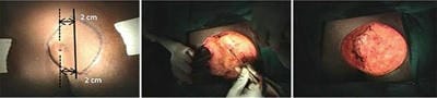
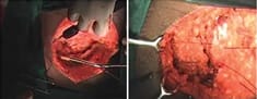
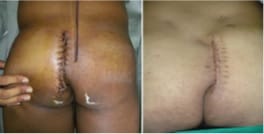
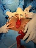
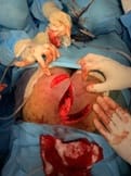
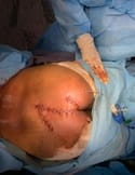
Aim: The purpose of this study was to evaluate patients with sacrococcygeal pilonidal sinus disease (SPSD) who underwent the Karydakis procedure and Z plasty at our centre concerning the perioperative findings, late postoperative results and recurrence. Patients and Methods: A total of 30 patients presenting with SPSD at our centre underwent Karydakis flap repair and Z plasty from May 2019 to June 2021. These patients were then followed up and evaluated concerning operative time, drain use, hospital stay, suture removal, complications, and recurrence. The adipocutaneous flap of Karydakis was devised to shift the natal cleft, while Z-plasty involves a fasciocutaneous flap. Results: The mean operative time was 60 min with a median hospital stay of 4 days. Drains were removed at a median of 5 days and sutures at a median of 15 days. The duration of hospitalisation for the Karydakis procedure was found significantly lesser than that for Z-plasty Patients who were followed up for a median of 12 months. The overall complications were more in Z-plasty. Flap necrosis developed in 30 % of the cases in the Z-plasty group, comparable to no recurrence seen in the Karydakis procedure. Conclusion: Karydakis flap was found superior to Z-plasty, having less seroma formation, no flap necrosis and no local hematoma Karydakis flap procedure is a relatively simple procedure for SPSD and has advantages over Z-plasty technique like keeping scar away from the midline and flattening of the natal cleft, thus reducing local recurrence rates.

Keywords: Complications, Karydakis flap repair, Z-plasty technique, Pilonidal sinus recurrence

Corresponding Author How to Cite this Article To Browse
R Niranjan Kumar, M.S Gen surgery, D.Ortho, Institute of General Surgery, MMC, Chennai, Tamilnadu, India.
Email:
SP Gayathre, R Niranjan Kumar, MJ Prabu, R Kannan M.S., A Comparative Study on flap necrosis following Karydakis Flap Reconstruction and Z-Plasty in Patients with Sacrococcygeal Pilonidal Disease – Our experience. Surgical Rev Int J Surg Trauma Orthoped. 2021;7(4):85-90.
Available From
https://surgical.medresearch.in/index.php/ijoso/article/view/240

Manuscript Received Review Round 1 Review Round 2 Review Round 3 Accepted
2021-07-27 2021-08-04 2021-08-14 2021-08-24 2021-08-31
Conflict of Interest Funding Ethical Approval Plagiarism X-checker Note
No Nil Yes 6%

© 2021 by SP Gayathre, R Niranjan Kumar, MJ Prabu, R Kannan M.S. and Published by Siddharth Health Research and Social Welfare Society. This is an Open Access article licensed under a Creative Commons Attribution 4.0 International License https://creativecommons.org/licenses/by/4.0/ unported [CC BY 4.0].

Introduction

Pilonidal sinus disease (SPSD) is a global problem with a propensity for recurrence that causes significant problems if not dealt with appropriately in the primary setting. A higher incidence of the disease is noted among males and army recruits.[1] Early pilonidal changes are amplified due to deep tissue disruption from moisture, anaerobic conditions, hairs, and bacteria.[2,3].

The focus of treatment should be actions to change the conditions that attack epidermis, rather than wide excision, which attacks deep and healable tissue excision with midline closure, oblique excision and asymmetric closure, Karydakis and z plasty procedures. Karydakis procedure is a gluteal advancement flap [2]. Z-plasty is a fasciocutaneous flap; Karydakis flap is an adipocutaneous flap; it is, therefore, technically easier, less bloody, and less time consuming; it has a better cosmetic outcome, a single, lateral, longitudinal scar; and it requires a significantly shorter hospital stay.

It has been proved that flap procedures are superior to traditional methods of excision of the tract to prevent recurrence of the lesion.[1,4]. The procedure of choice, however, remains debatable [4]. The principle of the Karydakis procedure being flattening of the natal cleft and lateral shift of midline (scar), which prevents lodging of hair in the natal cleft and hence recurrence [5,6]. Flap procedures are often accused of being time-consuming and technically demanding.

Methods and Materials

The present study was undertaken as a prospective study from May 2019 to June 2021. All patients subjected to the Karydakis procedure, Z plasty into two divided groups for PSD at our centre were included in the study whether presenting with primary or recurrent disease. A single surgical unit performed all procedures. The sample size was decided according to the experience of cases of pilonidal sinuses and local guidelines. The cases are randomised based on the hospital registration number by the initial part. The screening personnel did not get involved in the operative procedure.

Inclusion Criteria: Patients aged between 20 to 40yrs Includes both males and females

Exclusion Criteria: It doesn't include pregnant women.

Osteomyelitis of the underlying bone, Fistula in ano Perianal abscess, Sebaceous cyst, Dermoid cyst, Hydradinitis, Tubercular and Syphilitic granuloma

All procedures were done under spinal anaesthesia. Patients were placed in a prone jackknife position. A quantity of dilute methylene blue is instilled into the sinus tracts. An asymmetrical ellipse was marked to encompass the pilonidal complex. The upper and lower ends of the ellipse, thus marked, were at least 2 cm away from the midline marked area was then excised full-thickness up to the sacral fascia with a straight edge on the side of flap mobilisation a sloping edge on the other side.

This is followed by mobilisation of the flap across the midline. A layer of 1-0 polyglactin sutures was placed, the needle was passed into the sacral fascia in the midline and then into the V junction of the flap and secured. A suction drain was placed and brought out well laterally. The second layer of polyglactin sutures was then put to secure the flap to the lateral edge of the wound. The skin was approximated using 2-0 nylon mattress sutures, and a pressure dressing was applied.

Wound inspection was done on the 2nd postoperative day, and the patient was discharged, usually on the 4th or 5th postoperative day, sometimes with drains in situ. Drains were removed 7 to 8 days later. Following suture removal, patients were followed-up once in 2 weeks for the next one and a half months, followed by once every two months for the next six months and then once in 6 months

Stages of Surgery

surgery_240_01.jpg

(a) marking incision(b) excision(c)post excision

surgery_240_02.jpg

(d) raising the flap(e)securing flap


surgery_240_03.jpg

Fig: Immediate postop After suture removal

The Z-plasty involves the transposition of two triangular flaps, A and B. The limbs of the Z must be equal in length to the central limb but can extend at varying angles (30–90 °) depending on how much length is required. The classic Z-plasty has an angle of 60 ° and provides a 75% theoretical gain in length of the central limb by recruiting lateral tissue. The depth of the incision is to the fascia, to allow a pre fascial cutaneous and subcutaneous flap. The flaps are suitable interdigitated and sutured in three layers according to the depth of the wound to avoid negative spacing. MacDermott proposed that the suture line be shifted laterally away from the midline gluteal fold to reduce the recurrence rate by modifying local conditions.[7] All patients were asked to pay attention to hygiene rules, not to sit or use a semi-sitting position for 15 days, and to depilate hairs around their gluteal region as necessary every 3months beginning after hospital discharge.

surgery_240_04.jpg

surgery_240_05.jpg surgery_240_06.jpg

The surgical procedure was an early failure if the patient experienced purulent discharge, abscess, or complete wound dehiscence, for which further treatment was required within 30 days of the operation.


Results and Analysis

The age of patients ranged from 18 to 56 years with a mean age of 26.45 years, with the majority (18 out of 30-75.72%) in the 21to 40 year's age group. The incidence in male patients was nine times more than that in females. In both groups, the incidence was more common in patients with coarse body hair density (80–84%) and patients with a deep natal cleft (88%). Grossly, the primary disease was present in 82% of the patients, among which 87.8% Recurrent disease was present in 20% of patients, and all of them are males, and all these patients have a deep natal cleft.

Similar to our study, Chintapatla et al. showed a male preponderance of this disease [8]. Awad and Saad reported that 56.3 % of pilonidal disease patients had coarse hair density and deep natal cleft [9]. This may be mainly due to their more hirsute nature. There were four recurrent cases in group 2 and Nil in group 1. In both groups, the common clinical presentations were small pus discharging sinus (56%), multiple discharging sinuses (28%), and swelling with pus discharging sinus (16%).

Four of these patients had recurrent disease and had undergone a primary procedure like excision and curettage or excision and primary midline closure. All cases were found to have these openings within 4 cm cranial or caudal to the primary opening, and secondary openings situated laterally were within 2 cm on one side of the midline. In patients with the serous collection, the collection was let out, following which wounds healed within two weeks of regular dressings. In patients with wound infection, pus was drained, and they were administered appropriate antibiotics and underwent regular dressing.

In five patients, the wounds healed by secondary intention within 3-4 weeks. Only one patient in the above group had to be taken up for secondary suturing at the end of 4 weeks, and healing was complete in another three weeks. This was an obese patient who had recurrence twice following excision and primary midline closure and underwent the Karydakis procedure the 3rd time.

Table 1: Complications observed in both groups

Z-plasty Karydakis procedure X2 P significance
Infection of wound (48%), Infection (12%), 10.272 <0.01 S

Necrosis (56%), - 15.790 <0.001 HS
Hematoma (52%), - 17.567 <0.001 HS
Seroma (48%), seroma (12%) 10.784 <0.01 S
wound dehiscence (48%) - 15.790 <0.001 HS
Loss of sensation (40%). Loss of sensation (4%). 9.441 <0.01 S

There was an absence of hematoma, necrosis, and wound dehiscence in the Karydakis procedure. Recurrence developed in 32% of the cases in the Z-plasty group, comparable to no recurrence seen in the Karydakis procedure, but the patients were followed up until 6months. 85% of complications occur in male patients and the remaining 15% in females.

Discussion

Procedures like excision with primary midline closure though simple, pose a greater risk of recurrence (up to 16%) than flap procedures. These patients underwent the Karydakis procedure. Complications noted were serous wound collection in 4 patients and wound infection in 2 patients. Most of the patients were discharged within five days, with only eight patients staying >10 days. Other studies have quoted hospital stays ranging from 1 to 4 days. The more prolonged hospital stay for the patients with complications sacrococcygeal pilonidal sinus disease is a health issue affecting young adults worldwide that requires surgical intervention. Recurrences have led to various procedures being practised, from simple excision to complicated wound infection. Those who were reluctant to go home with drain in-situ stayed back for more extended periods.

Postoperative comparison: wound healing, and work off periods are relative measures of outcome. They are strongly related to personal, sociocultural, and socioeconomic levels, type of job, social assurance and behavioural patterns. A study by Awad et al. [10] put forward a scoring system to classify the cases into three groups. Depending on the scores, the patients underwent excision with secondary healing, excision with primary midline closure or flap procedure.

We observed a shorter hospitalisation with the Karydakis flap technique, around five days, compared to Z-plasty, around 13days. In the study, the mean duration of wound healing was approximately 14 days, the number of days in which pain lasted postoperatively was 12 days, and work off periods were 12 days in the Karydakis flap procedure.


In contrast, in the Z-plasty technique, the mean days of wound healing were 30 days the number of days in which pain lasted postoperatively was 37 days, and work off periods were 40 days; hence the meantime of healing in the Karydakis flap was less than 15 days Complications: the overall complication rates were maximum for the Z-plasty group commonest were tip necrosis (56%), loss of sensation 50%, Infection (8%), seroma formation (4%), and wound dehiscence (4%). Loss of local sensation is a known complication following Z-plasty because, in this technique, flaps are transposed in such a way as to avoid a suture line in the midline. [11,12]. To achieve this, we have to make an incision, thereby cutting cutaneous nerves.

In Z-plasty, to minimise the tip necrosis, we have observed and advise to do the following:

(i) the incision to achieve transposition of flaps should be in full depth, having deeper tissues along with the skin;

(ii) approximation of soft tissue (subcutaneous and deeper) should be meticulous by vicryl suture;

(iii) the cautery should be minimally used.

Similarly, the experience of 15 cases with the Karydakis procedure, reported among early complications loss of local sensation in 4%, seroma in 12%, wound infection in 16.7% and wound dehiscence in 0.3%. flap necrosis and wound dehiscence were significantly less common.[7]. None of the patients had any recurrence, which is comparable to other studies like Kitchen (4%),[13] Anyanwuet al. (0%)[14]. and Amir (0%).[15].

In both groups, wound dehiscence and infection were more common in cases having deep natal cleft and coarse body hair density. This is because, as the perineum to be the most contaminated area of our body, with the mid natal cleft being the bacterial harbour where maximum bacterial colonisation occurs, dense body hair adversely affects the maintenance of proper hygiene, thus increasing the infection rate. So, it is evident that not only are risk factors for the causation of the disease but also complicate the procedure done, increasing the infection, necrosis, and wound dehiscence rate.

Conclusion

Recurrences have led to a variety of procedures being practised, from simple excision to complicated flap procedures.

Procedures like wide excision or excision with primary midline closure through simple pose a greater risk of recurrence (up to 16%). Karydakis flap was found superior to Z-plasty, having less seroma formation, no local hematoma, and no flap necrosis. Statistically, this comparison was highly significant.

Karydakis flap has advantages over Z-plasty technique like keeping scar away from the midline and flattening of the natal cleft, thus reducing local recurrence rates. No recurrence proves the superiority of the Karydakis flap procedure over the Z-plasty technique for the treatment of pilonidal disease.

Hence, we use of Karydakis flap technique on a more significant number of patients. Though it was a novel attempt to address this issue, simple wide excision is seldom practised in centres dealing with pilonidal sinus regularly given the longer time taken for healing and also propensity to recur.

Hence, the procedure of choice remains controversial, often led by the merits of the individual case and the surgeon's school of training and experience.

Author's contribution

PROF Dr SP Gayathre formulated the aims & objectives with study design and supervised for study.

DR R Niranjan Kumar contributed to the study methodology, the patient follows up, literature review and data analysis.

DR M J Prabu contributed to the preparation of the manuscript, clinical photos and statistics analysis.

PROF DR Kannan R for his invaluable help in conducting this study and permission in the department.

This new study adds to existing knowledge

Even though more unique Flap techniques are available in pilonidal sinus treatment protocol, a decade-old Karydakis flap procedure has minor complications, is less time consuming, is easier to do and with almost no reoccurrences. Hence we recommend the Karydakis flap procedure for pilonidal sinus.


Reference

01. Bascom JU. Procedures for pilonidal disease, In: Carter D, Russell RCG, Pitt HA (eds) Atlas of general surgery, 3rd edn. Chapman and Hall, London. 1997;862–872. [Crossref][PubMed][Google Scholar]

02. Da Silva JH. Pilonidal cyst: cause and treatment. Dis Colon Rectum. 2000 Aug;43(8):1146-56. doi: 10.1007/BF02236564 [Crossref][PubMed][Google Scholar]

03. Søndenaa K, Andersen E, Nesvik I, Søreide JA. Patient characteristics and symptoms in chronic pilonidal sinus disease. Int J Colorectal Dis. 1995;10(1):39-42. doi: 10.1007/BF00337585 [Crossref][PubMed][Google Scholar]

04. Hull TL, Wu J. Pilonidal disease. Surg Clin North Am. 2002 Dec;82(6):1169-85. doi: 10.1016/s0039-6109(02)00062-2. [Crossref][PubMed][Google Scholar]

05. Bendewald FP, Cima RR. Pilonidal disease. Clin Colon Rectal Surg. 2007;20(2):86-95. doi: 10.1055/ s-2007-977486 [Crossref][PubMed][Google Scholar]

06. Karydakis GE. Easy and successful treatment of pilonidal sinus after explanation of its causative process. Aust N Z J Surg. 1992 May;62(5):385-9. doi: 10.1111/j.1445-2197.1992.tb07208.x [Crossref][PubMed][Google Scholar]

07. McDermott FT. Pilonidal sinus treated by Z-plasty. Aust N Z J Surg. 1967 Aug 1;37(1):64-9. doi: 10.1111/j.1445-2197.1967.tb06989.x. [Crossref][PubMed][Google Scholar]

08. Chintapatla S, Safarani N, Kumar S, Haboubi N. Sacrococcygeal pilonidal sinus: historical review, pathological insight and surgical options. Tech Coloproctol. 2003 Apr;7(1):3-8. doi: 10.1007/s101 510300001 [Crossref][PubMed][Google Scholar]

09. Khaled, M Saad, and MS Awad Mohamed. Does closure of chronic pilonidal sinus still remain a matter of debate after bilateral rotation flap? (N-shaped closure technique). Indian Journal of Plastic Surgery. 39;02(2006): 157-162. [Crossref][PubMed][Google Scholar]

10. Awad MM, Elbaset AA, Ebraheem S, Tantawy E, Elhafez MA, Elsayed AM. A scoring system as a method to evaluate pilonidal sinus disease to make an easy decision for its management. Indian J Plast Surg. 2009 Jan-Jun;42(1):43-8. doi: 10.4103/0970-0358.53011 [Crossref][PubMed][Google Scholar]

11. Monro Rs, Mcdermott Ft. The Elimination of Causal Factors In Pilonidal Sinus Treated By Z-Plasty. Br J Surg. 1965 Mar;52:177-81. doi: 10.1002/bjs.1800520306. PMID: 14261120 [Crossref][PubMed][Google Scholar]

12. Senapati A, Cripps NP, Thompson MR. Bascom's operation in the day-surgical management of symptomatic pilonidal sinus. Br J Surg. 2000 Aug;87(8):1067-70. doi: 10.1046/j.1365-2168.2000.01472.x [Crossref][PubMed][Google Scholar]

13. Kitchen PR. Pilonidal sinus: excision and primary closure with a lateralised wound - the Karydakis operation. Aust N Z J Surg. 1982 Jun;52(3):302-5. doi: 10.1111/j.1445-2197.1982.tb05405.x [Crossref][PubMed][Google Scholar]

14. Anyanwu AC, Hossain S, Williams A, Montgomery AC. Karydakis operation for sacrococcygeal pilonidal sinus disease: experience in a district general hospital. Ann R Coll Surg Engl. 1998 May;80(3):197-9. [Crossref][PubMed][Google Scholar]

15. Karydakis GE. New approach to the problem of pilonidal sinus. Lancet. 1973 Dec 22;2(7843):1414-5. doi: 10.1016/s0140-6736(73)92803-1 [Crossref][PubMed][Google Scholar]