E-ISSN:2455-5436
P-ISSN:2456-9518
RNI:MPENG/2017/70870

Research Article

Disease

Surgical Review: International Journal of Surgery Trauma and Orthopedics

2021 Volume 7 Number 4 July-August
Publisherwww.medresearch.in

A retrospective cohort study of diabetic foot disease during the covid pandemic in a tertiary care hospital in Kerala

Mohandas P.1, Vasu Keloth A.2*, G Krishnan D.3, Rahman N.4
DOI: https://doi.org/10.17511/ijoso.2021.i04.01

1 P.G. Mohandas, Professor, Department of General Surgery, KMCT Medical College, Kozhikode, Kerala, India.

2* Anukesh Vasu Keloth, Assistant Professor, Department of General Surgery, KMCT Medical College, Kozhikode, Kerala, India.

3 Divya G Krishnan, Associate Professor, Department of Pharmacology, KMCT Medical College, Kozhikode, Kerala, India.

4 Neema Rahman, Junior Resident, Department of General Surgery, KMCT Medical College, Kozhikode, Kerala, India.

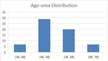
Introduction: Diabetes affects 88 million people in south-east Asia with a regional prevalence of 8.8%, and of them, 77 million live in India. Diabetic Foot Disease is an important complication that accounts for significant morbidity and mortality in a diabetic individual. Aim: This study was conducted to study the presentation stage of diabetic foot disease during the pandemic period as seen at a tertiary care hospital in North Kerala. Methods: This was a retrospective cohort study carried out on patients diagnosed with diabetic foot and admitted to the Department of Surgery of a tertiary care hospital in North Kerala during the study period of 6months from July 2020 to December 2020. A sample size of 63 was calculated. Data were collected from the case records of the patients, and the stages of presentation, relevant blood investigations, culture reports, radiological imaging and arterial doppler ultrasound findings were noted. Results: In this study, a total of 63 patients were included. Of the 63 patients, 51(80.9%) were males, and 12 (19.0%) were females with a male to female ratio of 4.25:1. The mean age of the patients was 57.74 years. In this study, as per the Wagner-Meggitt Classification of diabetic foot, the most common stage of presentation of diabetic foot encountered was Grade III (42.9%) followed by Grade IV (28.6%), Grade II (19.0%) and Grade V (9.5%). Conclusion: This is in part may be due to financial constraints of the patient's families due to lack of job, transportation difficulties, shortage of medicines, shutting down of small scale health care facilities and pharmacies, prolonged neglect of wounds, discontinuing of regular medications and poor home wound care which in turn are due to the frequent regional lockdowns as a result of the covid pandemic.

Keywords: Diabetic Foot Disease, Foot Ulcer, Wagner-Meggitt Classification

Corresponding Author How to Cite this Article To Browse
Anukesh Vasu Keloth, Assistant Professor, Department of General Surgery, KMCT Medical College, Kozhikode, Kerala, India.
Email:
P.G. Mohandas, Anukesh Vasu Keloth, Divya G Krishnan, Neema Rahman, A retrospective cohort study of diabetic foot disease during the covid pandemic in a tertiary care hospital in Kerala. Surgical Rev Int J Surg Trauma Orthoped. 2021;7(4):78-84.
Available From
https://surgical.medresearch.in/index.php/ijoso/article/view/239

Manuscript Received Review Round 1 Review Round 2 Review Round 3 Accepted
2021-07-25 2021-08-09 2021-08-17 2021-08-24 2021-08-30
Conflict of Interest Funding Ethical Approval Plagiarism X-checker Note
No Nil Yes 4%

© 2021 by P.G. Mohandas, Anukesh Vasu Keloth, Divya G Krishnan, Neema Rahman and Published by Siddharth Health Research and Social Welfare Society. This is an Open Access article licensed under a Creative Commons Attribution 4.0 International License https://creativecommons.org/licenses/by/4.0/ unported [CC BY 4.0].

Introduction

Diabetes affects 88 million people in south-east Asia with a regional prevalence of 8.8%, and of them, 77 million live in India. [1]. The age-adjusted comparative prevalence of Diabetes in India is 10.4%. [2] Diabetic Foot Disease is an important complication that accounts for significant morbidity and mortality in a diabetic individual. [3]. The global prevalence of diabetic foot ulcers was estimated to be 6.3% and is more prevalent in those who have Type 2 Diabetes and in males.[4]. The prevalence of diabetic foot disease is expected to rise even more in the coming decade . [5,6]. This rise is not only due to poor glycaemic control, poor foot care, lack of education, lack of proper healthcare facilities, trauma and secondary infections but also due to the co-existence of predisposing factors like Peripheral Neuropathy, Peripheral Vascular Disease and Previous History of Diabetic Foot Disease. [7,8]. In a developing country like India, the health care system has greatly improved and have become modernized to provide better care to the large population of the country especially in states like Kerala, over the last decade. However, the covid pandemic and the consequent frequent regional lockdowns have severely hindered the access of patients to nearby hospitals, in addition to crippling the already overburdened healthcare system. Besides, there are only a few published studies from India on Diabetic Foot Disease. Hence the objective of conducting this study was to determine the stage of presentation of diabetic foot disease during the pandemic period as seen at a tertiary care hospital in North Kerala.

Materials and Methods

Setting: This study was carried out on patients diagnosed with diabetic foot and admitted to the Department of Surgery of a tertiary care hospital in North Kerala.

Duration and Type of Study: The was a retrospective cohort study conducted on patients admitted during the period of 6months from July 2020 to December 2020.

Sampling Methods: Consecutive Sampling method was used to include all patients that met the inclusion criteria.

Sample Size Calculation: A sample size of 63 was calculated. The following formula was used for calculating the adequate sample size

- n=Z2P(1−P)/d2, where

- n is the sample size

- Z is the statistic corresponding to the level of confidence

- P is expected prevalence (that can be obtained from the same studies or a pilot study conducted by the researchers)

- d is precision (corresponding to effect size)

Inclusion Criteria: All patients presenting to Surgery OPD/Casualty with diabetic foot who have been admitted for inpatient care and had adequate data were included in the study.

Exclusion Criteria: All patients presenting to Surgery OPD/Casualty with diabetic foot admitted for inpatient care but were later referred to a higher centre or had inadequate data were excluded from the study.

Data Collection Procedure: Data were collected from the case records of the patients. The previous history of Diabetic Foot Disease, the stages of presentation, Glycosylated Hemoglobin Levels, Fasting Blood Sugar Levels, Culture reports of wound discharge, digital radiographic imaging of the foot, Blood Urea Levels, Serum Creatinine levels and arterial Doppler ultrasound findings of the affected lower limb on admission were noted.

The presence of the pathogenic organism in culture was used to record the presence of infection. The company of bony changes adjacent to the ulcer in the Digital radiograph was used to record the presence of Bony involvement and osteomyelitis. The presence of monophasic and biphasic flow in arterial doppler ultrasound was used to record the presence of Peripheral Vascular Disease. The status of the patient at the time of discharge was noted. The collected data were entered into the study proforma.

Scoring System: The Wagner Meggitt Classification of Diabetic Foot Ulcers was used to stage the Diabetic Foot of each patient. The Staging system categorizes Foot Ulcers into six grades as follows-

Grade 0 - Pre- or post-ulcerative lesion completely epithelialized
Grade 1 - Partial/full-thickness ulcer confined to the dermis, not extending to the subcutis
Grade 2 - Ulcer of the skin extending through the subcutis with exposed tendon or bone; No abscess formation or osteomyelitis


Grade 3 - Deep ulcer with abscess formation or osteomyelitis

Grade 4 - Localised gangrene of the toes or partial foot gangrene

Grade 5 - Whole foot gangrene

Surgical Procedures: Surgical intervention as necessitated by the patient's condition was done during the inpatient stay and included Wound Debridement, Fasciotomy, Incision and Drainage and amputations of the toe, foot and leg. This retrospective study, however, did not have the type of procedures that were done as part of the treatment of the patient.

Ethical Considerations and Permissions: The study was conducted after obtaining Institutional Ethics Committee approval, and the patient's treatment was not altered in any form for this study.

Statistical Analysis: The collected data was analyzed using Frequency, Mean and Standard Deviation.

Results

In this study, a total of 63 patients were included. Of the 63 patients, 51(80.9%) were males, and 12 (19.0%) were females with a male to female ratio of 4.25:1. The mean age of the patients was 57.74 years, with a standard deviation of 8.37 (Table/Chart 1). In this study, as per the Wagner-Meggitt Classification of diabetic foot, the most common stage of presentation of diabetic foot encountered was Grade III (42.9%) followed by Grade IV (28.6%), Grade II (19.0%) and Grade V (9.5%) (Table/Chart 2). All of the 63 patients admitted had poor glycemic control with an HbA1C value of at least 7%, and of them, 12 patients had an HbA1c value of more than 13% (Table/Chart 3). The mean value of HbA1C was 11.71%, with a standard deviation of 1.90. Out of the 63 patients, 57 had an FBS value of more than 120mg/dl, and of them, 27 had a value more than 250mg/dl. Out of the 63 patients, 57 patients showed pathogenic bacteria in the culture and sensitivity study of discharge from the ulcer. Eighteen patients had elevated serum creatinine values. A serum creatinine value of more than 2mg/dl was present in 6 patients. The presence of bone involvement was detected in 19% of the patients. 33 patients out of the 63 had coexisting peripheral vascular disease. There was a previous history of diabetic foot disease requiring treatment in 24 patients (Table/Chart 4).

Of the 63 patients, three died during hospitalization due to septic shock. Of the remaining 60 patients, all underwent some form of surgical procedure and were discharged with healing wounds. Of them, 50(83.3%) patients continued to follow up visits at the surgical out-patient department at this institute and 10 (16.7%) patients discontinued after the initial 1month as they opted for further treatment at local peripheral centres.

Chart 1

surgery_239_01.jpg

Table 2. Stage of presentation of Diabetic Foot

Stage of presentation Number Percentage
Grade 0 0 0
Grade I 0 0
Grade II 12 19.0
Grade III 27 42.9
Grade IV 18 28.6
Grade V 6 9.5
Total 63 100.0

Chart 3. Distribution of HbA1C Values

surgery_239_02.jpg

Table 4. Frequency of Coexisting Diseases

Coexisting Diseases Number (Out of 63) Percentage (Out of 63)
Previous Diabetic Foot Disease 24 38.0
Secondary Infection 57 90.5
Bone Involvement/Osteomyelitis 12 19.0

Altered Renal Parameters 18 28.6
Peripheral Vascular Disease 33 52.4

Discussion

Globally, Diabetes mellitus affected approximately 463 million people in 2019 and is estimated to affect up to 700 million people by 2045.[9] In India, this number is expected to rise from 77 million in 2019 to approximately 134 million by 2045.[2]. Diabetes increases the risk of several diseases like atherosclerosis and coronary artery disease and leads to several life-disabling complications. The complications associated with Diabetes is responsible for the increased morbidity and mortality in people with Diabetes. Diabetes can result in a spectrum of complications that include Cardiovascular Disease, Diabetic Eye Disease, Diabetic Kidney Disease, Peripheral Neuropathy, Peripheral Vascular Disease and Diabetic Foot Disease.[8].

The study by Yazdanpanah L et al. has shown that the annual incidence of Diabetic Foot Disease is 2.8%.[10]. Several studies and past literature have revealed that the etiology of Diabetic Foot Disease is multifactorial and is closely associated with three main components- Peripheral neuropathy (motor, sensory and autonomous), Peripheral vascular disease and Immunodeficiency. [5,8,11,12]. Altered foot biomechanics and gait, along with the above three components, play an important role in the pathophysiology of Diabetic Foot Ulceration. [11]. Other causes include poor glycemic control, calluses, foot deformities, improper foot care and ill-fitting footwear.[7].

Diabetes-related microvasculopathy and Immunodeficiency predispose and eventually result in multi-floral soft tissue infections and ulcerations, which may locally advance to involve deeper tissues like bone. Persistent infection and underlying osteomyelitis along with Peripheral arterial occlusive disease and varying degrees of peripheral neuropathy together lead to gangrene of the foot. [11,13]. Several studies have shown that Diabetic Foot Disease is responsible for a significant proportion of non-traumatic lower-limb amputations.[11,12,14]. The Wagner Meggitt Classification System is one of the most commonly used systems to classify Diabetic Foot Ulcers. It classifies foot ulcers into six grades (0-5) and is based on the depth of the ulcer, presence of abscess, osteomyelitis or gangrene and the extent of involvement of foot. [15,16].

The management of Diabetic Foot Disease is based on the severity of the foot ulcer, presence of infection, vascularity of the affected limb and glycaemic control. [11,16]. The gender distribution of Diabetic Foot Disease showed a male to female ratio of 4.25:1 in this study. This finding is much higher than those published in recent literature.[4]. However, the study by Thanh Dinh et al. revealed no relationship between gender and foot ulceration, especially when associated with peripheral neuropathy and ischaemic disease.[17]. The mean age of patients in this study was 57.74 years which is comparable to recent studies. [18,19]. The study by Al-Rubeaan K et al. showed that the prevalence of Diabetic Foot Disease increased with age.[19].

The most frequent stage of presentation of diabetic foot in the study group was Grade III foot ulcers. These are comparable to findings in the other studies. [20] In this study, 80.9% of the patients had a foot ulcer complicated with an abscess, osteomyelitis or gangrene. The higher frequency of these complications is possibly due to untreated or undetected Diabetes, poor glycaemic control, neglected wound care and untreated infections in the wound, in addition to the presence of associated pre-existing diseases in these patients. This scenario has risen in turn due to lack of easy access to hospital care as a result of regional lockdowns and shutting down of nearby clinics and dispensaries as a consequence of the covid pandemic.

All of the patients included in this study had poor glycaemic control as all had elevated Glycosylated Hemoglobin (HbA1C) values. The mean value of HbA1C in this study group was 11.71% which is much higher than previous studies done during the pre-pandemic era.[19,21]. Worsening of HbA1C values has been known to correlate with worsening diabetic foot disease and increases the risk of secondary infection and likelihood of amputations.[22]. This lack of proper glycaemic control is again likely to be due to lack of regular visits to the family physician or local health care facility on account of the covid pandemic and also due to stoppage of regular medications or inadequate dosage of medications leading to poor control of Hyperglycaemia.

The previous history of Diabetic Foot Disease was present in 38% of the patients in this study, which is in accordance with common literature. [11]. The presence of secondary infection was seen in 90.5% of the patients.


Radiographs of the foot revealed osteomyelitis in 19% of the patients. These two findings suggest that proper and timely wound care was not provided to these patients leading to worsening of the foot. This study revealed that 28.6% of the patients had altered renal parameters at the time of admission. This may be related to worsening infection or the onset of Diabetic Nephropathy in these patients. The presence of monophasic or biphasic waveform with loss of reverse flow Arterial Doppler Ultrasound Imaging is regarded as a reliable indicator of significant ischaemic disease in the lower limbs.[23]. In this study, 52.4% of the patients had Peripheral Vascular Disease in their arterial doppler findings. The frequency of coexisting peripheral vascular disease was higher than that in previous literature. [7,19,24]. This may be related to the higher mean value of HbA1C in this study group, as stated in a study by Selvin E et al., which found a positive association between the HbA1C value and the risk of Peripheral Vascular Disease in diabetic patients.[25].

Conclusion

Though the distribution of frequencies of different stages of presentation of diabetic foot disease is comparable to other studies, the higher proportion of Grade III, grade IV and grade V diabetic feet indicates the co-existence of secondary infections, deep-seated abscesses, osteomyelitis and gangrene in the affected limb. This rise is due to poor glycaemic control and undetected or untreated coexisting diseases that predispose to Diabetic foot disease. This is in part may be due to financial constraints of the patient's families due to lack of job, transportation difficulties, shortage of medicines, shutting down of small scale health care facilities and pharmacies, prolonged neglect of wounds, discontinuing of regular medications and poor home wound care which in turn are due to the frequent regional lockdowns as a result of the covid pandemic.

What does this study add to present knowledge?

This study highlights a recent increase in the burden of Diabetes in Kerala on account of undetected or neglected life-disabling Diabetic Foot Disease despite the presence of a reasonably adequate and easily accessible health care system covering all parts of Kerala.

This study stresses the need to ensure and provide proper and timely delivery of primary health care facilities to patients with Diabetes amidst the ongoing covid pandemic. This study highlights the necessity to create Regional Registers for patients with Diabetes and Diabetic Foot Disease in Kerala and India and also to provide Video and Telemedicjne Facilities along with Mobile Home Care Units to permit Healthcare access to such patients even during the covid pandemic.

Author Contributions

The first author has conducted this study in a Tertiary Care Centre in a rural part of North Kerala. Each of the co-authors has provided valuable contributions in the literature review, data collection, data analysis and references for discussion in this study.

Limitations of the Study

The limitation of this study is that the analysis was performed in a single centre during the covid pandemic. So the number of patients in the study group was small, and the duration of the study was less compared to other studies. More sensitive and advanced tests to detect Osteomyelitis, Renal Disease and Peripheral Vascular Disease were not used in this study. Hence, the presence of coexisting diseases, though documented, was not studied for the significance of association in this study.

Reference

01. International Diabetes Federation. IDF Diabetes Atlas 9th Edition [Internet]. Brussels, Belgium: International Diabetes Federation. 2019. Available from: [Article][Crossref][PubMed][Google Scholar]

02. International Diabetes Federation. IDF Diabetes Atlas 9th Edition [Internet]. Brussels, Belgium: International Diabetes Federation. 2019. Available from: [Article][Crossref][PubMed][Google Scholar]

03. Moulik PK, Mtonga R, Gill GV. Amputation and mortality in new-onset diabetic foot ulcers stratified by etiology. Diabetes Care. 2003 Feb;26(2):491-4. doi: 10.2337/diacare.26.2.491 [Crossref][PubMed][Google Scholar]

04. Zhang P, Lu J, Jing Y, Tang S, Zhu D, Bi Y. Global epidemiology of diabetic foot ulceration: a systematic review and meta-analysis. Ann Med. 2017 Mar;49(2):106-116. doi: 10.1080/07853890. 2016.1231932 [Crossref][PubMed][Google Scholar]


05. Amin N, Doupis J. Diabetic foot disease: From the evaluation of the "foot at risk" to the novel diabetic ulcer treatment modalities. World J Diabetes. 2016 Apr 10;7(7):153-64. doi: 10.4239/wjd.v7.i7.153 [Crossref][PubMed][Google Scholar]

06. Boulton AJ, Vileikyte L, Ragnarson-Tennvall G, Apelqvist J. The global burden of diabetic foot disease. Lancet. 2005 Nov 12;366(9498):1719-24. doi: 10.1016/S0140-6736(05)67698-2 [Crossref][PubMed][Google Scholar]

07. Unnikrishnan R, Anjana RM, Mohan V. Diabetes mellitus and its complications in India. Nat Rev Endocrinol. 2016 Jun;12(6):357-70. doi: 10.1038/nrendo.2016.53 [Crossref][PubMed][Google Scholar]

08. International Diabetes Federation. Clinical Practice Recommendation on the Diabetic Foot: A guide for health care professionals [Internet]. Brussels, Belgium: International Diabetes Federation. 2017. Available from: [Article][Crossref][PubMed][Google Scholar]

09. International Diabetes Federation. IDF Diabetes Atlas 9th Edition [Internet]. Brussels, Belgium: International Diabetes Federation. 2019. Available from: [Article][Crossref][PubMed][Google Scholar]

10. Yazdanpanah L, Shahbazian H, Nazari I, Arti HR, Ahmadi F, Mohammadianinejad SE, et al. Incidence and Risk Factors of Diabetic Foot Ulcer: A Population-Based Diabetic Foot Cohort (ADFC Study)-Two-Year Follow-Up Study. Int J Endocrinol. 2018 Mar 15;2018:7631659. doi: 10.1155/2018/7631659 [Crossref][PubMed][Google Scholar]

11. Joseph EL, Michael LG, Rubinn JP, Christopher GW, Fu-Chan W. Plastic and Reconstructive Surgery, In: Brunicardi FC, Andersen DK, Billiar TB, Dunn DL, Hunter JG, Mathews JB, Pollock RE, editors. Schwartz's Principles of Surgery, 10th ed New York: McGraw Hill. 2015;1877-79. [Crossref][PubMed][Google Scholar]

12. Musa IR, Ahmed MON, Sabir EI, Alsheneber IF, Ibrahim EME, Mohamed GB, et al. Factors associated with amputation among patients with diabetic foot ulcers in a Saudi population. BMC Res Notes. 2018 Apr 27;11(1):260. doi: 10.1186/s13104-018-3372-z [Crossref][PubMed][Google Scholar]

13. Ugwu E, Adeleye O, Gezawa I, Okpe I, Enamino M, Ezeani I. Predictors of lower extremity amputation in patients with diabetic foot ulcer: findings from MEDFUN, a multi-center observational study. J Foot Ankle Res. 2019 Jun 14;12:34. doi: 10.1186/s13047-019-0345-y [Crossref][PubMed][Google Scholar]

14. Deerochanawong C, Home PD, Alberti KG. A survey of lower limb amputation in diabetic patients. Diabet Med. 1992 Dec;9(10):942-6. doi: 10.1111/j.1464-5491.1992.tb01736.x [Crossref][PubMed][Google Scholar]

15. Wagner FW Jr. The dysvascular foot: a system for diagnosis and treatment. Foot Ankle. 1981 Sep;2(2):64-122. doi: 10.1177/107110078100200202 [Crossref][PubMed][Google Scholar]

16. Frykberg RG. Diabetic foot ulcers: pathogenesis and management. Am Fam Physician. 2002 Nov 1;66(9):1655-62. [Crossref][PubMed][Google Scholar]

17. Dinh T, Veves A. The influence of gender as a risk factor in diabetic foot ulceration. Wounds. 2008 May;20(5):127-31. [Crossref][PubMed][Google Scholar]

18. Abbott CA, Carrington AL, Ashe H, Bath S, Every LC, Griffiths J, et al. North-West Diabetes Foot Care Study, The North-West Diabetes Foot Care Study: incidence of, and risk factors for, new diabetic foot ulceration in a community-based patient cohort. Diabet Med. 2002 May;19(5):377-84. doi: 10.1046/j.1464-5491.2002.00698.x [Crossref][PubMed][Google Scholar]

19. Al-Rubeaan K, Al Derwish M, Ouizi S, Youssef AM, Subhani SN, Ibrahim HM, et al. Diabetic foot complications and their risk factors from a large retrospective cohort study. PLoS One. 2015 May 6;10(5):e0124446. doi: 10.1371/journal. pone.0124446 [Crossref][PubMed][Google Scholar]

20. Hasan F A I Z U L, et al. "Diabetic foot-assessment and management of 100 cases". Pak J Med Health Sci. 5(2011): 677-681. [Crossref][PubMed][Google Scholar]

21. Farooque U, Lohano AK, Hussain Rind S, Rind MS Sr, Karimi S, Jaan A, et al. Correlation of Hemoglobin A1c With Wagner Classification in Patients With Diabetic Foot. Cureus. 2020 Jul 15;12(7):e9199. doi: 10.7759/cureus.9199 [Crossref][PubMed][Google Scholar]


22. Farooque U, Lohano AK, Hussain Rind S, Rind MS Sr, Karimi S, Jaan A, et al. Correlation of Hemoglobin A1c With Wagner Classification in Patients With Diabetic Foot. Cureus. 2020 Jul 15;12(7):e9199. doi: 10.7759/cureus.9199 [Crossref][PubMed][Google Scholar]

23. Forsythe RO, Hinchliffe RJ. Assessment of foot perfusion in patients with a diabetic foot ulcer. Diabetes Metab Res Rev. 2016 Jan;32 Suppl 1:232-8. doi: 10.1002/dmrr.2756 [Crossref][PubMed][Google Scholar]

24. Bajaj S, Mahajan A, Grover S, Mahajan V, Goyal P, Gupta VK. Peripheral Vascular Disease in Patients with Diabetic Foot Ulcers - An Emerging Trend: A Prospective Study from North India. J Assoc Physicians India. 2017 May;65(5):14-17. [Crossref][PubMed][Google Scholar]

25. Selvin E, Wattanakit K, Steffes MW, Coresh J, Sharrett AR. HbA1c and peripheral arterial disease in Diabetes: the Atherosclerosis Risk in Communities study. Diabetes Care. 2006 Apr;29(4):877-82. doi: 10.2337/diacare.29.04.06.dc05-2018 [Crossref][PubMed][Google Scholar]