E-ISSN:2455-5436
P-ISSN:2456-9518
RNI:MPENG/2017/70870

Research Article

Retrospective

Surgical Review: International Journal of Surgery Trauma and Orthopedics

2021 Volume 7 Number 2 March-April
Publisherwww.medresearch.in

Retrospective analysis of Intramedullary K-Wire fixation of Metacarpal Fractures

Kumar Kirar S.1, Upadhyay S.2*, Singh S.3, Varshney A.4
DOI: https://doi.org/10.17511/ijoso.2021.i02.05

1 Sunil Kumar Kirar, Senior Resident, Department of Orthopaedics, ABV Government Medical College, Vidisha, Madhya Pradesh, India.

2* Sanjay Upadhyay, Assistant Professor, Department of Orthopaedics, ABV Government Medical College, Vidisha, Madhya Pradesh, India.

3 Sanat Singh, Associate Professor, Department of Orthopaedics, ABV Government Medical College, Vidisha, Madhya Pradesh, India.

4 Atul Varshney, Professor & HOD, Department of Orthopaedics, ABV Government Medical College, Vidisha, Madhya Prdaesh, India.

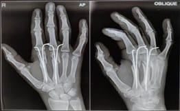
Introduction: The majority of fractures of the metacarpal bones occur at a young age.Most of the times these metacarpal fractures can be treated conservatively in a POP slab(cock up slab) producing good functional results.Surgery was indicated in patients with palmar dislocation of >30° and shortening of >5 mm.Our study aimed to evaluate the clinical results of all metacarpal fractures treated surgically by intramedullary Kirschner-wire fixation presented in our hospital.Materials and Methods: It was a retrospective study in which we included 50 patients with metacarpal fractures(both open andclosed) that came in our hospital, treated surgically by closed reduction and were fixed with two intramedullary k-wires. Result: K-wires were removed after 4 weeks postoperatively,under local anaesthesia in the OPD. Metacarpal joint functions (flexion, extension, rotation) were clinically followed up in all patients, on the median periodof6 months (3 months to 9 months). In our study, we found in all patients,flexion and extension were normal on both sides.Conclusion: Closed reduction and intramedullary k-wire fixation of metacarpal bone fractures produce good functional results in the longterm. We found a very low rate of complication and thus recommendthis surgical method for the stabilization of all these types of fractures.

Keywords: Metacarpal, Flexion, Extension, Rotation, K-wire

Corresponding Author How to Cite this Article To Browse
Sanjay Upadhyay, Assistant Professor, Department of Orthopaedics, ABV Government Medical College, Vidisha, Madhya Pradesh, India.
Email:
Kirar SK, Upadhyay S, Singh S, Varshney A. Retrospective analysis of Intramedullary K-Wire fixation of Metacarpal Fractures. Surgical Rev Int J Surg Trauma Orthoped. 2021;7(2):22-27.
Available From
https://surgical.medresearch.in/index.php/ijoso/article/view/228

Manuscript Received Review Round 1 Review Round 2 Review Round 3 Accepted
2021-04-04 2021-04-15 2021-04-19 2021-04-21
Conflict of Interest Funding Ethical Approval Plagiarism X-checker Note
No Nil Yes 5%

© 2021 by Sunil Kumar Kirar, Sanjay Upadhyay, Sanat Singh, Atul Varshney and Published by Siddharth Health Research and Social Welfare Society. This is an Open Access article licensed under a Creative Commons Attribution 4.0 International License https://creativecommons.org/licenses/by/4.0/ unported [CC BY 4.0].

Introduction

The majority of fractures of the metacarpal bones occur at a young age [1].Metacarpal fractures are among the most commonly treated upper extremity injuries in adults, and represent about 10% of all fractures [2].The most commonly affected metacarpal bone is the 5thmetacarpal bone and sub-capital fracture is more common than a diaphyseal one [3].Metacarpal fractures are most often caused by axial loads applied to the hand, frequently with the wrist flexed [4]. The usual etiologies are a direct blow during fisticuffs, or a fall [6]. Indirect torsional forces can also result in metacarpal fracture [4].With the pull of intrinsic muscles on the metacarpal capitulum, the distal fragment is dislodged in the palmar direction. Most of the times these metacarpal fractures can be treated conservatively in a POP slab (cock up slab) producing good functional results[5,6].

Surgery was indicated in the following cases [7]:

  • palmar dislocation of >30°
  • shortening of >5 mm.
  • open fractures
  • fractures with intra- articular displacements

Our study aimed to evaluate the clinical results of all metacarpal fractures treated surgically by intramedullary Kirschner-wire fixation, presented in our hospital.

Materials and Methods

It was a retrospective study in which we included 50 patients with metacarpal fractures(both open and closed)that came in our hospital, treated surgically by closed reduction and were fixed with two intramedullary k-wires. All patients between 1 January 2019 to 31 July 2020 who came to our hospital were included in the studyPreoperative X-rays of hand (AP and Oblique view) were taken to assess palmardeviation, shortening and rotatory deficiency.

The fracture was classified based on AO recommendation: A type: diaphyseal fractures, B type: epiphyseal and metaphyseal fractures and C type: intra-articular fracturesThere are various ways of fixations but we preferred intramedullary k-wire osteosynthesis of the metacarpal bones because of the simplicity of the method and also it puts minimum strain on the sliding tissue.

Intramedullary stabilization of sub-capital metacarpal fractures using Kirschner wire (k-wire) was introduced by Foucher [7,8]. After anesthesia (general/regional), painting and draping weredone and the fracture was reduced with k-wires under C-arm intensifier. P.O.P. cock-up/below elbowslab was applied after the surgeryAll patients were discharged the next day after the surgery after getting their postoperative x-rays. In our study, 12 cases were open fracture and 5 cases had associated injuries whose duration of stay was prolonged (mean duration was 4 days)

All patients were called for follow up at 15 days, 4th week (k-wire was removed and repeat x-ray wasdone), third month and sixth month postoperatively Metacarpal joint function (flexion/extension) and rotatory displacement was clinically assessed during follow-up examinations. After a radiological demonstration of bone healing normally after 4 weeks, the k-wires were removed under local anesthesia in OPD. Physiotherapy (all movements) was done under the supervision of the treating doctor and physiotherapist and the patient was advised to do the same at home.

Result

Most of the patients in our study were young male of the age group 20-30 years. There were 34 males (68%) and 16 females (32%) in our study. The dominant hand (right in all cases) was affected in about 56% (n=28) cases and the non-dominant hand (left hand in all cases) was affected in about 44%(n=22) cases.

The most common cause fell on hand (38%;n=19), followed by fight (assault ) in 12 cases, road traffic accident (RTA) in 08 cases, fall of the heavy object directly on the hand in 5 cases and other causes like hitting against any object like a wall, table etcwere seen in 6 cases. The most common fracture pattern was AO type B fracture (epiphyseal and metaphyseal) seen in 48% of cases. (n=24),there were 32 %(n=16) A type fractures and 20% (n=10)cases of type C fractures

After preoperative evaluation including an x-ray of the injured hand (AP and Oblique view), surgery was planned for patients having palmar dislocation of >30°,shortening of >5 mm. and cases with open fractures.

K-wires were removed after 4 weeks postoperatively, under local anaesthesia in the OPD.


Metacarpal joint functions (flexion, extension, rotation) were clinically followed up in all patients at15 days, 4th week (k-wire was removed), third month and sixth month postoperatively. In our study, we found in all patients, flexion and extension was normal on both sides at the end of the sixth month

Table 1: Gender distribution of patients

Sex Cases
Male 34
Female 16
Total 50

Table 2: Distribution based on side involved

Side Total
Right 28
Left 22
Total 50

Table 3:Distribution based on AO classification:

AO classification Cases
Type A 16
Type B 24
Type C 10
Total 50

Table 4: Distribution based onaetiology:

Etiology Cases
RTA (roadtrafiic accident) 08
Assault 12
Fall on hand 19
Fall of a heavy object 5
Other causes 6
Total 50

surgery_228_01.jpg

Fig 1: Post Op Xray

Preoperative evaluation of hand function of all patients was done and range of motion in terms of flexion, extension and rotation was noted of all patients.

None of the patients was found to have any pre-existing functional deficits. Preoperative X-rays were done of all patients to note down palmer dislocation and shortening.

In all patients, when compared with the normal opposite side, the hand functions was not found to be impaired.

Discussion

Most of the times these metacarpal fractures can be treated conservatively in a POP slab(cock up slab) producing good functional results [7]. Surgery was indicated in patients with palmar dislocation of >30° and shortening of >5 mm. [7]. In 1939, Kuntscher et al. first described intramedullary [10]. Fixation of long bones [11,12]. In the study,it was found that intramedullary fixation decreased infection rates, shortened hospital stays, and allowed for rapid return to function.

Intra medullary fixation was first used for metacarpal fractures by a military surgeon, Lord [13]. In 1975, Foucher et al. [8]. Introduced the “bouquet” method in which he used to pass three Kirschner wires (K-wire) longitudinally into the medullary in a divergent fashion. Bouquet osteosynthesis gained popularity through Europe thereafter [7]. and since then many articles with different variations in techniques were published with good results [10,14,23]. Also various retrospective and case series studies with good results are published in favour of intramedullary fixation of metacarpal fractures[10,13,17,22,24].

This technique of intramedullary fixation was found to be technically easy with a lesser complications rate and shorter hospital stay. In a study by Wong et al., they noted while using intramedullary fixation there was K-wire migration and distal perforation of the metacarpal head which can be avoided by using Foucher’s bouquet technique [25]. Since then,the treatment of metacarpal fractures had been extensively discussed and several guidelines are now available [26,27,28].

In a cadaver study by Low et al. (9), palmar dislocation of >30° and shortening of >5 mm resulted in considerable impairment of flexion and extension. This is the reason that we considered surgery for all such cases [29,30,31].

Even though there arereports of very good metacarpal joint functionality after meta carpal fracture healingin an extreme false position [6,29].


Intra medullary fixation of metacarpal fractures can be done on an outpatient basis under general anesthesia [10,16,17,18,22]. a regional block [10,13,15,18,19,22]. or local anesthetic [10]. A tourniquet may [13,18,19]. or may not be applied [16,17]. A closed reduction is performed first before the procedure. Then surgeons may choose their approach based on the site of the fracture (i.e., a proximal incision for a distal fracture of distal incision for a proximal fracture) [15,20,22].

Depending on whether an antegrade or retrograde approach is taken, a small incision is then made over the affected metacarpal base [8,13,14,16,18,23]. or head (10,17,21), respectively. The K-wires are cut to approximate the metacarpal length, and then bent according to the surgeon’s preferences, which usually involves making a small curve in the wire and sharp bends (13) at the blunt end to act as a handle [19]. An awl is used to open the medullary canal and the appropriate placement is confirmed using fluoroscopy.

Imaging is routinely used for guiding K-wire insertion, although Foucher originally argued it is not necessary [14]. The number of K-wires can vary from one [14,15]. to four (18,20), although greater than one K-wire provides better rotational stability [23]. The number of K-wires used depends on surgeon experience, size of the medullary canal, K-wire diameter, and fracture stability [14,18]. The K-wires can be cut to lie subcutaneously [14,22,23]. entirely within the IM canal [14,19,20]. or with a short protruding portion to allow for easy removal [10,13,16,17]. K-wires were removed after 4 weeks postoperatively, under local anaesthesia in the OPD.

At the end of the sixth months, we found complete restoration of all hand functions in our patients.The recent evidence-based review by Friedrich and Vedder (28) suggested that an intramedullary fixation is an attractive option for metacarpal fracture treatment. Intramedullary fixation of metacarpal fractures was first introduced in 1957, and its efficacy has been demonstrated in multiple case series and observational studies [10,14-23]. Foucher et al. (7,8) introduced the bouquet osteosynthesis technique in 1976.

The technique is relatively quick and reproducible, while allowing for early mobilization. Rhee et al. (10) recently published a large prospective series metacarpal neck and shaft fractures with excellent functional and cosmetic results. Compared with

the other available surgical methods (open reduction with subsequent screw or plate osteosynthesis; closed reduction with external fixator), intramedullary splinting of metacarpal bones is the mostsimple method, lesser duration of stay and it does not harm the sliding tissue [3,29,30]. Closed reduction with subsequent intramedullary k-wire stabilization of metacarpal bone fractures produces good functional results in the longterm. With a low rate of complications, the method can be recommended for the stabilization of such fractures. The intraoperative need fora C-arm intensifier is the only drawback. Implant removal at the outpatient department is a further advantage.

Conclusion

Closed reduction and intramedullary k-wire fixation of metacarpal bone fractures produce good functional results in the longterm. We found a very low rate of complications and thus recommendthis surgical method for the stabilization of all these types of fractures.

What does this study add to present knowledge?

Closed reduction with subsequent intramedullary k-wire stabilization of metacarpal bone fractures produces good functional results in the longterm.

Author contribution

SKK, SU: conceptual framework, data collection.

SU, SS, AV: a review of literature, methodology review.

SKK, SU: manuscript writing and editing.

Reference

01. de Jonge JJ, Kingma J, van der Lei B, Klasen HJ. Fractures of the metacarpals- A retrospective analysis of incidence and aetiology and a review of the English-language literature. Injury. 1994 Aug;25(6)365-9. doi: 10.1016/0020-1383(94)90127-9 [Crossref][PubMed][Google Scholar]

02. Bucholz R W. "Chapter 3- The epidemiology of fractures. Rockwood and Green's fractures in adults". 2009. [Crossref][PubMed][Google Scholar]


03. Schlageter M, Winkel R, Porcher R, Haas HG. Die intramedulläre Osteosyn these distaler Metakarpal frakturen mitgebogenen Drähten [Intramedullary osteosynthesis of distal metacarpal fractures with curved wires]. Handchir Mikrochir Plast Chir. 1997 Jul;29(4)197-203. [Crossref][PubMed][Google Scholar]

04. Dye TM. Metacarpal fractures. Medscape. 2012, April 29th, 2012. Available at: [Article][Crossref][PubMed][Google Scholar]

05. Abdon P, Mühlow A, Stigsson L, Thorngren KG, Werner CO, Westman L. Subcapital fractures of the fifth metacarpal bone. Arch Orthop Trauma Surg. 1984;103(4)231-4. doi: 10.1007/BF00387327 [Crossref][PubMed][Google Scholar]

06. Lowdon IM. Fractures of the metacarpal neck of the little finger. Injury. 1986 May;17(3)189-92. doi: 10.1016/0020-1383(86)90332-3 [Crossref][PubMed][Google Scholar]

07. Foucher G. "Bouquet" osteosynthesis in metacarpal neck fractures- a series of 66 patients. J Hand Surg Am. 1995 May;20(3 Pt 2)S86-90. doi: 10.1016/s0363-5023(95)80176-6 [Crossref][PubMed][Google Scholar]

08. Foucher G, Chemorin C, Sibilly A. Nouveau procédéd'ostéosynthèse original dans les fractures du tiers distal du cinquièmemétacarpien [A new technic of osteosynthesis in fractures of the distal 3d of the 5th metacarpus]. Nouv Presse Med. 1976 Apr 24;5(17)1139-40. French [Crossref][PubMed][Google Scholar]

09. Low CK, Wong HC, Low YP, Wong HP. A cadaver study of the effects of dorsal angulation and shortening of the metacarpal shaft on the extension and flexion force ratios of the index and little fingers. J Hand Surg Br. 1995 Oct;20(5)609-13. doi: 10.1016/s0266-7681(05)80120-2 [Crossref][PubMed][Google Scholar]

10. Rhee SH, Lee SK, Lee SL, Kim J, Baek GH, Lee YH. Prospective multicenter trial of modified retrograde percutaneous intramedullary Kirschner wire fixation for displaced metacarpal neck and shaft fractures. Plast Reconstr Surg. 2012 Mar;129(3)694-703. doi: 10.1097/PRS.0b013e3182402e6a [Crossref][PubMed][Google Scholar]

11. Itadera E, Hiwatari R, Moriya H, Ono Y. Closed intramedullary fixation for metacarpal fractures using J-shaped nail. Hand Surg. 2008;13(3)139-45. doi: 10.1142/S0218810408003980 [Crossref][PubMed][Google Scholar]

12. Kuntscher G. Di Marknagelung von Knockenbruchen Langenbeck's Archiv far Klinische Chirugievereinigtmit Deutsche Zeitschrift fur Chirugie. 1940;200;443–9. [Crossref][PubMed][Google Scholar]

13. LORD RE. Intramedullary fixation of metacarpal fractures. J Am Med Assoc. 1957 Aug 17;164(16)1746-9. doi: 10.1001/jama.1957.02980160018005 [Crossref][PubMed][Google Scholar]

14. Balfour GW. Minimally invasive intramedullary rod fixation of multiple metacarpal shaft fractures. Tech Hand Up Extrem Surg. 2008 Mar;12(1)43-5. doi: 10.1097/BTH.0b013e31815678ef [Crossref][PubMed][Google Scholar]

15. Blazar PE, Leven D. Intramedullary nail fixation for metacarpal fractures. Hand Clin. 2010 Aug;26(3)321-5, v. doi: 10.1016/j.hcl.2010.05.005 [Crossref][PubMed][Google Scholar]

16. Calder JD, O'Leary S, Evans SC. Antegrade intramedullary fixation of displaced fifth metacarpal fractures. Injury. 2000 Jan;31(1)47-50. doi: 10.1016/s0020-1383(99)00201-6 [Crossref][PubMed][Google Scholar]

17. Chammaa RH, Thomas PB, Khalil A. Single retrograde intramedullary wire fixation of metacarpal shaft fractures. Acta Orthop Belg. 2010 Dec;76(6)751-7. [Crossref][PubMed][Google Scholar]

18. Downing ND, Davis TR. Intramedullary fixation of unstable metacarpal fractures. Hand Clin. 2006 Aug;22(3)269-77. doi: 10.1016/j.hcl.2006.02.016 [Crossref][PubMed][Google Scholar]

19. Faraj AA, Davis TR. Percutaneous intramedullary fixation of metacarpal shaft fractures. J Hand Surg Br. 1999 Feb;24(1)76-9. doi: 10.1016/s0266-7681(99)90039-6 [Crossref][PubMed][Google Scholar]

20. Gonzalez MH, Igram CM, Hall RF Jr. Flexible intramedullary nailing for metacarpal fractures. J Hand Surg Am. 1995 May;20(3)382-7. doi: 10.1016/S0363-5023(05)80091-7 [Crossref][PubMed][Google Scholar]


21. KarbelnigMj. Fracture of The Metacarpal Shaft. A Method of Treatment. Calif Med. 1963 May;98(5):269-70 [Crossref][PubMed][Google Scholar]

22. Liew KH, Chan BK, Low CO. Metacarpal and proximal phalangeal fractures-fixation with multiple intramedullary Kirschner wires. Hand Surg. 2000 Dec;5(2)125-30. doi: 10.1142/s0218810400000314 [Crossref][PubMed][Google Scholar]

23. Manueddu CA, Della Santa D. Fasciculated intramedullary pinning of metacarpal fractures. J Hand Surg Br. 1996 Apr;21(2)230-6. doi: 10.1016/s0266-7681(96)80104-5 [Crossref][PubMed][Google Scholar]

24. Kelsch G, Ulrich C. Intramedullary k-wire fixation of metacarpal fractures. Arch Orthop Trauma Surg. 2004 Oct;124(8)523-6. doi: 10.1007/s00402-004-0706-1 [Crossref][PubMed][Google Scholar]

25. Wong TC, Ip FK, Yeung SH. Comparison between percutaneous transverse fixation and intramedullary K-wires in treating closed fractures of the metacarpal neck of the little finger. J Hand Surg Br. 2006 Feb;31(1)61-5. doi: 10.1016/j.jhsb.2005.06.022 [Crossref][PubMed][Google Scholar]

26. Chin SH, Vedder NB. MOC-PSSM CME article- Metacarpal fractures. Plast Reconstr Surg. 2008 Jan;121(1 Suppl)1-13. doi: 10.1097/01.prs.0000294704.48126.8c [Crossref][PubMed][Google Scholar]

27. Friedrich JB, Vedder NB. An evidence-based approach to metacarpal fractures. Plast Reconstr Surg. 2010 Dec;126(6)2205-2209. doi: 10.1097/PRS.0b013e3181f830ad [Crossref][PubMed][Google Scholar]

28. McNemar TB, Howell JW, Chang E. Management of metacarpal fractures. J Hand Ther. 2003 Apr-Jun;16(2)143-51. doi: 10.1016/s0894-1130(03)80009-1 [Crossref][PubMed][Google Scholar]

29. Larkin G, Brüser P, Safi A. Die Möglichkeiten und Grenzen der intramedullären Kirschner-DrahtosteosynthesezurBehandlung der Metakarpalfrakturen [Possibilities and limits of intramedullary Kirschner wire osteosynthesis in treatment of metacarpal fractures]. Handchir Mikrochir PlastChir. 1997 Jul;29(4)192-6. [Crossref][PubMed][Google Scholar]

30. Prokop A, Kulus S, Helling HJ, Burger C, Rehm KE. Gibt es RichtlinienzurBehandlung von Mittelhandfrakturen? EigeneErgebnisse und eineLiteraturanalyse der letzten 12 Jahre [Are there guidelines for treatment of metacarpal fractures? Personal results and literature analysis of the last 12 years]. Unfallchirurg. 1999 Jan;102(1)50-8. doi: 10.1007/s001130050372 [Crossref][PubMed][Google Scholar]

31. Manner M, Roesler B. Die orthograde Kirschnerdrahtosteosynthese, Erfahrungenmit der intramedullärenSchienungbei der distalenMetacarpale V-Fraktur [Orthograde Kirschner wire osteosynthesis- Experiences with intramedullary fixation of the distal metacarpal V fracture]. Chirurg. 2000 Mar;71(3)326-30. doi: 10.1007/s001040050054 [Crossref][PubMed][Google Scholar]