E-ISSN:2455-5436
P-ISSN:2456-9518
RNI:MPENG/2017/70870

Review Article

Granulomatous

Surgical Review: International Journal of Surgery Trauma and Orthopedics

2021 Volume 7 Number 5 September October
Publisherwww.medresearch.in

Idiopathic Granulomatous Mastitis – Still an Enigma

Thomas V.1, George R.2, Nair L.3, Jojo J.4*
DOI: https://doi.org/10.17511/ijoso.2021.i05.05

1 Vijy Paul Thomas, MS, MRCSED, Professor, Department of General Surgery, MOSC Medical College and Hospital, Kolenchery, Ernakulam, Kerala, India.

2 Rahul George, MS, Assistant Professor, Department of General Surgery, MOSC Medical College and Hospital, Kolenchery, Ernakulam, Kerala, India.

3 Lakshmi Malavika Nair, MBBS, Junior Resident, Department of General Surgery, MOSC Medical College and Hospital, Kolenchery, Ernakulam, Kerala, India.

4* Joicy Els Jojo, MBBS, Junior Resident, Department of General Surgery, MOSC Medical College and Hospital, Kolenchery, Ernakulam, Kerala, India.

Introduction: Idiopathic granulomatous mastitis (IGM) is a chronic inflammatory disease of the breast, the aetiology of which has, as yet, not been fully established. The disease granulomatous mastitis was first described in the year 1972. Purpose: To date, there have been numerous reports regarding the symptoms, clinical examination, imaging findings and treatment options; however no protocol for evaluation and treatment has been established. Methods: A systematic search of the literature was performed using a defined search strategy and articles selected after adequate independent screening for suitability. The general understanding of the pathophysiology of the disease as well as the various methods of investigation and treatment employed in the various studies were analysed. Results: The most common modalities used for evaluation were ultrasound scans and mammograms with some role for MRI scans too. Core biopsy is ideal to obtain a tissue diagnosis. Regarding the treatment, many options have been successfully used of which antibiotics, steroids and surgery have been the most common. Conclusion: Consensus for an investigation and treatment protocol will require more studies in future.

Keywords: Mastitis, Granulomatous, Idiopathic

Corresponding Author How to Cite this Article To Browse
Joicy Els Jojo, MBBS, Junior Resident, Department of General Surgery, MOSC Medical College and Hospital, Kolenchery, Ernakulam, Kerala, India.
Email:
Vijy Paul Thomas, Rahul George, Lakshmi Malavika Nair, Joicy Els Jojo, Idiopathic Granulomatous Mastitis – Still an Enigma. Surgical Rev Int J Surg Trauma Orthoped. 2021;7(5):123-129.
Available From
https://surgical.medresearch.in/index.php/ijoso/article/view/219

Manuscript Received Review Round 1 Review Round 2 Review Round 3 Accepted
2021-07-17 2021-07-19 2021-07-26 2021-08-02 2021-08-08
Conflict of Interest Funding Ethical Approval Plagiarism X-checker Note
Nil Nil Yes

© 2021by Vijy Paul Thomas, Rahul George, Lakshmi Malavika Nair, Joicy Els Jojoand Published by Siddharth Health Research and Social Welfare Society. This is an Open Access article licensed under a Creative Commons Attribution 4.0 International License https://creativecommons.org/licenses/by/4.0/ unported [CC BY 4.0].

Introduction

The disease granulomatous mastitis was first described by Kessler and Wolloch in the year 1972 [1]. It is a chronic inflammatory disease affecting the breast with the aetiology not having been clearly defined as yet [2]. The disease can occur in males as well as females but is predominantly seen in women of the childbearing age group [3]. The exact aetiology of the disease has not been identified, but autoimmunity, hyperprolactinemia, pregnancy or oral contraceptive pill intake have all been proposed as possible causative factors [2]. Although numerous reports exist regarding the various aspects of the disease, no definite scheme for investigation or treatment exists. The difficulty in the treatment of this disease is the similarity to breast malignancy in the presentation, making investigation and treatment often difficult [4]. The chronicity of the disease as well as its varied presentation make it a diagnostic dilemma for many a treating physician. It can have a varied presentation, most commonly as a breast lump or as an abscess. However, it may also present with nipple retraction, peau de orange or even axillary lymphadenopathy grossly mimicking carcinoma of the breast [5]. Also, the prolonged course and presentations like recurrent abscesses and sinus formation make it very traumatic and distressing for patients. What is further surprising about this disease is that despite having been identified about 50 years ago, there still is no definite investigation or treatment protocol described. The imaging to be done as part of the evaluation of this disease is not yet delineated mainly because such imaging is often inconclusive. Core needle biopsy is probably the ideal method to make a diagnosis of this disease [6]. The purpose of this review is to examine the literature to date regarding idiopathic granulomatous mastitis to get a greater understanding of the disease and try to evolve a management protocol.

Materials & Methods

This review aimed to analyse available literature regarding the various forms of clinical presentation, investigations and management of idiopathic granulomatous mastitis. The literature search was performed using the search criteria mastitis, granulomatous and idiopathic. All original articles, systematic reviews, observational

studies and case reports that reported on aetiology, clinical features, imaging, pathology and treatment options were included. All the articles studied were those available in the English language.  The abstract and title were independently screened by the authors and studies with ambiguous findings or opinions were included after discussion regarding suitability. The data extracted were independently cross-checked by the second, third and fourth authors.

Results

71 articles were selected after a systematic search of databases and subjected to detailed scrutiny, of which 26 were finally analysed in this review. Idiopathic Granulomatous Mastitis being a relatively rare disease, most were retrospective studies with study populations of less than 50 patients, some being very much fewer. Various aetiologies have been suggested in these studies of which autoimmunity is now considered the most likely aetiology [7]. The most common presentation is that of a unilateral breast mass in any quadrant, predominantly the upper outer quadrant [8]. Other features described include pain and erythema of the overlying skin as well as abscesses and draining tracts or fistulae.  All studies emphasise that a high index of suspicion is required to make a diagnosis especially because it can simulate malignancy and as there are no specific radiological findings. Diagnostic workup suggested is as for any breast mass. There seems to be a consensus among studies that a core biopsy is better than an FNAC to make the diagnosis.

surgery_219_01.JPG
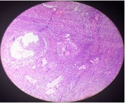
Figure 1:Low power view - granuloma H & E X40.


The various studies looked at plain observation, steroids, antibiotics, immunosuppressants like methotrexate and surgery as treatment options. Several studies found significant resolution (up to 80% in some cases) of the disease with steroids and hence the general opinion seems to be against wide local excision as the mainstay of treatment. The study by Hasan Karanlik et al in 2014 on 37 patients with one arm being treated with prednisolone followed by surgery and showing no recurrence merits attention.

surgery_219_02.JPG
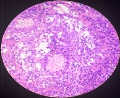
Figure 2: High power view - granuloma with cholesterol cleft and foreign body giant cell reaction H & E X100.

Discussion

Kessler and Wolloch, when they first published their findings of 5 cases of granulomatous mastitis in 1972, described it as follows - “clinically suspected of having malignant tumours and the lesions appearing to represent a well-defined entity which differed from the various other types of mastitis previously described [1]. Half a century later, we still describe it as a chronic inflammatory disease that is often misdiagnosed as cancer and the aetiology of which has not been elucidated [9]. The disease even as of today is an enigma, both in terms of diagnosis as well as response to the various forms of treatment. It is then no wonder that even fifty years after the original description of the disease we do not have a standard investigation or treatment protocol. It is postulated that IGM starts with ductal epithelial damage followed by the transition of luminal secretions to the lobular connective tissue, local inflammation in the connective

tissue, followed by macrophage-lymphocyte migration to the region and local granulomatous inflammatory response [10]. The exact aetiology of this disease is still considered unknown. The three most important factors considered in the pathogenesis of this disease are autoimmunity, infections and hormonal factors.  Autoimmunity is thought to be the most likely factor among these due to two findings – i) presence of T lymphocyte predominance on histopathologic examination and ii) the good response to steroids very often noted during treatment of this disease. It is postulated that the extravasation of milk protein within the interstitial tissues of the breast resulting from epithelial damage to the mammary ducts triggers the autoimmune response. Further damage to the lobules of the breast can occur due to chemical irritation or even due to trauma. The infection could also, by damaging the breast lobules, trigger the autoimmune reaction. Infection is considered as a possible aetiological factor because Corynebacterium, a normal skin commensal, is quite commonly found in idiopathic granulomatous mastitis, with C. kroppenstedtii, C.amycolatum and C. tuberculostearicum being isolated in decreasing order of frequency in these cases. There is a retrospective study done in 2002 that reports isolation of Corynebacterium kroppenstedtii in up to 40% of patients with IGM [11]. Idiopathic Granulomatous Mastitis (IGM) is mainly a disease of exclusion. Conditions that can mimic IGM include breast abscess, breast malignancies, tuberculous mastitis, fungal mastitis, foreign body granulomas, Wegener’s granulomatosis, sarcoidosis, mammary duct ectasia and fat necrosis [9]. Fungal agents like histoplasmosis or rarely paracoccidioidomycosis have been described as causative factors especially in the subset of immune-compromised patients like post-transplant patients, those on immunotherapy or chemotherapy, patients with malnutrition etc [12]. It has been suggested that all specimens should undergo Ziehl-Neelsen acid-fast staining to rule out tuberculosis as the cause of the mastitis because steroids used in the treatment of the disease can significantly worsen mycobacterial infections [9]. Also, in endemic regions, mycotic granulomatous mastitis should always be considered especially because the organisms are slow-growing and difficult to grow in culture, thus giving a false negative result [12].

IGM can have varied clinical presentations, which is


one of the main reasons that diagnosis becomes difficult. It could take the form of a peripheral inflammatory breast mass or as multiple abscesses with inflammation and ulceration or even present with nipple retraction and peau de orange [13]. Extramammary presentations include axillary lymphadenopathy, arthritis and erythema nodosum [9]. Rami Yaghan et al proposed a clinically based classification that considers these varied presentations which are as follows - A. hard painless breast mass B. hard painful breast mass with gross inflammation C. breast abscess like presentation and D. subacute presentation with ulceration, sinus or fistula formation. Higher grades in this classification, they postulated, would suggest an increased risk of recurrence [14]. Imaging evaluation of a case of IGM almost always is similar to that of any breast lesion. A mammogram is advised in ladies above 30 years of age and ultrasound for lesser age groups. The usual findings on mammography include thickening of the skin, focal or global asymmetry, irregular focal mass, trabecular coarseness or distortion in the parenchyma, smooth-edged mass, calcification and lymph node enlargement [15]. Ultrasound scans show skin thickening, hypoechoic mass with tubular extensions, heterogeneous parenchyma, acoustic shadowing, abscess, sinus tract formation and lymph node enlargement. On Doppler scanning, prominent arterial and venous signals can be found in the inflamed parenchyma. Compressive elastography usually shows soft properties with low elasticity scores and strain ratios. Acoustic radiation force impulse imaging shows that compared to malignant lesions, IGM tends to have low median marginal and internal velocities. On MRI scanning, skin alterations owing to inflammation are noted with T1, T2 and STIR intensity changes and intense contrast uptake with a progressive plateau or washout pattern. Other findings include mass lesions with ring enhancement, segmental regional non-mass enhancement, diffusion changes, necrosis and abscess, fistula tracts, dilated ducts with dense content, enhanced ductal walls and lymphadenopathy. non-mass enhancement is the most common finding followed by enhancing masses [10].

It must however be understood that imaging findings alone may not be able to distinguish IGM from malignancy. Hence early pathologic confirmation is mandatory especially

when antibiotics do not result in clinical improvement in a lesion of the breast showing any of the clinical characteristics of IGM described earlier. Response to treatment can be monitored using ultrasound scan or even by MRI scan, especially in aggressive, diffuse and unresponsive diseases. As with any other breast lump, FNAC or core biopsy can be used to make a diagnosis. The limitation in the use of FNAC is that it is seen to be diagnostic in only 39% of patients, whereas core biopsy is diagnostic in about 94.5% of patients. In addition, FNAC has difficulty differentiating granulomatous mastitis from other granulomatous diseases [10]. No definite treatment guidelines exist for the management of this disease although various treatment options have been tried ranging from plain observation at one end of the spectrum to surgical excision at the other. Even in recent times, recurrence rates after treatment have been noted to be in the range of about 50% suggesting that optimal treatment plans are not in place [10].

Lai et al in their case series were able to demonstrate spontaneous resolution in about 50% of cases and stable disease in the other half while keeping these patients under observation without any intervention. The period of observation in this study was approximately two years [16]. Scoglietti et al have also made a similar observation that granulomatous mastitis will usually resolve over 1-2 years if left untreated [17]. This is the basis of plain observation as a mode of treatment, at least initially, in patients with the first episode of IGM. However what makes this method of treatment unsuccessful often is the prolonged period of observation that is required, as well as the subset of patients with IGM who may be symptomatic [16].

Antibiotics are another option in the treatment of cases of IGM. The role of antibiotics is controversial because of the lack of establishment of a direct link between bacterial infiltration and the disease. When given, initial treatment should be against gram-positive bacteria because Corynebacteria is the most common species of bacteria isolated. However antibiotic administration becomes inevitable when abscesses form and incision and drainage is done. In such cases, antibiotics will need to be administered as per microbiological data.

Corticosteroids form the mainstay of treatment in IGM as of today. In a prospective study by Pandey et al, complete resolution of disease


was noted in 35 out of 44 patients treated with steroids with a median time to resolution of 5.3 months. Of the 23% had a recurrence of disease and all of them resolved with the second course of steroids [18]. In a retrospective study by Oran et al, out of 25 patients from a group of 46 who were treated with steroids, only 3 (7%) failed to respond and required surgical excision. It is suggested that doses as high as 60 mg of prednisone daily may be administered gradually tapering down the dose, with a duration of treatment of 3 to 6 months [2]. However, the side effects of long term steroid therapy in the form of weight gain, hypertension, glucose intolerance, Cushing’s syndrome and steroid myopathy are a problem [19]. Immunosuppressive agents like methotrexate and azathioprine have a role in the treatment of these cases, both as steroid-sparing agents as well as in those cases resistant to steroids [20].

Before the introduction of steroids, IGM was primarily managed with surgery. In a retrospective study by Oran et al, wide local excision with negative margins was performed in 18 patients out of 46 being followed up by the same surgical team [21]. Out of them, 3 had a recurrence of disease and they were then treated successfully with steroids and re-excision.  However now the surgical treatment of IGM is gradually falling out of favour due mainly to the good response to steroid treatment as well as due to the high rate of disfigurement, fistula formation and poor wound healing [9]. Although no established marker exists to predict post-surgical recurrence of IGM, the most widely accepted factor is the failure of the surgical excision margin. Recurrence rates described are in the range of 5 – 50% despite wide surgical margins.

Lai et al in a systematic review and meta-analysis noted that the complete remission rate on oral steroids at 71.8% was much lower than that with surgical management at 90.6% and that with oral steroids and surgery at 94.5%. Also, it was reported that the recovery period on steroids was much longer than that of surgical excision. Several births > 2, duration of lactation over 18 months, BMI of more than 31, having fistula on physical examination, abscess collection on ultrasound scan and luminal inflammatory score more than 2 were all predictors of higher risk of recurrence [22].

In a study published in 2014 by Hasan Karanlik et al, 37 patients were treated with 0.5mg/kg/day

of methylprednisolone followed by wide excision and compared with 23 patients treated with steroid alone [23]. The second group showed a 30% recurrence versus no recurrence in the first group. The interesting finding here was that histopathologic examination of resected specimens showed evidence of IGM in all these patients, despite complete radiological response confirmed by ultrasound scan or MRI scans. It is thus hypothesised that the residual area or previous lesion bed was probably the area responsible for the autoimmune process in the first place. So it is suggested that steroid therapy followed by surgery should be considered as the first-line treatment of IGM [23]. At the other extreme, there is also a contradictory view that this condition can be managed by observation and thorough drainage as the treatment modality of choice thus saving patients from the side effects of high dose steroids [24]. Also, based on a study on 14 patients, it has been suggested that the addition of azathioprine to glucocorticoid treatment initially itself will help in quick tapering of steroid doses and help increase chances of success in obtaining cure of the disease [25].

Conclusion

Idiopathic granulomatous mastitis is a relatively rare disease affecting the breasts in women of reproductive age group, which due to its varied clinical presentations, unpredictable response to treatment and similarity to malignancy remains an enigma even today. The exact aetiology of the disease has not been established as yet although autoimmunity, infections and hormonal factors are considered most likely. Imaging of the lesions using mammograms and ultrasound are the most common modalities used although MRI scanning is also being used often. There are certain features described with each of these modalities that are likely to suggest the diagnosis of IGM but none are so specific as to diagnose the disease with certainty. Hence core biopsy remains the procedure of choice to make the diagnosis, especially since FNAC has poor sensitivity.

The treatment of IGM has not yet been standardised due mainly to the lack of a substantial number of cases that could facilitate establishing treatment protocols. Various methods of treatment ranging from plain observation and


waiting for spontaneous resolution, antibiotics and steroids to surgery have been tried with varying degrees of success. Of the treatment options, steroids are the favourite due to the significant response obtained without the morbidity of surgery, although this will need further validation. The addition of azathioprine for its steroid-sparing effect as well as to avoid the adverse effects of prolonged high dose steroids has also been suggested. An interesting observation that the use of oral steroids followed by surgery can give almost complete cure needs to be studied further. It is hoped that with more cases with their clinical features, investigation reports and treatment responses being reported, a uniform investigation and treatment protocol will be put in place which will help treat surgeons as well as patients suffering from the disease immensely.

Reference

01. Kessler E, Wolloch Y. Granulomatous mastitis: a lesion clinically simulating carcinoma. Am J Clin Pathol. 1972 Dec;58(6):642-6. doi: 10.1093/ajcp/58.6.642 [Crossref][PubMed][Google Scholar]

02. Benson, John R. , and Dorin Dumitru. "Idiopathic granulomatous mastitis: presentation, investigation and management. " Future Oncology 12. 11 (2016): 1381-1394 [Crossref][PubMed][Google Scholar]

03. Gautier N, Lalonde L, Tran-Thanh D, El Khoury M, David J, Labelle M, et al. Chronic granulomatous mastitis: Imaging, pathology and management. Eur J Radiol. 2013 Apr;82(4):e165-75. doi: 10.1016/j.ejrad.2012.11.010 [Crossref][PubMed][Google Scholar]

04. Erhan Y, Veral A, Kara E, Ozdemir N, Kapkac M, Ozdedeli E, et al. A clinicopthologic study of a rare clinical entity mimicking breast carcinoma: idiopathic granulomatous mastitis. Breast. 2000 Feb;9(1):52-6. doi: 10.1054/brst.1999.0072 [Crossref][PubMed][Google Scholar]

05. Kiyak G, Dumlu EG, Kilinc I, Tokaç M, Akbaba S, Gurer A, et al. Management of idiopathic granulomatous mastitis: dilemmas in diagnosis and treatment. BMC Surg. 2014 Sep 4;14:66. doi: 10.1186/1471-2482-14-66 [Crossref][PubMed][Google Scholar]

06. Pandey TS, Mackinnon JC, Bressler L, Millar

A, Marcus EE, Ganschow PS. Idiopathic granulomatous mastitis--a prospective study of 49 women and treatment outcomes with steroid therapy. Breast J. 2014 May-Jun;20(3):258-66. doi: 10.1111/tbj.12263 [Crossref][PubMed][Google Scholar]

07. Imoto S, Kitaya T, Kodama T, Hasebe T, Mukai K. Idiopathic granulomatous mastitis: case report and review of the literature. Jpn J Clin Oncol. 1997 Aug;27(4):274-7. doi: 10.1093/jjco/27.4.274 [Crossref][PubMed][Google Scholar]

08. Baslaim MM, Khayat HA, Al-Amoudi SA. Idiopathic granulomatous mastitis: a heterogeneous disease with variable clinical presentation. World J Surg. 2007 Aug;31(8):1677-81. doi: 10.1007/s00268-007-9116-1 [Crossref][PubMed][Google Scholar]

09. Mathew Thomas V, Alexander SA, Bindal P, Vredenburgh J. Idiopathic Granulomatous Mastitis-A Mystery Yet to be Unraveled: A Case Series and Review of Literature. Cureus. 2020 Feb 5;12(2):e6895. doi: 10.7759/cureus.6895 [Crossref][PubMed][Google Scholar]

10. Halim NA, Uthman I, Rammal R, Assi HI. Idiopathic Granulomatous Mastitis Presenting as a Breast Pseudotumor: Case Reports with Review of the Literature. Case Rep Rheumatol. 2018 Dec 27;2018:4264012. doi: 10.1155/2018/4264012 [Crossref][PubMed][Google Scholar]

11. Paviour S, Musaad S, Roberts S, Taylor G, Taylor S, Shore K, et al. Corynebacterium species isolated from patients with mastitis. Clin Infect Dis. 2002 Dec 1;35(11):1434-40. doi: 10.1086/344463 [Crossref][PubMed][Google Scholar]

12. Merino-Alado R, Pineda J, Rasquin JH, Landaeta ME, Mata-Essayag S. Granulomatous mastitis due to coinfection with Histoplasma sp. and Paracoccidioides sp. : A case report. Med Mycol Case Rep. 2020 Jan 16;27:52-54. doi: 10.1016/j.mmcr.2020.01.006 [Crossref][PubMed][Google Scholar]

13. Maione C, Palumbo VD, Maffongelli A, Damiano G, Buscemi S, Spinelli G, et al. Diagnostic techniques and multidisciplinary approach in idiopathic granulomatous mastitis: a revision of the literature. Acta Biomed. 2019 Jan 23;90(1):11-15. doi: 10.23750/abm.v90i1.6607 [Crossref][PubMed][Google Scholar]


14. Yaghan R, Hamouri S, Ayoub NM, Yaghan L, Mazahreh T. A Proposal of a Clinically Based Classification for Idiopathic Granulomatous Mastitis. Asian Pac J Cancer Prev. 2019 Mar 26;20(3):929-934. doi: 10.31557/APJCP.2019.20.3.929 [Crossref][PubMed][Google Scholar]

15. Durur-Subasi I. Diagnostic and Interventional Radiology in Idiopathic Granulomatous Mastitis. Eurasian J Med. 2019 Oct;51(3):293-297. doi: 10.5152/eurasianjmed.2019.19211 [Crossref][PubMed][Google Scholar]

16. Lai EC, Chan WC, Ma TK, Tang AP, Poon CS, Leong HT. The role of conservative treatment in idiopathic granulomatous mastitis. Breast J. 2005 Nov-Dec;11(6):454-6. doi: 10.1111/j.1075-122X.2005.00127.x [Crossref][PubMed][Google Scholar]

17. Scoglietti V, Woodridge R, Leitch M et al. Development and initiation of a clinical protocol for the treatment of idiopathic granulomatous mastitis. J Rare Disord. 2014;2(1):1-6. [Crossref][PubMed][Google Scholar]

18. Pandey TS, Mackinnon JC, Bressler L, Millar A, Marcus EE, Ganschow PS. Idiopathic granulomatous mastitis--a prospective study of 49 women and treatment outcomes with steroid therapy. Breast J. 2014 May-Jun;20(3):258-66. doi: 10.1111/tbj.12263 [Crossref][PubMed][Google Scholar]

19. Moris D, Damaskos C, Davakis S, Vailas M, Garmpis N, Spartalis E, et al. Is idiopathic granulomatous mastitis a surgical disease? The jury is still out. Ann Transl Med. 2017 Aug;5(15):309. doi: 10.21037/atm.2017.05.24 [Crossref][PubMed][Google Scholar]

20. Yılmaz TU, Gürel B, Güler SA, Baran MA, Erşan B, Duman S, Utkan Z. Scoring Idiopathic Granulomatous Mastitis: An Effective System for Predicting Recurrence? Eur J Breast Health. 2018 Apr 1;14(2):112-116. doi: 10. 5152/ejbh.2018.3709 [Crossref][PubMed][Google Scholar]

21. Baslaim MM, Khayat HA, Al-Amoudi SA. Idiopathic granulomatous mastitis: a heterogeneous disease with variable clinical presentation. World J Surg. 2007 Aug;31(8):1677-81. doi: 10.1007/s00268-007-9116-1 [Crossref][PubMed][Google Scholar]

22. Zhou F, Yu LX, Ma ZB, Yu ZG. Granulomatous lobular mastitis. Chronic Dis Transl Med. 2016 Apr 22;2(1):17-21. doi: 10.1016/j.cdtm.2016.02.004 [Crossref][PubMed][Google Scholar]

23. Karanlik H, Ozgur I, Simsek S, Fathalizadeh A, Tukenmez M, Sahin D, et al. Can Steroids plus Surgery Become a First-Line Treatment of Idiopathic Granulomatous Mastitis? Breast Care (Basel). 2014 Oct;9(5):338-42. doi: 10. 1159/000366437 [Crossref][PubMed][Google Scholar]

24. Yukawa M, Watatani M, Isono S, Fujiwara Y, Tsujie M, Kitani K, et al. Management of granulomatous mastitis: a series of 13 patients who were evaluated for treatment without corticosteroids. Int Surg. 2015 May;100(5):774-82. doi: 10.9738/INTSURG-D-14-00231.1 [Crossref][PubMed][Google Scholar]

25. Konan A, Kalyoncu U, Dogan I, Kiliç YA, Karakoç D, Akdogan A, et al. Combined long-term steroid and immunosuppressive treatment regimen in granulomatous mastitis. Breast Care (Basel). 2012 Aug;7(4):297-301. doi: 10.1159/000341388 [Crossref][PubMed][Google Scholar]