E-ISSN:2455-5436
P-ISSN:2456-9518
RNI:MPENG/2017/70870

Research Article

Prospective

Surgical Review: International Journal of Surgery Trauma and Orthopedics

2020 Volume 6 Number 6 November-December
Publisherwww.medresearch.in

Clinicopathological features of Pseudocyst Pancreas and its Management: A Prospective and Retrospective Study

Koshariya M.1, Sharma A.2*, Gupta B.3, Suroshe T.4, Singour J.5, Songra M C.6
DOI: https://doi.org/10.17511/ijoso.2020.i06.01

1 Mahim Koshariya, Professor, Department of Surgery, Gandhi Medical College, Bhopal, Madhya Pradesh, India.

2* Agam Sharma, Senior Resident, Department of Surgery, Government Medical College, Shivpuri, Madhya Pradesh, India.

3 Brahmanand Gupta, Senior Resident, Department of Surgery, Government Medical College, Shivpuri, Madhya Pradesh, India.

4 Tushar Suroshe, Senior Resident, Department of Surgery, Gandhi Medical College, Bhopal, Madhya Pradesh, India.

5 Jaiprakash Singour, Senior Resident, Department of Surgery, Gandhi Medical College, Bhopal, Madhya Pradesh, India.

6 Songra M C, Professor and Head of Department, Senior Resident, Department of Surgery, Gandhi Medical College, Bhopal, Madhya Pradesh, India.

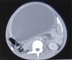
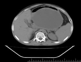
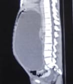
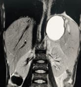
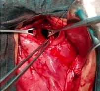
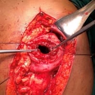
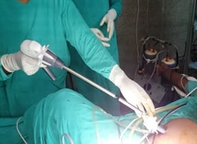
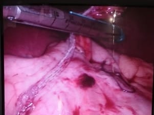
Background: Revised Atlanta Classification has veered a change in our understanding of pseudocyst pancreas which mandates renewed inquiry into pseudocysts defined as per new criteria. The present study provides an overview of experience with Pseudocyst Pancreas for over a decade. Methodology: 100 cases of pseudocysts diagnosed over the last 10 years at GMC, Bhopal, conforming to the present definition were reviewed. Cysts with the inhomogeneous collection, debris, necrosis, or any other non-liquid component, specifically in those diagnosed before 2012 were excluded. Relevant data were analyzed. Results: The majority were male (85%) in the age group of 40-50 years with alcohol-induced chronic pancreatitis (77%) being the most frequent etiology. Abdominal pain (40%), lump (30%), and abdominal tenderness (59%) were common at presentation. 58% were in the Head of the pancreas, 29% in the Neck and Body, and 13% in the Tail and surrounding areas. Mean cyst diameter was 8.6cm and volume 252cc. 85% were managed surgically and 40% of those managed conservatively also needed surgical intervention eventually due to complications. History of chronic alcoholic pancreatitis, the large size of the cyst (≥6cm and ≥60cc), and communication with the main pancreatic duct were highly predictive of surgical intervention. Conclusion: Radiological characteristics along with the clinical picture may suggest appropriate intervention. Surgery remains the principal modality of treatment, with high success rates.

Keywords: Pseudocyst, Pancreas, Pancreatitis

Corresponding Author How to Cite this Article To Browse
Agam Sharma, Senior Resident, Department of Surgery, Government Medical College, Shivpuri, Madhya Pradesh, India.
Email:
Koshariya M, Sharma A, Gupta B, Suroshe T, Singour J, Songra M C. Clinicopathological features of Pseudocyst Pancreas and its Management: A Prospective and Retrospective Study. Surgical Rev Int J Surg Trauma Orthoped. 2020;6(6):338-346.
Available From
https://surgical.medresearch.in/index.php/ijoso/article/view/200

Manuscript Received Review Round 1 Review Round 2 Review Round 3 Accepted
2020-08-26 2020-11-03 2020-11-19 2020-12-12
Conflict of Interest Funding Ethical Approval Plagiarism X-checker Note
No Nil Yes 6%

© 2020 by Mahim Koshariya, Agam Sharma, Brahmanand Gupta, Tushar Suroshe, Jaiprakash Singour, Songra M C and Published by Siddharth Health Research and Social Welfare Society. This is an Open Access article licensed under a Creative Commons Attribution 4.0 International License https://creativecommons.org/licenses/by/4.0/ unported [CC BY 4.0].

Introduction

Pancreatic pathologies include the diverse entities of pancreatic fluid collections and cystic pancreatic lesions of which the pseudocyst pancreas embodies an important component. Accounting for about 75% of cystic lesions of the pancreas [1], they are classically differentiated from other peripancreatic fluid collections (cystic neoplasms and congenital, parasitic, and extrapancreatic cysts) by their lack of an epithelial lining, high concentration of pancreatic enzymes within the pseudo pancreatic cyst commonly forming after an episode of pancreatitis or pancreatic trauma.

The revised Atlanta classification in 2012 has improved standardized reporting and effective communication between radiologists and clinicians by the first categorization of the type of acute pancreatitis into necrotizing pancreatitis and interstitial edematous pancreatitis (IEP), based on the presence or absence of necrosis, respectively, and further identifying distinct pancreatic collection subtype according to the time elapsed since the onset of pancreatitis. Such precise description of pancreatitis pseudocysts can now be defined as cystic collections after Interstitial Edematous Pancreatitis (IEP) in more than 4 weeks with a well-defined wall and absence of any solid component or necrotic debris [2].

The revision has maneuvered a rational change in the operational use of the term pseudocyst pancreas. A better understanding of the natural course of Pseudocyst Pancreas and an increasing focus on minimally invasive procedures has also led to a dramatic shift from an unquestioned aggressive approach to the exploration of cautious conservative options. In the last two decades, diagnosis and management have increasingly been supplemented by endoscopy, mostly reducing open surgery to a salvage intervention in case of failure and complication. However, the lack of endoscopic facilities and dedicated gastroenterology units especially in developing countries with the high patient load still necessitates the utilization of open techniques and their role as such cannot be overlooked.

But overall treatment planning remains variable, and apart from a few clear indications like infection of the cyst or biliary obstruction, intervention, and management strategies are neither well defined nor standardized in the published data [3].

A clear lack of global consensus on an optimal management strategy warrants further scientific inquiry, as the question of when and whom to treat remains widely unsettled. The revision of the Atlanta Classification has also rendered the already limited number of retrospective studies focusing on pseudocysts of the pancreas obsolete [4]. The present study aims to provide a descriptive overview of experience in the management of pseudocyst pancreas in the last decade at a tertiary care center in a developing country with a large catchment area catering to almost 10 million people and evaluate the same especially in the light of newer understanding of this entity.

Materials and methods

Study Setting: The study was conducted in the Department of Surgery, Gandhi Medical College and Hospital, a tertiary referral center in Bhopal.

Duration and type of study: The actual study was conducted from 2017-2019. It is descriptive, with retrospective and prospective data collected on patients treated for pseudocyst pancreas at GMC, Bhopal from 2009-2019. Retrospective data collection was for patients presenting with pancreatic pseudocysts between 2009 and 2017 and prospective from 2017 to 2019.

Sampling methods: Non-probability purposive sampling technique

Sample size calculation: A sample size of 96 was established considering a 95% confidence interval with a 10% margin of error and an engaging round figure of 100 was determinatively assumed.

Inclusion criteria: All patients with pseudocyst pancreas diagnosed with pseudocyst pancreas were included. In patients with pseudocysts diagnosed before 2012, only those with clinical and radiological features conforming with the current guidelines adapted from the Revised Atlanta Classification were selected for the study.

Exclusion criteria: Cysts with the inhomogeneous collection, debris, necrosis, or any non-liquid component, specifically in those diagnosed before 2012 were excluded.

Data collection procedure: After proper permissions, case files of patients with pseudocyst pancreas from January 2009- January 2017 were drawn from the Records Section and clinical notes and investigation reports were evaluated.


Patients admitted from Feb 2017- Jan 2019 were closely followed and evaluated prospectively.

Case data included detailed clinical history including evaluation of precipitating Acute or Chronic Pancreatitis based on presentation, the pattern of a single episode or recurrent acute exacerbation of symptoms, and time since the first onset. Radiological characteristics of the pseudocyst were assessed and re-evaluated in the light of newer criteria for the diagnosis of pseudocysts especially in those diagnosed before 2012.

Ethical consideration and permission: Institute’s Ethical Committee approval and permission to undertake the study were taken through the proper protocol. Informed consent from patients included in the prospective part of the study was obtained and permission for waiver of consent for cases collected retrospectively was granted by the board.

Statistical Analysis: The statistical analyses of the data was performed using SPSS version 25.0. The graphs were plotted using Sigma Plot 12.0 and MS Excel. The qualitative characteristics were analyzed using Chi-Square and Fisher’s Exact test. The results on quantitative parameters were computed and their mean and standard deviations were obtained. Variables were assessed for their effect on the clinical outcome using Statistical models to evaluate factors predictive of therapeutic decisions.

Results

The majority of patients were 30-50 years of age with a mean age of 38.9 years. Overall affection in males was considerably high (male/female: 85/15). 78% of patients were from Low and Upper Lower socio-economic class as per Modified Kuppuswamy Scale. Rest belonged to the lower middle class.

Various clinical, etiological, radiological, and biochemical parameters are summarised in Table 1. The most common presenting complaint was abdominal pain (40%) associated with an array of upper gastrointestinal symptoms like nausea, bloating, indigestion, etc. Clinical jaundice, palpable lump, and subjective weight loss were also reported.

Table-1: Clinical, etiological, radiological, and biochemical parameters.

Clinical, etiological, radiological, and biochemical parameters Total N=100
Chief Symptom
Abdominal pain
Lump
Jaundice
Nausea/Vomiting
Weight Loss
Low-Grade Fever
40%
30%
6%
18%
2%
4%
Etiology
Alcoholic pancreatitis
Biliary pancreatitis
Trauma
Idiopathic
77%
13%
9%
1%
Radiological Features
Mean Volume of Cyst at Imaging (cubic cm)
Average of Maximum Cyst Diameter (in cm)
Ascites
CBD Dilation
Cyst Communication with Main Pancreatic Duct
252.5 ± 274.1
8.6 ± 2.6
4%
6%
34%
Biochemical lab values (Average)
Serum Amylase
Serum Lipase
Serum Bilirubin (Direct)
SGOT
SGPT
Alkaline Phosphatase
125 IU/l
103 IU/l
0.33 mg/dl
42 IU/l
46 IU/l
62 IU/l

20% of patients had pseudocyst following Acute Pancreatitis and 80% had Chronic Pancreatitis. Alcoholic pancreatitis was the most common etiology (77%), followed by Biliary pancreatitis (13%), Trauma(9%), and Idiopathic(1%). 48.05% of the patients with alcoholic etiology were also smokers.

21% of the patients had an associated medical illness. 12% of the patients had diabetes, 11% had hypertension and 6% had obesity with diabetes and hypertension. 2 patients had active Tuberculosis for which they were on DOTS, and 1 patient had chronic ITP who was on prednisolone.

Extremely wide variation was noted in individual cyst characteristics on imaging (Figures 1-3). Overall 58% of the cysts were found in the Head of the pancreas, 29% in the Neck and Body, and 13% in the Tail and surrounding areas.

However, the location of cysts in the surgical intervention group was more than three times more frequent at the head as compared to that in the conservative group. The most common location of cyst in the conservative group was the body of the pancreas.


surgical_200_01.jpg

surgical_200_03.jpgsurgical_200_02.jpg

Fig-1: CECT Scan images: A) Well defined, homogenously hypodense lesion about the pancreatic neck, body, and tail, extending into the lesser sac, displacing stomach anteriorly) in Axial View. B) Well defined, a homogenously hypodense lesion about the pancreatic neck, body, and tail, extending into the lesser sac, compressing stomach anteriorly

surgical_200_04.jpg

Fig-2: CT Scan image of pseudocyst pancreas in the sagittal plane.

surgical_200_05.jpg

Fig-3: MRCP showing pseudocyst pancreas.

Radiological characteristics were evaluated for two treatment groups, surgical intervention, and conservative, and cyst size exhibited significant variation between the two. The average size of the cyst in its largest diameter was 8.6 cm and volume 252 cc.

The maximum diameter and volume of the cyst were tested for significance in the conservative and surgical group and were found to be significantly larger in the surgical group (p=0.000 each).

A larger size of the cyst(>6cm) predicted a tendency for surgical intervention. (p=0.0001). The volumes of pseudocyst ranged from 10-130cc in the conservative group, while in the surgical group-wide variation in the recorded volume of pseudocysts was noted (1.09-1600 cc).

The average time in months between the presentation of the patient with pseudocyst and assumed precipitating factor was comparable in the two groups (5.23 months in the conservative group vs 6.34 months in the surgical group). Communication with Main Pancreatic Duct was significantly more in the surgical group (P = 0.002)

Treatment leaned more towards intervention via the surgical approach, the ratio being similar in males and females. Surgical intervention was undertaken in 85% of the cases.


In those managed conservatively, 40% of patients required an eventual intervention and 60% underwent complete resolution.

The mean time of follow-up was 6 weeks. Management Schemes in our patients are represented in Table 2.

Table-2: Management strategies (n=100).

Surgical intervention Conservative
Complete Resolution Did Not Resolve (Needed Eventual Intervention)
85 9 6

Of 85 patients who underwent primary surgical intervention, Cystogastrostomy (Figures 4-8) was the most commonly performed surgical intervention done in 52 (61.1%) patients of which 48 were performed with an open approach and the rest were done laparoscopically (Figures 7-8). Cholecystectomy was done simultaneously in 3 patients.

Cystojejunostomy was done in 11(12.9%) patients and cystoduodenostomy in 3(3.5%). Open External Drainage was done in 14(16.5%) where intraperitoneal adhesions, infected pseudocysts, and thin immature pseudocyst wall prohibited internal drainage.

4(4.8%) patients with poor general condition underwent Percutaneous Ultrasound-guided External Drainage. Recurrence was noted in 2 of the patients after percutaneous aspiration and cystogastrostomy was performed eventually.

surgical_200_06.jpg

Fig-4: Patient with pseudocyst pancreas presenting with a lump in the upper abdomen.

surgical_200_07.jpg

Fig-5: Intraoperative transgastric pseudocyst aspiration and confirmation.

surgical_200_08.jpg

surgical_200_09.jpg

Fig-6 (a) and (b): Pseudocystogastric anastomosis


surgical_200_10.jpg

Fig-7: 12mm umbilical port with insertion of Endo GIA Echelon flex 60 mm Stapler for Laparoscopic cystogastrostomy.

surgical_200_11.jpg

Fig-8: Intraoperative picture Showing closure of anterior gastric wall using Endo GIA Stapler during of Laparoscopic Pseudocystogastrostomy.

1(1.2%) had pseudocyst complicated with hemorrhage and was clinically unstable at presentation; emergency laparotomy was performed in this patient and distal pancreatectomy with splenectomy done.

In those managed conservatively, 6 patients had the increasing size of cyst on follow up which was managed with internal drainage eventually. During follow-up in the surgical intervention group, complications included postoperative surgical site infection in 8, recurrent pain episodes in 6, and pleural effusion in 4 patients.

Pseudocyst content and wall biopsy were sent for cytological and histopathological evaluation. Mean Cyst Fluid amylase was 715 IU/L. CEA (Carcino-Embryonic Antigen) values were available for only

24 patients with a mean of 0.48 ng/ml. The cyst wall was negative for malignancy in all cases.

Table-3: Surgical procedures.

Intervention N=85
Cystogastrostomy 61.1%
Cystojejunostomy 12.9%
Cystoduodenostomy 3.5%
Open External Drainage 16.5%
Percutaneous Drainage(Pigtail) 4.8%
Distal Pancreatectomy 1.2%

Table-4: post-operative complications

Complications N=85
Surgical Site Infection 9.41%
Pain 7%
Black Stool 3.5%
Recurrence 2.3%
Pleural Effusion 4.7%
Post-op Pancreatitis 1.1%
Pancreatic Abscess 2.3%

Discussion

Many older studies have extensively evaluated the incidence of pseudocysts in both acute and chronic pancreatitis. The relative incidence of pseudocysts in an acute or chronic setting in these studies varied and depended on how pancreatic pseudocysts were defined and by what means they were detected at the time. With the change in the operational definition of the pseudocyst, however, pseudocysts after acute pancreatitis are now hardly diagnosed.

Overall, the highest incidence of pancreatic pseudocysts has commonly been reported in alcoholic patients with chronic pancreatitis [5,6,7,8]. In the present study, pseudocysts most frequently developed in male patients with chronic alcohol-induced pancreatitis, consistent with the literature.

Alcohol metabolism has been understood to cause oxidative injury and thus disruptions of the pancreatic duct followed by extravasation of pancreatic secretions [9,10]. Chronic inflammation may lead to the establishment of a connection between the pseudocyst and the main pancreatic duct. The current study noted a demonstrable communication in almost one third (34%) of our patients.

The average diameter of the cyst and mean volume of cyst in these patients were quite large, 9.3 cm and 280.70 cc respectively.


The larger size and volume of the pseudocyst is attributable to its connection with the main pancreatic duct which acts as a conduit for continuous drainage of pancreatic secretions, preventing its spontaneous regression. Interestingly, 100% of these patients underwent surgical intervention which was statistically significant (p=0.002).

The current study evaluated our data for the two lines of management: Conservative or Interventional (Surgical drainage). 85% of the patients underwent procedures for surgical drainage, rest were given conservative support. All patients were evaluated for resolution at 6 weeks. In those managed conservatively, 60% underwent complete resolution, and 40% of patients required an eventual intervention for increasing the size of the cyst.

Current therapeutic strategies include endoscopic transpapillary or transmural drainage, percutaneous catheter drainage, or open surgery. In a large systematic review and meta-analysis published in 2019, Galileu et al [11] analyzed data from numerous studies comparing endoscopic versus surgical treatment for pancreatic pseudocysts and reported no significant difference between surgical and endoscopic treatment success rates, adverse events, and recurrence for pseudocyst.

Laparoscopy has contributed enormously in the management of pseudocysts and excellent outcomes are achieved with minimally invasive procedures. The present study notes the lack of endoscopic procedures in the management of pseudocyst at our center, however, in most centers throughout the developing world surgery remains the primary intervention technique and its importance cannot be disregarded as endoscopic treatment facilities are either non-existent or lack specialists prevent its widespread use.

The spectrum of presentation of pancreatic pseudocysts ranges from completely asymptomatic lesions to multiple pseudocysts with pancreatic and bile duct obstruction. The latter are traditionally considered to require immediate endoscopic or surgical intervention to prevent secondary complications. Up to 50% of pancreatic pseudocysts resolve without interventions [12].

However, the selection of the right patient for conservative management is important as the prediction of the timepoint of the development of complications is uncertain.

Delay in decision making may be catastrophically contributing to higher incidences of postoperative complications, readmission, morbidity, and mortality as noted by Ito K et al [13] in their study evaluating delayed surgical intervention and adverse outcomes.

Overall rates of intervention were significantly more in those with larger cysts especially in patients with a cyst size of >6 cm (OR,56; CI, 95%; p=0.0001). Radiologically, pseudocyst was localized to the head of the pancreas 3.4 times more frequently in patients who underwent intervention. However radiological appearance of pseudocyst in areas other than the head of the pancreas (neck, body, tail, and surrounding areas) was a more significant indicator of surgical intervention (OR, 5.78; CI 95%; p=0.021).

Recurrence after surgical drainage occurred in 2 patients and abscess developed in 2, all of whom required re-intervention (surgery). In the conservative group, the additional therapeutic intervention was required in 6 patients due to non-resolution or other complications. Comparison of data on patients requiring a second or additional surgical intervention was significantly higher in the conservative group (OR 13.5; CI 95%; p=0.0006). Compelling to note, in all patients with failure of conservative management, the mean diameter of the cyst was 6.5 cm and the volume of pseudocyst was 60 cc and above (p=0.0002).

Jai-Hui Tan et al [14] evaluated potential risk factors for pseudocysts, with regards to their formation and need for intervention and noted only alcohol and chronic pancreatitis to be the common denominator. However, their comparative radiological variables between interventional and observational groups consisted of size, cyst location, and several cysts of which only size > 6cm was significant. The present study explored other radiological variables and found that a cyst size of ≥6cm, a volume of ≥60cc, and communication with the main pancreatic duct was predictive of an eventual surgical intervention even in asymptomatic patients.

Thus, primary intervention in such patients, even if asymptomatic at presentation, may be considered as a definitive plan of treatment. Though the overall location of the cyst was more common in the head (in both surgical and additional intervention groups), the mere presence of a cyst in the head of the pancreas could not predict a final surgical intervention (p=0.0752).


The major limitation of the present study is the retrospective nature for collection of a major portion of data and assessment of optimal management, whether conservative or interventional, relying on the availability of follow up case notes. Further prospective studies with a larger sample size considering the new operational definition for pseudocysts will help evaluate the objective utilization of clinical and radiological features alone at the time of presentation for deciding the best course of management.

Conclusion

The introduction of new and sensitive imaging techniques permits the detection of more pancreatic cystic lesions with better evaluation of adjacent structures and therefore, better treatment planning. Radiological characteristics are objective and should be considered in addition to the clinical picture to decide the time point of intervention for minimal complications. The risk of complications increases with time and drainage of large pseudocysts (≥6 cm and ≥60cc volume) especially with the communication channel connecting the cyst with the main pancreatic duct should not be postponed. Thus, surgical management of pseudocysts remains an important component especially if the pseudocyst is part of a more complex pathology. Endoscopic drainage being minimally invasive should be considered as the first line of management however the choice between surgical and endoscopic therapy remains to be made based on resource availability, individual experience, and specific characteristics of the pancreatic pseudocyst.

What does the study add to the existing knowledge?

The study proposes correlative use of radiological findings of pseudocyst volume (≥60cc) and communication with the main pancreatic duct in addition to cyst diameter (≥6 cm) to decide the time point of intervention in the pseudocyst pancreas when the clinical picture is equivocal. An early intervention even in an asymptomatic patient with the above radiological parameters will reduce the risk of further morbidity and complications.

Author’s Contribution

Dr. MC Songra and Dr. Mahim Koshariya conceived the idea, designed and supervised the overall project.

Dr. Agam Sharma took the lead in manuscript preparation and contributed along with Dr. Brahmanand Gupta, Dr. Tushar Suroshe, and Dr. Jai Prakash Singour in data collection and analysis. All authors provided critical feedback and helped shape the research, analysis, and manuscript.

Reference

  1. Costa P, Rego A, Araujo-Filho I. Pancreatic cystic lesions- classification, diagnosis and treatment. Int Surg J. 2016;3(2)443-451.
  2. Banks PA, Bollen TL, Dervenis C, Gooszen HG, Johnson CD, Sarr MG, et al. Classification of acute pancreatitis—2012- revision of the Atlanta classification and definitions by international consensus. Gut. 2013;62(1)102-111.
  3. Tan J, Zhou L, Cao R, Zhang G. Identification of risk factors for pancreatic pseudocysts formation, intervention and recurrence- a 15-year retrospective analysis in a tertiary hospital in China. BMC Gastroenterology. 2018;18(1)1-9.
  4. Yip H, Teoh A. Endoscopic Management of Peri-Pancreatic Fluid Collections. Gut Liver. 2017;11(5)604-611.
  5. Usatoff V, Brancatisano R, Williamson R. Operative treatment of pseudocysts in patients with chronic pancreatitis. Brit J Surg. 2000;87(11)1494-1499.
  6. Aghdassi AA, Mayerle J, Kraft M, Sielenkämper AW, Heidecke CD, Lerch MM. Pancreatic pseudocysts – when and how to treat?. HPB. 2006;8(6)432-441.
  7. Habashi S, Draganov P. Pancreatic pseudocyst. World J Gastroenterol. 2009;15(1)38.

  1. Guardado-Bermúdez F, Azuara-Turrubiates AJ, Ardisson-Zamora FJ, Guerrero-Silva LA, Villanueva-Rodríguez E, Gómez-de Leija NA, et al. Pancreatic pseudocyst- Case report and literature review. Cir. 2014;82(4)425-431.
  2. Samokhvalov AV, Rehm J, Roerecke M. Alcohol consumption as a risk factor for acute and chronic pancreatitis- a systematic review and a series of meta-analyses. E Bio Medicine. 2015;2(12)1996-2002.
  3. Aghdassi A, Mayerle J, Kraft M, Sielenkämper AW, Heidecke CD, Lerch MM. Diagnosis and treatment of pancreatic pseudocysts in chronic pancreatitis. Pancreas. 2008;36(2)105-112.
  4. Farias GFA, Bernardo WM, De Moura DTH, et al. Endoscopic versus surgical treatment for pancreatic pseudocysts- Systematic review and meta-analysis. Med (Baltimore). 2019;98(8)e14255.
  1. Almaihan A, Matar A, Murshid E, Al-Ostaz S, Shebly A, Miftah M, et al. Conservative management versus surgical drainage in pancreatic pseudocyst. Int Surg J. 2018;5(7)2383.
  2. Ito K, Perez A, Ito H, Whang EE. Pancreatic pseudocysts- is delayed surgical intervention associated with adverse outcomes. J Gastrointest Surg. 2007;11(10)1317–1321.
  3. Tan JH, Zhou L, Cao RC, Zhang GW. Identification of risk factors for pancreatic pseudocysts formation, intervention and recurrence- a 15-year retrospective analysis in a tertiary hospital in China. BMC Gastroenterology. 2018;18(1):1-9.