E-ISSN:2455-5436
P-ISSN:2456-9518
RNI:MPENG/2017/70870

Research Article

Fracture

Surgical Review: International Journal of Surgery Trauma and Orthopedics

2020 Volume 6 Number 5 September-October
Publisherwww.medresearch.in

Comparison of clinical, radiological, and functional outcome of closed fracture of distal third tibia treated with nailing and plate osteosynthesis

Vora J.1, Jetpariya D.2*, Desai K.3
DOI: https://doi.org/10.17511/ijoso.2020.i05.02

1 Jinesh Vora, Associate Professor, C.U Shah Medical College and Hospital, Surendranagar, Gujarat, India.

2* Divyesh Jetpariya, Postgraduate Student, C.U Shah Medical College and Hospital, Surendranagar, Gujarat, India.

3 Kabir Desai, Senior Resident, C.U Shah Medical College and Hospital, Surendranagar, Gujarat, India.

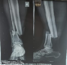
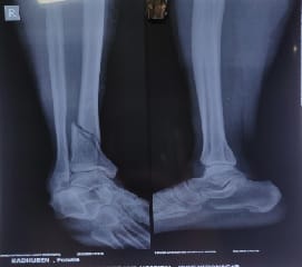
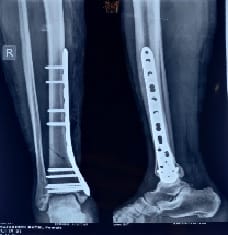
Aim: This is a prospective study of 30 patients with distal tibia fracture (Closed extra-articular distal third tibia fractures - 4 to 11cm from tibial plafond) who underwent surgical fixation were included in this study after excluding compound, pathological and pediatric fractures. Materials and Methods: 15 underwent closed intramedullary interlocking nail and 15 were treated with plate osteosynthesis (MIPO). Results: The age distribution ranged from 23 to 68 years with the mean age of 39.4 years. The mode of injury in all patients was due to vehicle accidents. All 30 patients were classified according to AO classification of which 15 belonged to A1, 14 belonged to A2, and 1 belonged to A3. The time of fixation from injury varied from 6 hours to 18 days. Conclusion: Plate osteosynthesis by minimally invasive technique and Intramedullary interlocking nailing is an equally effective method of stabilization for distal tibia fracture when considering the union rates and final functional outcome. However, malunion, nonunion and secondary procedures were more frequent after intramedullary interlocking nail. In MIPO platting Infection followed by an exposed plate occurs in 2 patients. Randomized prospective evaluation of distal tibia fractures may clarify the efficacy of plate versus nail treatment and optimize patient care.

Keywords: Distal tibial locking plate, Tibial nailing, MIPPO

Corresponding Author How to Cite this Article To Browse
Divyesh Jetpariya, Postgraduate Student, C.U Shah Medical College and Hospital, Surendranagar, Gujarat, India.
Email:
Vora J, Jetpariya D, Desai K. Comparison of clinical, radiological, and functional outcome of closed fracture of distal third tibia treated with nailing and plate osteosynthesis. Surgical Rev Int J Surg Trauma Orthoped. 2020;6(5):298-304.
Available From
https://surgical.medresearch.in/index.php/ijoso/article/view/199

Manuscript Received Review Round 1 Review Round 2 Review Round 3 Accepted
2020-08-18 2020-08-28 2020-08-31 2020-09-02
Conflict of Interest Funding Ethical Approval Plagiarism X-checker Note
No Nil Yes 7

© 2020 by Jinesh Vora, Divyesh Jetpariya, Kabir Desai and Published by Siddharth Health Research and Social Welfare Society. This is an Open Access article licensed under a Creative Commons Attribution 4.0 International License https://creativecommons.org/licenses/by/4.0/ unported [CC BY 4.0].

Introduction

The mode of treatment of distal third tibial fractures is still controversial. Distal third tibial fractures differ from proximal third fractures by their difference in anatomy and difference in healing potential [1]. In current orthopedic practice, minimally invasive plating osteosynthesis (MIPO) and interlocking nailing are the preferred techniques for fractures of the distal third tibia. The intramedullary nail spares the extraosseous blood supply, allows load sharing, and avoids extensive soft tissue dissection [2-3]. However, proximal and distal shaft fractures can be difficult to control with an intramedullary device, increasing the frequency of malalignment [4]. Concerns regarding difficulties with reduction/loss of reduction, inappropriate fixation in fractures with articular extension, anterior knee pain [5], and hardware failure have slowed the acceptance of intramedullary nailing as a treatment of fractures of the distal tibia. The recent innovation of nails with tip locking is a testimony that earlier nails were insufficient fixation tools for the distal tibia; however, tip locking is technically difficult and fractures that require it are essentially difficult to fix with nails [2-6].

Aim

A comparative study was conducted on the clinical and radiological union in either of the closed IM interlocking nail or plate and screw fixation. The advantages, disadvantages, follow‑up, complications if any and overall functional outcome will be evaluated in these patients.

Materials and Methods

This is a prospective study of 30 patients with distal tibia fracture (closed extra-articular distal third tibia fractures 4–11 cm from tibial plafond) who underwent surgical fixation were included in this study after excluding compound, pathological, and pediatric fractures. Fifteen underwent closed IM interlocking nail and 15 were treated with minimally invasive plate osteosynthesis (MIPO).

The age distribution ranged from 23 to 68 years with the mean age of 39.4 years. The mode of injury in all patients was due to vehicle accidents. All 30 patients were classified according to A0 classification of which 15 belonged to A1, 14 belonged to A2, 1 belonged to A3. The time of fixation from injury varied from 6 hours to 18 days.

All 15 patients who underwent IM interlocking nail were operated on under regional anesthesia with the patient’s supine on a standard radiolucent table by the patellar splitting approach. Nailing was done using the standard technique and all fractures were fixed with two proximal and three distal locking screws.

All 15 patients who underwent plate osteosynthesis by the MIPO technique were operated on under regional anesthesia with the patient supine on a standard radiolucent table. Through medial approach reduction of the fracture site was achieved and fixed with pre-contoured plates and appropriate screws by the minimally invasive technique under the guidance of an image intensifier.

The decision for adjunctive fibular stabilization as well as the number of the orientation of distal locking bolts was

made at the surgeon’s discretion. Duration of surgery and blood loss intra-operatively was estimated for all 30 patients. None of the patients were operated on under tourniquet control.

Duration and type of study: 1/8/2018 to 30/7/2020, observational

Sampling technique: non-probability

Data Collection method: Semi-structured and Questionnaire rating scale

Inclusion criteria:

  1. Closed fractures of the distal 1/3rd shaft of tibia
  2. Age Limit: 18 years and above

Exclusion criteria:

  1. Previous or existing infection in the involved leg
  2. Compound fractures with extensive soft tissue damage where the plate cannot be covered with soft tissue
  3. Pathological fractures other than osteoporosis
  4. Patients with Neurovascular deficits.

Radiographic evaluation was done with standard anteroposterior and lateral view of the tibia with knee and ankle joint. The active range of movements of knee and ankle joint along with quadriceps strengthening exercises was started on the next day of surgery. All patients were given 2 - 3 days of broad-spectrum intravenous antibiotics. Wound inspection was done on the 2nd, 5th, 10th postoperative day. Suture removal was done on 10-14 postoperative days.


Patients were maintained non- or toe-touch followed by partial weight bearing until clinical and radiographic signs of healing were seen after which full weight-bearing was allowed. Secondary surgeries like bone grafting, dynamization, implant removal, implant exchange were performed as determined by the surgeon for the failure of progression of healing, loss of fracture fixation, or infection.

Radiographic and clinical evaluation:

All 30 patients were followed-up for clinical and radiological evaluation using modified Klemn and Borner scoring system at 6 weeks, 3 months, 6 months, 1 year [Table 1].

Table-1: Modified Klemm and Borner scoring system.

Final Score ROM Knee and Ankle Muscle Atrophy Alignment Pain Union
Excellent 4 3 4 4 4
Good 3 2 3 3 3
Fair 2 1 2 2 2
Poor 1 0 1 1 1

Excellent: 15-19, Fair: 5-9

Good: 10-14, Poor: 4-8

surgical_199_01.jpg

Fig-1: Pre-Operative.

surgical_199_02.jpg

Fig-2: Post-operative.

surgical_199_03.jpg

Fig-3: Pre-Operative.

surgical_199_04.jpg

Fig-4: Post-operative.


Results

Fracture union was defined as the healing of at least 3 of 4 cortices on a biplanar plain radiograph. Delayed union was defined as a lack of any healing on plain radiograph within 3 months. Nonunion was defined as a lack of any healing on plain radiograph within 6 months. Malunion was defined as more than 5° of angular deformity or shortening of more than 1 cm.

All the fractures united solidly with a mean union time of 24.5 weeks ranging from 18 to 38 weeks. The patient who underwent nailing showed a mean healing time of 24.9 weeks which has not shown any significant advantage over the healing time of patients treated with plate osteosynthesis by MIPO technique which is 24.2 weeks [Table 2].

All patients who underwent nailing shows a good regain of a range of movements in both knee and ankle. Two of the patients in the plating group had ankle stiffness which compromised the overall functional outcome. In our series, none of the patients had significant limb length discrepancy and no note of limping gait was made.

Of 30 patients with distal tibia fractures, 25 patients were associated with distal fibula fractures. There were 13 and 12 patients in the plating and nailing group, respectively. Fixation of fibula fractures usually results in better alignment. In our series, there were 11 out of 13, and 6 out of 12 patients underwent fibula fixation in the plate and nailing group, respectively.

In the plating group, 84.6% of patients underwent fibula fixation out of which 9 underwent fibula platting and 2 underwent fibula rush pin. In this group, malalignment has resulted in 2 patients (anteroposterior deformity).

In the nailing group, only 40% of patients were treated with simultaneous fibula fixation out of which 5 underwent fibula platting and 1 underwent fibula rush pin but the fractures were united in valgus/varus malalignment in 5 cases [Table 3]. Even though fibula plating in the nailing group will prevent malalignment sometimes, it affects the fracture union rate and results in nonunion.

In our series two patients in the nailing group who had fibula fixation resulted in nonunion. These two patients had secondary procedures like implant exchange and bone grafting. In the nailing group, the infection rate was 6.6 which is significantly lower when compared to patients who were treated with plate osteosynthesis is 20% [Table 4].

4 out of 3 patients develop superficial infection treated with injectable higher antibiotics and regular dressing while 1 patient with MIPO develop deep infection underwent removal of implant and debridement. The final clinical and radiological outcome using the modified Klemn and Borner scoring system in the present study majority of patients had ended in excellent and good results.

Table-2: Fracture healing time.

Our series
Nailing Plating
24.9 24.2

Table 3: Number of cases malalignment.

Our series Number of cases Number of patients with fibula fractures Number of patients with fibula plating (%) Number of patients with fibula rushpin (%) Malalignment (%)
Plate 15 13 09 (60) 2 (13.3) 2 (13.3)
Nail 15 12 5 (33.33) 1 (6.6) 5 (33.3)

Table 4: Rate of infection

  Plate (%) Nail
Infection 3 (20) 1 (6.6)
Nonunion 1 (6.6) 2 (13.3)

Table 5: Final outcome.

  Nailing (%) Plating (%)
Excellent 3 (20) 5 (33.3)
Good 7 (46.6) 6(40)
Fair 2 (13.3) 2 (13.3)
Poor 3 (20) 2 (13.3)

Discussion

Distal tibial fractures that were treated with plate osteosynthesis especially after high energy injuries had encountered higher complication rates, because of which many surgeons preferred IM interlocking nailing techniques to minimize surgical insult to the fracture and adjacent soft tissue. However, proximal and distal tibia fractures can be difficult to control with IM device leading to malunion rates of 5–58%


[7-9].

Minimally invasive plating technique reduces surgical trauma and maintains a more biological environment for fracture healing. Despite the recently reported success of locking plates using MIPO for distal tibia fracture the optimal treatment for these remain controversial [10-13].

In the present study, to analyze these two techniques various parameters like union rate, the incidence of malalignment, rate of complications, and functional outcome using Klemn and Borner scoring system were used [14-16].

In the present study, there were 11 male and 4 female patients among the nailing group and 10 male and 5 female patients among the plating group. The mean average age incidence was 39 years. The youngest individual was 23 years, and the oldest was 68 years. In the nailing group, eight cases were of A0 type A2 comprising the majority.

In the plating group, there were eight cases of A0 type A1 comprising the majority. In the present study, the average period of the radiological union was found to be 24.2 weeks for the plating group and 24.9 weeks among the nailing group.

In Tzeng et al. study, the mean union times were 22.6 weeks in the nailing group and 27.8 weeks in the plating group. It has been found that the preservation of soft tissue envelope and blood supply surrounding the fractures by using indirect reduction techniques would promote fracture healing regardless of the type of fixation [15] [Table 6].

Table 6: Comparison with other studies.

Study Nailing (Weeks) Plating (Weeks)
Present study 24.9 24.2
Collinge C et al. study [8] 22.6 27.8
Im and Tae study [12] 18 20

In the present study, six cases in the nailing group showed higher than acceptable range of angulation, malalignment or loss of reduction may occur after nailing mostly due to improper reduction, poor nail position (e.g., not centering the nail in the distal fragment), or loose fixation.

Nonetheless, the correct nail position, good contact of the nail to the endplate, and rigid fixation of the nail‑screw‑bone construct should certainly be established [14] [Table 7].

Table 7: Nailing group complications.

  Present Im and Tae [12] Vallier et al [16]
Malalignment 5 (33.3) 4 (11.7) 22 (29)
Infection 1 (6.6) 1 (2.9) 4 (5.3)
Nonunion 2 (13.3) 0 5 (6.6)

Among the plating group, only two cases had a significant rotational deformity. It has been found that indirect fracture reduction and fixation with medial plate were effective at restoring and maintaining alignment in high energy, mechanically unstable fractures of the distal tibia.

Malalignment has been recognized previously as a potential pitfall when using minimally invasive plating techniques because the bone is not directly visualized. Reports of 7–35% of distal tibia fractures treated with MIPO have had problems with malalignment [Table 8].

Table 8: Plating group complications.

  Present Im and Tae [12] Vallier et al [16]
Infection 3 (20) 6 (20) 1 (2.7)
Malalignment 2 (13.3) 0 2 (5.4)
Nonunion 1(6.6) 2 (6) 1 (2.7)

There was one case of superficial infection in the nailing group, which healed on oral antibiotics. There were three cases of infection among the plating group which required debridement, removal of the implant, and external fixation was done.

The mechanism resulting in distal tibial metaphyseal fractures usually consists of a high energy trauma that not only creates complicated fracture but also extends the injuries to the soft tissues. This extended soft tissue injury could be further compromised by open reduction and internal fixation [14].

Teeny et al., reported as many as 50 patients with at least one major complications such as skin slough, wound dehiscence, infection, nonunion, and malunion or implant failure [Table 7]. Furthermore, in the case of recurrent osteomyelitis or soft tissue dehiscence, 16.6% of these patients eventually go on to amputation [12-13].

Recently, minimally invasive plating of injuries in this area has demonstrated less soft tissue problems and reasonably lessens the risk of infection [14]. It has been noted that concurrent fibula fixation among the patients treated by IM interlocking nail will prevent malalignment but sometimes, it affects the fracture union rate and results in nonunion [15].


In our series, two patients who were treated by IM interlocking nail, and concurrent fibula fixation resulted in nonunion. Mosheiff et al. and Schmidt et al. recommended routine fixation of the concomitant fibular fractures to lessen the risk of malalignment.

However, the essential benefit of IM nailing in avoiding soft tissue dissection might be compromised in this way. No patient suffered from the loss of reduction in the series conducted by them demonstrating that it is not necessary to fix the fibula in the fractures of distal tibial metaphysis [14].

Fibula fixation may improve tibia alignment in some cases, but that increased potential for delayed healing of the tibia may be seen when the fibula is stabilized concurrently [12]. It has been noted angular malalignment of 5° or more after nailing in 33.3% of patients, although malunion was not associated with the presence of fibula fracture or fixation of the fibula [17].

A recent comparative study has also described more malalignment after nailing (33.3% vs. 13.3% after plating), suggesting that the plating of complex fractures may result in less malalignment [15].

Percutaneous plating has had rates of secondary surgery between 0% and 20% for delayed fracture healing, compared with rates up to 42% after nailing suggesting that plating may be more efficacious in achieving timely fracture union without any secondary procedures [15].

The present study consisted of four patients among the nailing group who had undergone secondary procedures such as dynamization, bone grafting, and implant exchange.1 patient in the plating group had undergone any secondary surgery.

Recently, minimally invasive plating of injuries in this area has demonstrated excellent healing rates, and only a few required bone grafts. Recently reviewed literature founded 115 cases of minimally invasive plating for distal tibia fractures, and only 3% of these resulted in nonunion.

After reviewing various articles, the present study shows that plating by the MIPO technique has given an equally good result when comparing with the patients who had undergone IM interlocking nail.

In recent times, plate fixations by the MIPO technique for distal tibia fractures show the minimal incidence of wound gaping and implant failure.

These may be due to the evolution of minimally invasive surgical technique and better anatomical profile plates (thin profile plates) and locking plate technique.

Conclusion

  • Plate osteosynthesis by minimally invasive technique and IM interlocking nailing is an equally effective method of stabilization for distal tibia fracture when considering the union rates and final functional outcome
  • However, malunion, nonunion, and secondary procedures were more frequent after IM interlocking nail

What does the study add to the existing knowledge

Concurrent fibula fixation will minimize the incidence of malunion after stabilization with IM interlocking nail for distal tibia fracture. Randomized prospective evaluation of distal tibia fractures may clarify the efficacy of plate versus nail treatment and optimize patient care.

Auther’s contribution

Dr. Jinesh Vora: Study design, concept

Dr. Divyesh Jetpariya: Statistical analysis

Dr. Kabir Desai: Manuscript preparation

Reference

  1. Lau TW, Leung F, Chan CF. Campbell’s Opertive Orthopaedics. 9th ed. 2001, Chapter 34, p-2675‑2678.
  2. Nork SE, Schwartz AK, Agel J, Holt SK, Schrick JL, Winquist RA. Intramedullary nailing of distal metaphyseal tibial fractures. J Bone Joint Surg Am. 2005;87(6)1213-1221.
  3. Guo JJ, Tang N, Yang HL, Tang TS. A prospective, randomised trial comparing closed intramedullary nailing with percutaneous plating in the treatment of distal metaphyseal fractures of the tibia. J Bone Joint Surg Br. 2010;92(7)984-988.

  1. Dogra AS, Ruiz AL, Thompson NS, Nolan PC. Dia-metaphyseal distal tibial fractures—treatment with a shortened intramedullary nail- a review of 15 cases. Injury. 2000;31(10)799-804.
  2. Gustilo T, Shaw AD. Knee pain after intramedullary tibial nailing- its incidence, etiology, and outcome. J Orthop Trauma. 1997;11(2)103-105.
  3. Mohammed A, Saravanan R, Zammit J, King R. Intramedullary tibial nailing in distal third tibial fractures distal locking screws and fracture non-union. Int Orthop. 2008;32(4)547-549.
  4. Collinge C, Kuper M, Larson K, Protzman R. Minimally invasive plating of high‑energy metaphyseal distal tibia fractures. J Orthop Trauma. 2007;21(6)355-361.
  5. Collinge C, Protzman R. Outcomes of minimally invasive plate osteosynthesis for metaphyseal distal tibia fractures. J Orthop Trauma. 2010;24(1)24-29.
  6. Megas P, Zouboulis P, Papadopoulos AX, Karageorgos A, Lambiris E. Distal tibial fractures and non-unions treated with shortened intramedullary nail. Int Orthop. 2003;27(6)348-351.
  7. Egol KA, Weisz R, Hiebert R, Tejwani NC, Koval KJ, Sanders RW. Does fibular plating improve alignment after intramedullary nailing of distal metaphyseal tibia fractures?. J Orthop Trauma. 2006;20(2)94-103.
  8. Freedman EL, Johnson EE. Radiographic analysis of tibial fracture malalignment following intramedullary nailing. Clin Orthop Relat Res. 1995;(315)25-33.
  1. Im GI, Tae SK. Distal metaphyseal fractures of tibia- A prospective randomized trial of closed reduction and intramedullary nail versus open reduction and plate and screws fixation. J Trauma. 2005;59(5)1219-1223; discussion 1223.
  2. Iqbal HJ, Pidikiti P. Treatment of distal tibia metaphyseal fractures plating versus intramedullary nailing- a systematic review of recent evidence. Foot Ankle Surg. 2013;19(3)143-147.
  3. Pugh KJ, Wolinsky PR, McAndrew MP, Johnson KD. Tibial pilon fractures- A comparison of treatment methods. J Trauma. 1999;47(5)937-941.
  4. McFerran MA, Smith SW, Boulas HJ, Schwartz HS. Complications encountered in the treatment of pilon fractures. J Orthop Trauma. 1992;6(2)195-200.
  5. Vallier HA, Le TT, Bedi A. Radiographic and clinical comparisons of distal tibia shaft fractures (4 to 11 cm proximal to the plafond)- Plating versus intramedullary nailing. J Orthop Trauma. 2008;22(5)307-311.
  6. Tyllianakis M, Megas P, Giannikas D, Lambiris E. Interlocking intramedullary nailing in distal tibial fractures. Orthoped. 2000;23(8)805-808.