E-ISSN:2455-5436
P-ISSN:2456-9518
RNI:MPENG/2017/70870

Case Report

Recess

Surgical Review: International Journal of Surgery, Trauma and Orthopedics

2020 Volume 6 Number 4 July-August
Publisherwww.medresearch.in

A rare case of superior ileocecal recess hernia

M. Illangovan1, Kumar K.2, Misra S.3*
DOI: https://doi.org/10.17511/ijoso.2020.i04.11

1 Illangovan M., Professor, Department of Surgery, Government Theni Medical College, Theni, Tamil Nadu, India.

2 K. Ashok Kumar, Assistant Professor, Department of Surgery, Government Theni Medical College, Theni, Tamil Nadu, India.

3* Shivanshu Misra, Consultant Gastro And Laparoscopic Surgeon, Shivani hospital and IVF Center, Kanpur, Uttar Pradesh, India.

Internal hernias are a rare cause of small bowel obstruction, and one such internal hernia is superior ileocecal recess hernia, the subtype of pericycle hernia. The present study reports a case of small bowel obstruction due to superior ileocecal hernia for which laparotomy was performed. The surgery achieved a good outcome.

Keywords: Small bowel obstruction, Superior ileocecal recess hernia, small bowel obstruction, Internal hernia

Corresponding Author How to Cite this Article To Browse
Shivanshu Misra, Consultant Gastro And Laparoscopic Surgeon, Shivani hospital and IVF Center, Kanpur, Uttar Pradesh, India.
Email:
Illangovan M, Kumar KA, Misra S. A rare case of superior ileocecal recess hernia. Surgical Rev Int J Surg Trauma Orthoped. 2020;6(4):286-289.
Available From
https://surgical.medresearch.in/index.php/ijoso/article/view/198

Introduction

Internal hernia is an infrequent cause of small bowel obstruction (SBO), with a reported autopsy incidence of 0.2 to 0.9%, and is the cause of small-bowel obstruction in 0.6 to 5.8% of the cases. Internal hernias are protrusions of the viscera through the peritoneum or mesentery but remaining within the abdominal cavity [1-2]. Types of internal hernias include: Paraduodenal (MC) 53% , Pericaecal 13%, Foramen of Winslow 8%, Transmesenteric 8%, Intersigmoid 6%, Supravesical/pelvic 6%, Retroanastamotic 5%, Transomental 4%. The preoperative diagnosis of internal hernia is extremely difficult because of the nonspecific clinical presentation. Urgent surgical intervention to prevent strangulation is essential. The present

is reporting a case of small bowel obstruction due to superior ileocecal recess hernia.

Case report

A 50 years old male patient came to a casualty with H/o Diffuse abdominal pain days, Persistent vomiting, Bilious, projectile - 2 days, aggravated by food intake, Obstipation - 2 days, Breathlessness-1 day. On Examination, the Patient was conscious, oriented, tachypnoeic, dehydrated. Vitals were not recordable. P/A reveals distended abdomen with diffuse tenderness, guarding & rigidity were present. Patient was resuscitated with IV fluids and inotropes. After stabilizing the patient, the CT abdomen plain was taken, which shows dilated bowel loops and free fluid. Diagnosis of acute peritonitis/Septic


Manuscript Received Review Round 1 Review Round 2 Review Round 3 Accepted
2020-07-27 2020-08-18 2020-08-24 2020-08-28
Conflict of Interest Funding Ethical Approval Plagiarism X-checker Note
No Nil Yes 6%

© 2020 by Illangovan M., K. Ashok Kumar, Shivanshu Misra and Published by Siddharth Health Research and Social Welfare Society. This is an Open Access article licensed under a Creative Commons Attribution 4.0 International License https://creativecommons.org/licenses/by/4.0/ unported [CC BY 4.0].

shock was made. The process of emergency laparotomy with inotrope support.

Treatment: Surgical findings: Infected peritoneal fluid about 1 lit aspirated. A globular mass of 15*6 cm sized occupying Right Lumbar and iliac region which consists of the sac with gangrenous small bowel loops. Small bowel loops herniating through the defect in mesentery at the ileocecal region. Contents (distal jejunum and ileum) were reduced after opening the sac at the neck. Gangrene of whole ileum and jejunum sapring distal 10cm of ileum and proximal 30cm of jejunum. Resection of gangrenous segments with the closure of ileal and jejunal ends as a blind loop. Jejuno-transverse colic anastomosis done (side to side). Defect in the mesentery closed through which herniation occurred. 2 Drains kept (pelvic cavity & anastomotic site). Patient was kept under close observation in SICU and treated with broad-spectrum IV antibiotics. Oral fluids started at 5thday, after which he developed loose stools. 7th-day solid diet started after which the frequency of stools decreased. He was supplemented with albumin and essential amino acids. DT removed on the 9th POD. He was discharged on 12th POD. He was advised to take small frequent meals with low carbohydrate contents.

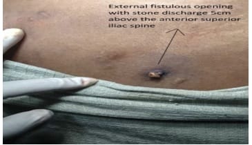
surgical_198_01.jpg

Fig-1: The patient presented with complaints of passing stones.

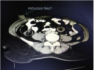
surgical_198_02.jpg

Fig-2: Computed tomography of the fistulous tract.

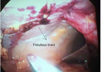
surgical_198_03.jpg

Fig-3: Fistulous tract adherent to the anterior abdominal wall.

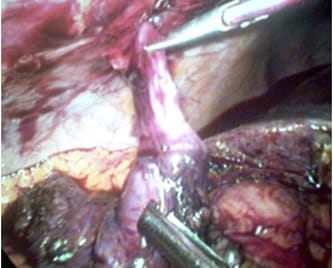
surgical_198_04.jpg

Fig-4: Gallbladder with the fistulous tract.

surgical_198_05.jpg

Fig-5: Fistulous tract removed anteriorly.

surgical_198_06.jpg

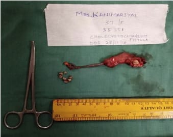
Fig-6: Gallbladder with fistulous tract along with gallbladder through skin incision extracted stones.


Discussion

Internal hernias may infrequently cause SBO, which may be fatal because of the risk of strangulation of the hernial content. Paracecal hernias account for a minority of internal hernia related SBOs. These hernias are the result of alterations in the normal process of intestinal rotation during embryonic development [2]. The embryological development of the cecum includes budding, exteriorization into the umbilicus, and subsequent retraction onto the posterior abdominal wall, which usually predisposes the paracecal fossa to the formation of a number of pockets or recesses.

Meyer classified boundaries of paracecal hernias as paracecal sulci, cecal fossa, cecal recess, superior ileocecal recess, inferior ileocecal recess, and retrocecal recess. Paracecal sulci are lateral depressions of the peritoneum invested on the cecum, but recesses may be absent. The cecal fossa is a groove that is formed by two peritoneal folds [3]. The lateral fold is a continuation of the white line of Toldt and the medial fold originating from the ileocecal angle, medial aspect of the cecum.

The clinical symptoms of internal hernias may range from intermittent mild digestive complaints to acute onset incarceration. The major symptoms are obstructive symptoms of abdominal pain, nausea, vomiting, constipation, and obstipation [5]. CECT abdomen is very useful in diagnosis as it detects an early or partial obstruction, closed-loop obstruction, and multiple segments of obstruction. Dilatation of small intestine loops with a transitional zone adjacent to the cecum or an edematous small bowel located lateral to the cecum allows a paracecal hernia to be diagnosed with high certainty [6]. Almost always the treatment for small bowel obstruction caused by a paracecal hernia is surgical intervention. Recently, the laparoscopic technique has been found to be useful for the diagnosis and treatment of bowel obstructions [7-9].

The cecal recess is formed by folds described for the cecal fossa, but in
In this instance, the cecum is entirely retroperitoneal. Superior and inferior ileocecal recesses are formed by a peritoneal fold originating from the terminal ileum to the cecum. A retrocecal recess is formed by the cecum anteriorly, the iliac fossa posteriorly, the right colic gutter laterally, and the mesentery medially [4]. In our case, jejunum and ileum herniated into the superior ileocecal recess through the defect in the mesentery.

Conclusion

Internal hernia is an infrequent cause of small bowel obstruction (SBO), with a reported incidence of 0.2 to 0.9%, and is the cause of small-bowel obstruction in 0.6 to 5.8% of the cases. Internal hernias are protrusions of the viscera through the peritoneum or mesentery but remaining within the abdominal cavity. Surgery achieved a good outcome.

Acknowledgments

The authors would like to thank the hospital surgical and pathology department as well as the patient for all their help in preparing this article.

Reference

  1. Choh NA, Rasheed M, Jehangir M. The computed tomography diagnosis of paracecal hernia. Hernia. 2010;14(5)527-529.
  2. Ogami T, Honjo H, Kusanagi H. Pericecal hernia manifesting as a small bowel obstruction successfully treated with laparoscopic surgery. J Surg Case Rep. 2016;2016(3)rjw020.
  3. Selcuk D, Kantarci F, Oğüt G, Korman U. Radiological evaluation of internal abdominal hernias. Turk J Gastroenterol. 2005;16(2)57-64.
  4. Lu HC, Wang J, Tsang YM, Tseng HS, Li YW. Pericecal hernia- a report of two cases and survey of the literature. Clinical Radiol. 2002;57(9)855-858.
  5. Kleyman S, Ashraf S, Daniel S, Ananthan D, Sanni A, Khan F. Pericecal hernia- a rare form of internal hernias. J Surg Case Rep. 2013;(2).
  6. Omori H, Asahi H, Inoue Y, Irinoda T, Saito K. Laparoscopic paracecal hernia repair. J Laparoendos Advan Surg Tech. 2003;13(1)55-57.
  7. Agha RA, Fowler AJ, Saeta A, Barai I, Rajmohan S, Orgill DP, et al. The SCARE statement: consensus-based surgical case report guidelines. Int J Surg. 2016;34;180-186.

  1. Sasaki K, Kawasaki H, Abe H, Nagai H, Yoshimi F. Retrocecal hernia successfully treated with laparoscopic surgery- a case report and literature review of 15 cases in Japan. Int J Surg Case Rep. 2016;18;45-47.
  1. Kabashima A, Ueda N, Yonemura Y, Mashino K, Fujii K, Ikeda T, et al. Laparoscopic surgery for the diagnosis and treatment of a paracecal hernia repair- report of a case. Surg. 2010;40(4)373-375.