E-ISSN:2455-5436
P-ISSN:2456-9518
RNI:MPENG/2017/70870

Research Article

Platelet

Surgical Review: International Journal of Surgery, Trauma and Orthopedics

2020 Volume 6 Number 4 July-August
Publisherwww.medresearch.in

The effect of Platelet-rich plasma on the repair of sports-related muscle, tendon and ligament injuries

Ligu L.1*, Jini M.2
DOI: https://doi.org/10.17511/ijoso.2020.i04.09

1* Lenin Ligu, Assistant Professor, Tomo Riba Institute of Health and Medical Sciences (TRIHMS), Naharlagun, Arunachal Pradesh, India.

2 Moji Jini, Director, Tomo Riba Institute of Health and Medical Sciences (TRIHMS), Naharlagun, Arunachal Pradesh, India.

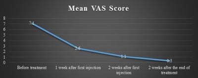
Introduction: Platelet-rich plasma (PRP) treatment is a developing technology that ambitions to recover the procedure of tissue overhaul via the distribution of bioactive representatives, which will deliver chemotactic, proliferative, and anabolic cellular retorts and improve retrieval of tissue purpose. Materials and methods: Fifty-three recreational athletes were registered in the study. The patients were enrolled from the Emergency ward in the University Hospital at Parma rendering to a pre-defined procedure. Each patient was evaluated by ultrasound imaging to assess the range and mark of muscle harm. Only grade II lesions were preserved with 3 ultrasound-guided vaccinations of autologous platelet-rich plasma every seven days. Platelet distillate was formed as per the standard methods, with a 10% erraticism in platelet count. The platelet gel for medical use was found by addition thrombin to the distillates beneath regular circumstances. Consequences measured were: pain reduction, muscle function retrieval, and reappearance to sports activity, ultrasound-imaging tissue remedial, relapses, local contagions, and any side effect through the action. Results: In all cases muscle lesions cured completely on ultrasound-imaging, the sting vanished, and muscle function retrieval was recognized with a reappearance to sports activity. A solo patient had a reversion 1 year after cure. Conclusion: Platelet-rich plasma vaccinated into the harm site is one of the greatest significant factors interpreting the behavior operative. Rendering to the present consequences, which document complete muscle retrieval and no reversion excluding for one patient, platelet-rich plasma ultrasound-guided vaccination signifies an effective mini-aggressive cure for muscle harm.

Keywords: Platelet-rich plasma, Sports-related injuries, Tendon injury, Muscle injury

Corresponding Author How to Cite this Article To Browse
Lenin Ligu, Assistant Professor, Tomo Riba Institute of Health and Medical Sciences (TRIHMS), Naharlagun, Arunachal Pradesh, India.
Email:
Ligu L Jini M. The effect of Platelet-rich plasma on the repair of sports-related muscle, tendon and ligament injuries. Surgical Rev Int J Surg Trauma Orthoped. 2020;6(4):270-275.
Available From
https://surgical.medresearch.in/index.php/ijoso/article/view/196

Manuscript Received Review Round 1 Review Round 2 Review Round 3 Accepted
2020-07-18 2020-08-05 2020-08-20 2020-08-28
Conflict of Interest Funding Ethical Approval Plagiarism X-checker Note
No Nil Yes 9%

© 2020 by Lenin Ligu, Moji Jini and Published by Siddharth Health Research and Social Welfare Society. This is an Open Access article licensed under a Creative Commons Attribution 4.0 International License https://creativecommons.org/licenses/by/4.0/ unported [CC BY 4.0].

Introduction

Platelet-rich plasma (PRP) treatment is a developing technology that ambitions to recover the procedure of tissue overhaul via the distribution of bioactive representatives, which will deliver chemotactic, proliferative, and anabolic cellular retorts and improve retrieval of tissue purpose [1].

Platelet-rich plasma goods are easily organized from the patients’ blood and typically include the native vaccination of a set volume of PRP or the submission of PRP gel form through operation directly at the place of the wound. The medical use of PRP for encouraging physiological wound remedial was introduced in the 1980s for the action of cutaneous ulcers [2].

Initial studies on PRP observed the curing effects of cleaned and quarantined recombinant development factors, such as platelet-derived growth factor-BB (PDGF-BB), as healing particles for wound heal­ing. Though, information on curative mechanisms has led to the conclusion that quarantined development factors cannot facilitate all biological features mandatory for tissue restoration.

Thus, an additional balanced policy would be the direction of a stable mixture of mediators that would perform in interaction to mimic the physical needs of the wounded tissue [3].

In the 1990s, advances in oral implantology were operated by the possible reformative properties of PRPs in bone tissue and perceiving the curative possessions in soft tissues. Since then, the usage of PRP has an extent to numerous additional medical areas, including ophthalmology, orthopedics, sports medicine, cardiology, dermatology, plastic surgery, and neurology [4].

The 1st conveyed submission of PRP in sports wounds was in the arthroscopic organization of an avulsion of articular gristle in a youth soccer player [5] Further expansions in PRP treatments have presented new prospects for tissue overhaul in sports remedy, such as original remedies for the organization of chronic pathologies (eg, tendinopathy and osteoarthritis) [6].

Platelet-rich plasma has the probable to accelerate the procedure of curing and tissue renewal in medical situations. In sports medicine, this might accelerate yield to play, mainly in best and specialized athletes.

Materials and Methods

Study Setting: The patients were hired from the Tomo Riba Institute of Health and Medical Sciences (TRIHMS).

Duration and type of study: Prospective, observational study was conducted over a 1-year period.

Sampling Method: Principles for enclosure in the study stood grade II muscular or myotendinous scratch according to the America Medicinal Association classification which happened within 3 days of admittance to the casualty. US imaging with a 7.5-12 MHz line investigation was continuously made by a skilled radiologist who resolute the precise size, extent, and location of the scratch. Only in a few nominated cases was magnetic resonance imaging (MRI) also carried out. At the same time, a medical reintegration expert assessed the clinical disorder of the patients. Seventy-three recreational athletes (46 men, 27 women) stood involved in the study.

Inclusion criteria: Included all the sports injured persons, willing to participate.

Exclusion criteria: Not willing to participate in the study.

Ethical consideration and permission: Once respectively patient had been registered in the study, an informed consent form signed was acquired and the course for the PRP treatment was recognized and the transfusion professional assessed the patient's suitability for autologous blood contribution according to the Provincial guidelines on autologous blood collection (Guidelines on Autologous Blood). Autologous blood modules underwent serological validation rendering to haemovigilance rules. Patients were treated with three PRP injections (one treatment every 7 days) and individually of them needed extemporaneous research of PRP.

The other authors measured as appropriate outcomes the following: pain reduction (evaluated with a visual analog scale, VAS), muscle role recovery, perceptible US-imaging tissue healing, lack of local infection throughout the treatment, and of any other side effects. These limitations were assessed before individual injection and 2 weeks after the end of the treatment. In all patients return to regular sports movement was also noted. All patients were analyzed 1 year after treatment in


order to detect reverts of the muscle lesion.

Preparation of the platelet-rich plasma: Platelets are key modules in hemostasis, and encourage the creation of new connective tissue and revascularization. They are obtaining from the division of originator megakaryocytes and have a lifespan of 5-9 days. Once initiated, by the act of thrombin, they conceal the substances of their important and solid particles which simplify altered phases of curing.

The autologous platelets were obtained from an autologous complete blood unit of 350 mL composed in a multiply blood bag and divided by centrifugation to extract 50-60 mL of PRP. The platelet units created were preserved under constant anxiety for 24 hours at 22 °C. Following serological endorsement, the platelet units were divided into three aliquots of 20 mL under a sterilized secure circuit with Sterile Tubing. Each aliquot was recognized with tags fulfilling with ethics and presented the script "autologous platelet concentrate" and frozen at −40 °C. The platelet content of individually aliquot was assessed in order to conclude whether the least healing dose of 1×10⁶ platelets/μL had been reached. The platelet counts in the aliquots of the platelet distillates used ranged from 960×105 to 1.35×10⁶ platelets/μL. [7]

Preparation of the thrombin: Autologous thrombin was found by collecting 20 mL of whole blood from the patient into four Vacutainer test-tubes. The test-tubes were centrifuged at 3200 g for 10 minutes, the serum was separated beneath a movement hood and 0.2 mL of 10% calcium gluconate were added before incubation at 37 °C for 15-30 minutes. Finally, the supernatant, enclosing thrombin precursors, was separated into two or three aliquots and categorized in order to make sure correspondence with the platelet distillates from the patient. The aliquots of thrombin were then stored at −40 °C.

Activation of the platelet-rich plasma: The aliquots of platelet distillate were melted at 37 °C for 15 minutes. The product was started at the patient's bedside. This was completed by extracting 5 mL of the platelet distillate with a syringe, then adding 1 mL of autologous thrombin and 1 mL of 10% calcium chloride. The result thus found was mixed mildly four or five times. Afterward, the activated PRP was vaccinated into the exact site of the tissue lesion under US guidance, using an 18-21 gauge needle.

Results

The nasty age of the 46 men involved in the study was around 26 years, whereas that of the 27 women was around 23 years. All of the applicants were recreational participants involved in various sports: volleyball, soccer, basketball, dancing, trekking, and skiing. All harms happened though the participants were performing their individual sport (competition or training activities). Table 1 shows which muscle was involved in relative to the sport they practiced when the muscle injury happened. Injuries treated in this study were 50 grade II muscle strains and three myotendinous lesions. Muscle injuries were categorized into grade I (mild wound), grade II (moderate wound), and grade III (severe wound) as shown in Table 2. [8]

The authors noted a liberal development of pain throughout treatment as shown in Figure 1. At baseline, the mean VAS score was 7.1 (range, 6-8). One week later the first vaccination the mean pain VAS score was 2.6 (range 2-4), 2 weeks later the first vaccination was 1.1 and 2 weeks later the end of the treatment the score was 0.3. Subsequently, each vaccination pain gradually disappeared within days and did not require nonsteroidal anti-inflammatory drugs except in two patients after the first vaccination.

All patients informed a reduction in pain later the first PRP vaccinated and in 62 patients (85%) progress of function was witnessed at the same time. After harm patients presented with restricted motion (flexion, internal and external rotation, abduction, and adduction) which was completely reestablished after treatment. Before the preliminary vaccination ultrasound inspections continuously showed muscle tissue failure, hemorrhagic formation, and hypoechoic gaps of numerous sizes at the site of harm or hematoma which penetrated the muscle or composed around the lesion.

surgical_196_01.jpg

Fig-1: Mean VAS score before treatment, 1 and 2 weeks after the first injection, and 2 weeks after the end of the treatment.


Therapeutic processes happening in a damaged muscle (necrosis/degeneration, inflammation, repair, and scar tissue formation) are all interconnected and time-dependent. Acute muscle collapse and irritation happen instantly after harm and last up to 7 days, while tissue propagation usually starts 7 to 10 days after the wound.

The proliferative procedure generally peaks at 2 weeks and transfers in the direction of mark evolution at 3 to 4 weeks after-harm and can last up to 1 year.

This mark tissue development (fibrosis) is the last invention of muscle repair which starts among the second and third weeks later the harm and upsurges in size over time [9].

In the present study, later vaccination of PRP it was continuously probable to evaluate the liberal parenchymal retrieval of the muscle, the growth of shallow hyperechoic mark tissue at the site of the harm, and the reabsorption of the nearby hematoma as determined. All participants started, at a mean of 20 days (±2 SD; range, 16-28) later the initial vaccination, a modified therapy and preparation program created on a physiatrist's physical valuation.

This program is contained in steady muscle consolidation primarily in some precise environs (a gym) and eventually in the field.

All patients reverted totally to their consistent sporting activity subsequently a mean period of 30 days (±1.2 SD; range, 28-35). Subsequently, there is restricted indication on the process to determine the effectiveness for recurring to sports activity, [10] the author's selected standards based on lack of pain on straight palpation and throughout muscular contraction with virtuous regular muscle purpose. No contagions, major side effects, or obstacles connected to the process were detected.

At the 1 year follow up 3 patients had new muscular harm in a diverse muscle and only one described new harm in the before treated muscle (all lesions of the hamstrings). This last patient re-injured himself playing soccer 5 months subsequently restarting his recreational sport.

The revert was a grade II lesion of the central third segment of the femoral biceps muscle although the initial harm was localized in the higher third section of the similar muscle. This patient was treated with PRP another time with virtuous outcomes.


Table-1: Localization of Lesions related to the type of sport.

Type of muscles involved Type of Sport Patients/Site of harm
Volley ball Soccer Basket ball Dancing Trekking Skiing
Femoral rectus 6 8 4 2 0 2 22
Femoral biceps 5 3 3 0 4 1 16
Calf muscles (medial and lateral gastrocnemius) 4 2 2 2 0 4 14
Long adductor 3 3 2 2 2 0 12
Abdominal rectus 0 0 0 0 0 1 1
Peroneal muscles 0 2 0 0 0 0 2
Femoral rectus myotendinous junction 2 2 0 0 0 1 5
Patients/sport 20 20 11 6 6 9 72

Table-2: Degree, description, and sonographic appearance of the muscle lesions.

Degree Description Sonographic appearance
Grade I Very slight laceration concerning < 5% of the side of muscles. No losses of strength or restriction in actions. Patchy zone with the hypoechoic area (diameter less than1 cm)
Grade II Cut with damage of muscle strength concerning 5 to 70% of muscle fibers. Oedematous imbibition and blood outpouring. Patchy zone with the hypoechoic area. (diameter less than 3 cm)
Grade III > 70% of muscle fibers complicated (subtotal lesion) or a whole rupture of muscle belly (total lesion). Muscle arrangement disturbance, with refutation and hypoechoic area. (diameter ≥3 cm)

Discussion

Though the purposes of all progression aspects complicated in tissue curing and restoration are not yet completely assumed possible profits of nearly have remained demonstrated [11] Plasma turn into a vehicle of development factors such as converting growing factors beta, platelet-resulting growth aspect, epidermal growth element, vascular endothelial growth factor, [12] platelet-derived epidermal development element, bone morphogenetic protein, insulin-like development element, endothelial cell development element and simple fibroblast growth factor (bFGF) [13]. These elements play key roles in most tissue curing processes, [14] allowing a further physiological and quick curing of muscle lesions.

Despite the great occurrence of muscle harm the greatest technique of their treatment has not yet been clearly defined when a rapid reoccurrence to sporting activity is the main goal [15].US-guided injection of PRP has been obtaining importance in the treatment of muscle injuries [16].

Considerate the physiological procedures of muscular tissue healing are important for creating a therapeutic treatment focused on accelerating healing. Nevertheless, there is presently very restricted technical confirmation of the clinical efficiency of PRP use for muscle stresses in athletes [17].

This study defines a procedure to appraise the clinical efficiency of 3 US-guided PRP vaccination after grade II muscle harm in recreational athletes in order to direct these patients' reappearance to involved their sporting activity care as soon as possible. The authors ruminate that the strengths of this study are the homogeneity of the trial (all grade II lesions treated with the same PRP treatment procedure) and a process that was always executed by the same physician under US-guide.

Even if various muscles were treated in this study and there was no control group, the authors ruminate the results to be valid and dependable. All patients had the whole curing of the muscular lesion without side effects, as testified by imaging, thus verifying the efficiency of PRP and representing that this procedure is well tolerated. Furthermore, the time of the reappearance to sports activity was alike to that in other intelligence in the literature in which PRP treatment was described [18-19].

Limitations

The limitations of this study include the lack of a randomized control group and the small number of patients. The present study could not measure the concentration of different growth factors present in the PRP.

Conclusion

Clinical interventions in musculoskeletal and sports medicine aim to achieve predictable, rapid tissue repair through the deposition of new, well-organized extracellular matrix to facilitate and enhance wound healing that will restore the high mechanical performance and functional levels of non-injured tissue in the shortest period. On the basis of the present outcomes, the researchers decided that US-guided PRP vaccinations allow physiological, quick, and enduring curing of muscle lesion and symbolize a valid and safe mini-invasive treatment for grade II muscles harms.

What does the study add to the existing knowledge

Platelet-rich plasma injected into the injury site is one of the most important factors rendering the treatment effective.

Author’s Contribution

Dr. Lenin Ligu: Prepared the manuscript, performed the procedure and data collection, verified the analytical methods, and supervised the findings of the work.

Dr. Moji Jini: Conceived the idea.

Reference

  1. Creaney L, Hamilton B. Growth factor delivery methods in the management of sports injuries- the state of the play. Br J Sports Med. 2008;42(5)314-320.
  2. Cyriax JH. The pathology and treatment of tennis elbow. J Bone Joint Surg. 1936;18(14)921-940.
  3. Sharma P, Maffulli N. Tendon injury and tendinopathy- healing and repair. J Bone Joint Surg. 2005;87(1)187-202.

  1. Landesberg R, Roy M, Glickman RS. Quantification of growth factor levels using a simplified method of platelet-rich plasma gel preparation. J Oral Maxillofac Surg. 2000;58(3)297-300.
  2. Kon E, Filardo G, Di Martino A, Marcacci M. PRP to treat sports injuries- evidence to support its use Knee. Surg Sports Traumatol Arthrosc. 2011;19(4)516-527.
  3. Engebretsen L, Steffen K, Alsousou J, Anitua E, Bachl N, Devilee R, et al. IOC consensus paper on the use of platelet-rich plasma in sports medicine. Br J Sports Med. 2010;44(15)1072-1081.
  4. Borrione P, Di Gianfrancesco A, Pereira MT, Pigozzi F. PRP in muscle healing. Am J Phys Med Rehabil. 2010;89(10)854-861.
  5. Kampa RJ, Connell DA. Treatment of tendinopathy- is there a role for autologous whole blood and PRP injection. Int J Clin Pract. 2010;64(13)1813-1823.
  6. Maffulli N, Longo UG, Denaro V. Novel approaches for the management of tendinopathy. J Bone Joint Surg. 2010;92(15)2604-2613.
  7. Andia I, Sanchez M, Maffulli N. Tendon healing and PRP therapies. Expert Opin Biol Ther. 2010;10(10)1415-1426.
  8. Gaweda K, Tarczynska M, Kryzanowski W. Treatment of Achilles tendinopathy with PRP. Int J Sports Med. 2010;31(8)577-583.
  1. Kraushaar BS, Nirschl RP. Tendinosis of the elbow (tennis elbow) Clinical features and findings of histological, immunohistochemical, and electron microscopy studies. J Bone Joint Surg Am. 1999;81(2)259-257.
  2. Bjordal JM, Couppe C, Ljunggren AE. Low level laser therapy for tendinopathy- evidence of a dose response. Phys Ther Rev. 2001;6(2)91-99.
  3. Kon E, Filardo G, Delcogliano M, Russo A, Bondi A, et al. PRP- new clinical application, A pilot study for treatment of jumper’s knee. Injury. 2009;40(6)598–603.
  4. Filardo G, Kon E, Della Villa S, Vincentelli F, Fornasari PM, Marcacci M. Use of PRP for the treatment of refractory jumper’s knee. Int Orthopaed. 2010;34(6)909-915.
  5. Vasseljen O, Jr, Høeg N, Kjeldstad B, Johnsson A, Larsen S. Low level laser versus placebo in the treatment of tennis elbow. Scand J Rehabil Med. 1992;24(1)37-42.
  6. Edwards SG, Calandruccio JH. Autologous blood injections for refractory lateral epicondylitis. J Hand Surg Am. 2003;28(2)272-278.
  7. Mishra A, Pavelko T. Treatment of elbow chronic tendinosis with buffered PRP. Am J Sports Med. 2006;34(11)1774-1778.
  8. Haake M, König IR, Decker T, Riedel C, Buch M, Müller HH, et al. Extra corporeal shock wave therapy in the treatment of lateral epicondylitis- A randomized multicenter trial. J Bone Joint Surg Am. 2002;84(11)1982-1991.