E-ISSN:2455-5436
P-ISSN:2456-9518
RNI:MPENG/2017/70870

Research Article

Osteosynthesis

Surgical Review: International Journal of Surgery, Trauma and Orthopedics

2020 Volume 6 Number 2 March-April
Publisherwww.medresearch.in

Functional outcome of minimally invasive plate osteosynthesis (MIPO) in proximal tibial fractures

Vora J.1, Shah J.2*, Vala G.3
DOI: https://doi.org/10.17511/ijoso.2020.i02.05

1 Jinesh Vora, Associate Professor, Department of Orthopaedics, C.U Shah Medical College and Hospital, Surendranagar, Gujarat, India.

2* Jay Shah, Postgraduate Student, Department of Orthopaedics, C.U Shah Medical College and Hospital, Surendranagar, Gujarat, India.

3 Gaurav Vala, Associate Professor, Department of Orthopaedics, C.U Shah Medical College and Hospital, Surendranagar, Gujarat, India.

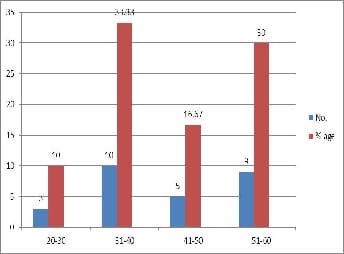
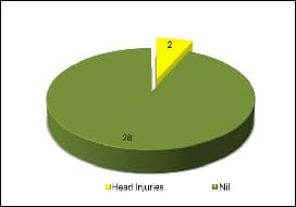
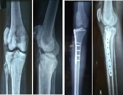
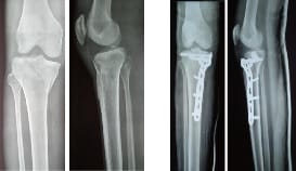
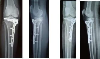
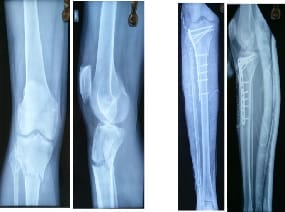
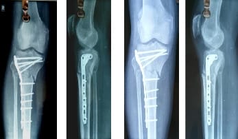
Introduction: This study is undertaken to evaluate the outcome of the MIPO technique in Proximal Tibial Condyle Fractures which is expected to provide a stable fixation with minimum exposure, early mobilization, fewer complications, and a better quality of life. Material and methods: Study design: Prospective cohort study carried out at C.U Shah Medical College. 30 cases of proximal tibia fractures (Schatzker type 1- VI) operated with the MIPO technique. Follow-up of these patients was done at 6 months and 9 months after surgery by using Sanders's 40-point functional scale. Results: There were 16 cases of Schatzker type I fractures, 5 cases of Type II, 4 cases of type III, 2 cases of type IV, 1 case of type V and 2 cases of type VI fractures. The healing process was determined by radiographically and functionally. Meantime to union was 17.6 weeks, with 50 % of fractures uniting in 14-16 weeks. Infection was observed in two cases (10%), two cases of collapse of fracture, and two cases of malunion. Sanders's score was applied to analyze the functional outcome among the cases. 63% of patients had excellent performance, 20% had good results, 13% had fair results and one patient performed poorly. Conclusion: MIPO is a good technique to stabilize the proximal tibia condyle fractures especially when used with meticulous intraoperative handling of soft tissues.

Keywords: MIPO, Proximal tibial condyle fractures, Invasive plate osteosynthesis

Corresponding Author How to Cite this Article To Browse
Jay Shah, Postgraduate Student, Department of Orthopaedics, C.U Shah Medical College and Hospital, Surendranagar, Gujarat, India.
Email:
Vora J, Shah J, Vala G. Functional outcome of minimally invasive plate osteosynthesis (MIPO) in proximal tibial fractures. Surgical Review Int J Surg Trauma Orthoped. 2020;6(2):110-120.
Available From
https://surgical.medresearch.in/index.php/ijoso/article/view/158

Manuscript Received Review Round 1 Review Round 2 Review Round 3 Accepted
16-03-2020 26-03-2020 02-04-2020 07-04-2020
Conflict of Interest Funding Ethical Approval Plagiarism X-checker Note
No Nil Yes 15%

© 2020 by Jinesh Vora, Jay Shah, Gaurav Vala and Published by Siddharth Health Research and Social Welfare Society. This is an Open Access article licensed under a Creative Commons Attribution 4.0 International License https://creativecommons.org/licenses/by/4.0/ unported [CC BY 4.0].

Introduction

Intraarticular fractures of the proximal tibia involve a major weight-bearing joint which if not managed well, results in functional impairment. Most of the injuries in 55-70% cases affect the lateral condyle. Isolated injuries to the medial condyle occur in 10-23% cases; whereas bicondylar fractures are seen in 10-30% cases [1].

High energy complex tibial condyle fractures being intra-articular are usually associated with injury to ligaments, capsule, and other soft tissues around the joint. Wedge failure, wedge depression, and pure depression type failures are typical of the lower energy spectra [2].

Various classification systems are available for classifying these fractures including the Schatzker classification system and AO classification [3]. The Schatzker classification system for proximal tibia fractures, which divides these fractures into six types, is widely recognized by orthopedic surgeons to assess the initial injury, plan management, and predict prognosis.

Non-operative procedures like cast, braces, or traction are complicated by intrinsic risks of poor functional results and extended hospital stay. Open reduction and internal fixation have their own complications. The middle path of the minimally invasive technique of closed reduction by ligamentotaxis and stabilizing the fracture by limited internal fixation is being developed.

This utilizes percutaneous screws and K-wires, external fixation frames, or a combination of external fixation with some degree of internal fixation to stabilize tibial condyle fractures. The minimally invasive technique attributes to operative and non-operative philosophies as it gives outstanding functional results [4].

With the use of the MIPO technique unilateral fixation of the condyles is possible as well as there is minimal handling of the soft tissue which further helps in achieving a good functional outcome [5].

Published reports have stated that the proximal tibial locking compression plate is based on the biomechanical principle of external fixators and internal fixators. The angle-stable interface between the screws and the plate allows placement of the plate without any contact to the bone with the advantage of preserving the periosteal blood supply and bone perfusion.

It is reported that locking plate systems have unique features such as outriggers, jigs, and blunted ends to improve the surgeon’s ability to pass the plate in a submuscular or subcutaneous manner for minimally invasive application.

Thus, there is significantly less soft tissue dissection resulting in the preservation of the hematoma and local blood supply that enhances the healing of the fracture [2].

Extensive surgery on a severely comminuted fracture may result in poor internal fixation and a need for postoperative immobilization, which often results in an unstable joint [6]. The present study aimed to evaluate the functional outcome of proximal tibial fractures treated with Minimally Invasive Plate Osteosynthesis (MIPO).

Materials and Methods

The functional outcome of Minimally Invasive Plate osteosynthesis (MIPO) using locking and non-locking compression plates in tibial condyle fractures was evaluated in this study. The study included 30 cases of fracture of tibial condyle presenting to the emergency. The results were compiled and analyzed using Sanders 40 Point Functional Evaluation Scale

Inclusion criteria

  1. Closed fractures of the tibial condyle ( Grade I to VI)
  2. Age Limit: 18 years and above
  3. Both males and females

Exclusion criteria

  1. Previous or existing infection in the involved leg
  2. Compound fractures with extensive soft tissue damage where the plate cannot be covered with soft tissue
  3. Pathological fractures other than osteoporosis
  4. Patients with Neurovascular deficits.

A complete and thorough examination of the patient along with the associated injuries performed. The distal neurovascular status of the limb was assessed. The necessary x-rays(AP/Lat/Oblique) were obtained and evaluated.

CT Scans were obtained whenever required. Intravenous fluids, analgesics, and antibiotics were administered as per protocol. Tetanus prophylaxis as per requirement was administered. Routine Pre-


op fitness has taken from physicians and anesthetists required.

On a radiolucent fracture table, the patient was placed in the supine position and a sponge pack/sandbag was kept under the affected gluteal region in order to prevent external rotation of the lower limb. A pneumatic tourniquet was applied. The affected limb was prepared and draped below the tourniquet.

Intraoperative antibiotics (Intravenous) were continued for 5 postoperative days and then on oral antibiotics till the sutures were removed.

Post-operative X-ray was done to document proper reduction and fixation of fracture fragments. Patients were started with Quadriceps exercises and ankle mobilization from 2nd or 3rd postoperative day according to the tolerance of patients.

Progressive weight-bearing was allowed according to the callus formation as assessed in follow up X Rays.

The patients were regularly followed-up on an OPD basis and were assessed both clinically and radiologically. Patients were allowed toe touch partial weight bearing from one month of surgery and full weight-bearing thereafter on the subsequent follow-ups.

All long term complications like non-union, malunion, angular deformity, implant breakage, shortening, or infection were recorded. The patients were under follow up to 9 months.

The final result was based on the functional and radiological outcomes. The outcome was evaluated on the basis of Sanders 40 point functional evaluation scale.

Table-1: Evaluation scale.

Function
Range of motion (0)
Result Points
Flexion
>125
Excellent 6
100-124 Good 4
90-99 Fair 2
<90 Poor 0
Extension
0
Excellent 3
<5 Good 2
6-10 Fair 1
>10 Poor 0
Deformation Angulation(0) Excellent 3
<10 Good 2
10-15 Fair 1
>15 Poor 0
Shortening (cm)
0
Excellent 3
<1.5 Good 2
1.5-2.5 Fair 1
>2.5 Poor 0
Pain
None
Excellent 3
Occasional or with changes in weather or Both Good 2
With fatigue Fair 1
Constant Poor 0
Walking Ability
Walking
Unrestricted
Excellent 6
>30 minutes to <60 minutes Good 4
<30 minutes Fair 2
Walks at home are confined    
To wheelchair or is bedridden Poor 0

Table-2: Evaluation scale.

Function Result Points
Stair climbing    
No limitation Excellent 3
Holds rail Good 2
One stair at a time Fair 1
Elevator only Poor 0
Return to work (A or B)    
A. Employed before injury    
Returned to preinjury job Excellent Good 6
   
Returned to the preinjury job with difficulty Altered full-time job Fair 4
   
A part-time job or unemployed Poor 2
   
    0
B. Retired before injury Returned to preinjury lifestyle    
Needs occasional help Excellent Good 6
   
Needs assistance at home with activities of daily living Fair 4
   
Moved in with family or nursing home   2
   
  Poor  

Excellent = 36-4 points .Good 26-35 points .Fair = 16-25 points. Poor = 0-15 points


Results

surgery_161_01.jpg

Fig-1: Age distribution.

The age distribution shows most patients in 31-40 years (33.33%) followed by 51-60 years (30%) and least was seen in 20-30 years (10%).

surgery_161_02.jpg

Fig-2: All the cases under consideration were closed fractures.

Table 3: Schatzker classification.

Schatzker Type No. % Age
I 16 53.33
II 5 16.67
III 4 13.33
IV 2 6.67
V 1 3.33
VI 2 6.67

Schatzker type I (53.33%) were the maximum while the least was Type V (3.33%)

surgery_161_03.jpg

Fig-3: Pre-Op complications.

28 (98.3%) patients had no pre-op complications while 2 (6.66%) patients had head Injury

Table 4: Early postoperative complications.

Complications No. %age
Infection 1 3.33
Wound Gaping 1 3.33
Nil 28 93.33

28 (93.33%) patients had no early post-op complications While 1 (3.33%) patient developed Infection at the local site and 1 (3.33%) patient developed wound gaping.

Table 5: Late Post-operative complications.

Complications No. %age
Collapse of fracture 2 6.67
Malunion 2 6.67
Nil 26 86.66

26 (86.66%) patients had no late post-op complications While 2 (6.67%) patients developed Malunion and 2 (6.67%) patients developed fracture collapse.

Table 6: Fracture union.

Time(weeks) No. %age
14-16 16 53.33
17-19 10 33.33
20-22 3 10
23-24 1 3.33

In the present study, the average time to union was calculated to be 17.6 weeks.

In almost 50% of cases fracture united in

Table 7: Pain.

Pain No. %
None 13 43.33
Occasional 10 33.33
In-certain Positions 5 16.67
After Activity 2 6.67
Night Pains - -

Table 8: Walking capacity.

Walking capacity No. %
Normal 18 60
At least 1 Hr 7 23.33
At least 15 mins 4 13.33
Indoors Only 0 -
WheelChair/Bed Ridden 1 3.33

Table 9: Extension Lag.

Extension Lag No. %
Normal (0 degree) 23 76.67
Extension(0-10 degree0 6 20
>10 degree 1 3.33

Table 10: Range of motion.

Flexion No. %
140 Degree 16 53.33
120 Degree 12 40
90 Degree 2 6.67
60 Degree - -
30 Degree - -

Table 11: Final score.

Score Results No. of patients %age
27-30 Excellent 19 63.33
20-26 Good 6 20
10-19 Fair 4 13.33
<10 Poor 1 3.33

Sanders 40 point functional evaluation scale in the present study was applied to analyze the functional outcome among the cases. Pain, range of motion at the knee, walking capacity, residual extension lag, and stability were the criteria that determined the final outcome. After evaluation, it was observed that 85% of patients had good to excellent performance (Excellent - 19 patients; good - 6 patients). 4 had fair results and 1 performed poorly. The average range of motion in the present study was 0 (range 0-15°) to 120° (range 60-140°).

Operative images

Clinical results

Excellent

Good

surgery_161_04.jpg

Fig-4: Pre-Operativ        Fig-5: Post-Operative

surgery_161_05.jpg

Fig-6: 6 Months         Fig-7: 9 Months

surgery_161_06.jpg

Fig-8: Pre-Operative       Fig-9: Post-Operative

Fair

surgery_161_07.jpg

Fig-10: 6 Months                 Fig-11: 9 Months

surgery_161_08.jpg

Fig-12: Pre-Operative    Fig-13: Post-Operative


surgery_161_09.jpg

Fig-14: 6 Months             Fig-15: 9 Months

Discussion

The fractures of the proximal tibia are high energy injuries most commonly due to road traffic accidents associated with other skeletal, ligamentous, and multi-system injuries. The results of the treatment of these injuries have often been poor with loss of motion, instability, posttraumatic osteoarthritis, wound breakdown, and infection as final outcomes [1,2,8], but with proper interventions and good surgical techniques, these injuries had better results. In the present study, the final follow up of 30 patients was done.

In this study, it was observed that fractures of the proximal tibia were more prevalent in younger and middle-aged population with the mean age being 40 (range 21-50 years). Males were more commonly affected than females (24: 6).In the study by Tul B Pun et al (2014) [12], the mean age of the patients was 43.85 years (range 22-61 years). There were 20 male patients and 1 female. The mean age of the patients was 41.4 years (range 19-83) in the study by R. Jiang et al [9]. There were 29 males and 12 females in the same study.

In another study by P Phisitkul et al [10] the study included 22 males and 15 females aged between 22 and 71 years (mean age of 45 years). In a study by Zura RD et al [6], there were 78 patients with an average age of 43 years (range 14 to 81 years). Which included 59 male patients and 19 female patients. Hence, it can be concluded that the fractures of the proximal tibia were found to be common in the younger and middle-aged population. Since this age group is involved in more outdoor activities.

In the present study of 30 cases, 83.33% of fractures were due to Road Traffic Accidents and 16.67% fractures were due to fall from height. In the study by Hak DJ et al [5], the cause of the injury was due to high energy trauma with 40%

patients were pedestrians hit by a motor vehicle and 40% patients were fall from height and 4% were due to sports injury and remaining were due to motorcycle collision. In the study by P Phisitkul et al, [10] 15 patients sustained a fracture after falling from a height, 17 patients through RTA. 2 patients in a crush injury, and 1 patient during the fight.

In the study by GT Prasad et al [11]. 23 cases were pedestrians hit by two-wheelers, 8 cases fell from a two-wheeler, 9 cases were accidents due to four-wheelers. Thus, proximal tibial fractures are more common after high energy trauma especially motor vehicular accidents. All the 30 fractures analyzed in this study were graded in accordance with the Schatzker classification. It was observed

that 16 (53.33%) typed I. The next common being type II 5 (16.67%) and type III 4 (13.33%). This signified that the majority of fractures in this study had intraarticular fractures. A higher grade of these fractures was attributed to high-velocity trauma. Hak DJ et al [5] have used the Schatzker classification to classify the fractures in their study. In another study by Rademakers et al [7].

70 patients had a fracture of the lateral condyle (Schatzker I/II/III) and 7 had a fracture of medial condyle (Schatzker IV). Fourteen (14%) had a Schatzker type VI fracture. In another study by P Phisitkul et al [10]. 17 patients were of type I fracture, 7 patients were of type II fracture, 7 patients were of Type III fracture, 5 patients were of type V, 30 patients were of type VI. 46 patients were included in the study by GT Prasad (2013)[11] and the fractures were classified by Schatzker classification.

This signified that the majority of fractures in the study had severely comminuted intra-articular fractures. In the present study, 70% of the patients returned home in a weeks’ time. The majority of the patients had a hospital stay of fewer than 2 weeks with average being 12 days. In a study done by GT Prasad et al [11] the hospital stay of the patients varied from 5 to 14 days with a mean of 6 days. Out of the 30 cases analyzed in this study, 18 (60%) had isolated proximal tibia fractures.

The association of blunt trauma, chest 5 (16.66%), abdomen 3 (10%) was another important finding. In a study by Lee et al (2007)[14] associated injuries like pneumothorax and rib fractures were present in 5 patients. In another study, 39 patients had multiple fractures and 163 patients had isolated fractures, Rademakers et al [7]. In the study by


Egol et al [3], 10 of the 38 patients sustained associated fractures. In a study, there were 10 cases of poly traumatized patients and 12 cases of mono-trauma, by Schutz et al [15].

Since patients of Road Traffic Accidents are often associated with multiple injuries hence a thorough assessment is necessary. In the present study, on the basis of radiological and clinical examination average time to union was calculated to be 17.6 weeks.

In almost 50% of cases fracture united in 14-16 weeks. The study by R. Jiang et al [9] showed a union in 41 fractures at a mean time of 14.1 weeks. In a study by Rademakers MV et al [7]. 95% of patients had a fracture union at 16 weeks.

In the study by Tul BP [12] the patients started walking with support at an average time period of 5.86 months from the day of surgery, (range 4 to 7 months). In this study, 47 of 52 fractures showed

union and one bicondylar fracture went into a delayed union which too healed at nine months by Haidukewych et al [13].

Conclusions

Males were more commonly affected than females (24 males and 6 females). In cases of Schatzker Type V and VI, fractures MIPO technique did not show good results and dual plating was applied in 2(6.67%) cases (Lateral L.C.P and Medial buttress plate). Among the 30 patients, more than 70% of patients had associated injuries, thus signifying the severity of trauma. In the present study, the meantime to union was 14.6 weeks, with 54% of fractures uniting in 14-16 weeks. In the present study complication in the form of infection was observed in two (6.67%) cases. Also, there were two (6.67%) cases of collapse of the reduced fracture and two (6.67%) cases of malunion.

What does the study add to the existing knowledge?

In the present study, it was observed that type V and type VI fractures required dual plating and unilateral MIPO alone was insufficient. Sanders's score was applied to analyze the functional outcome of our cases. After evaluation, it was observed that 83% of patients had good to excellent performance. Four patients had fair results and one performed poorly.

The average range of motion of the knee joint was 0° to 120°. Thus it can be concluded that: Minimally Invasive Plate Osteosynthesis is a good technique to stabilize the fractures of the proximal tibia (Intra Articular) with minimal soft tissue dissection, preservation of the fracture hematoma and early range of motion, thus facilitating patients in early recovery and active participation in the rehabilitation program.

Author’s contribution

Dr. Jinesh Vora: Study design, concept

Dr. Jay Shah: Statistical analysis

Dr. Gaurav Vala: Manuscript preparation

Reference

  1. Agnew SG. Tibial Plateau Fractures. Operative Tech Orthop. 1999;9(3)197-205.
  2. Gaston P, Will EM, Keating JF. Recovery of knee function following fracture of the tibial plateau. J Bone Joint Surg Br. 2005;87(9)1233-1236.
  3. Egol KA, Su E, Tejwani NC, Sims SH, Kummer FJ, Koval KJ. Treatment of complex tibial plateau fractures using the less invasive stabilization system plate- clinical experience and a laboratory comparison with double plating. J Trauma. 2004;57(2)340-346.
  4. Smith WR, Ziran BH, Anglen JO, Stahe PF. Locking Plates- Tips and Tricks. J Bone Joint Surg Am. 2007;89(10)2298-2307.
  5. Hak DJ, Lee M, Gotham DR. Influence of Prior Fasciotomy on Infection After Open Reduction and Internal Fixation of Tibial Plateau Fractures. J Trauma. 2010;69(4)886-888.

  1. Zura RD, Adams SB, Jeray KJ, Obremskey WT, Stinnett SS, Olson SA. Timing of definitive fixation of severe tibial plateau fractures with compartment syndrome does not have an effect on the rate of infection. J Trauma. 2010;69(6)1523-1526.
  2. Marti RK, Kerkhoffs GM, Rademakers MV. Correction of lateral tibial plateau depression and valgus malunion of the proximal tibia. Oper Orthop Traumatol. 2007;19(1)101-113.
  3. Mathur H, Acharya S, Nijhawan VK, Mandal SP. Operative results of closed tibial plateau fractures. Indian J Orthop. 2005;39(2)108-112.
  4. Jiang R, Luo CF, Wang MC, Yang TY, Zeng BF. A comparative study of Less Invasive Stabilization System (LISS) fixation and two-incision double plating for the treatment of bicondylar tibial plateau fractures. Knee. 2008;15(2)139-143.
  5. Phisitkul P, McKinley TO, Nepola JV, Marsh JL. Complications of locking plate fixation in complex proximal tibial injuries. J Orthop Trauma. 2007;21(2)83-91.
  1. Prasad GT, Kumar TS, Kumar RK, Murthy GK, Sundaram N. Functional outcome of Schatzker type V and VI tibial plateau fractures treated with dual plates. Indian J Orthop. 2013;47(2)188-194.
  2. Oh CW, Oh JK, Kyung HS, Jeon IH, Park BC, Min WK, et al. Double plating of unstable proximal tibial fractures using minimally invasive percutaneous osteosynthesis technique. Acta Orthop. 2006;77(3)524-530.
  3. Haidukewych G, Sems SA, Huebner D, Horwitz D, Levy B. Results of Polyaxial Locked-Plate Fixation of Periarticular Fractures of the Knee. J Bone Joint Surg Am. 2008;90;2(Part 1);117-134.
  4. Lee JA, Stamatios A, Papadakis, Moon C, Zalavras CG. Tibial plateau fractures treated with the less invasive stabilisation system. Int Orthop. 2007;31(3)415-418.
  5. Schutz M, Kaab MJ, Haas N. Stabilization of proximal tibial fractures with the LIS-System- Early clinical experience in Berlin. Injury. 2003;34(1)A30-A35.