E-ISSN:2455-5436
P-ISSN:2456-9518
RNI:MPENG/2017/70870

Research Article

Humerus

Surgical Review: International Journal of Surgery, Trauma and Orthopedics

2020 Volume 6 Number 2 March-April
Publisherwww.medresearch.in

A study to evaluate the functional outcome of displaced supracondylar humerus fracture in pediatrics treated with closed reduction and k-wire fixation

Vora J.1, Patel B.2*, Vala G.3
DOI: https://doi.org/10.17511/ijoso.2020.i02.05

1 Jinesh Vora, Associate Professor, Department of Orthopaedics, C U Shah Medical College and Hospital, Surendranagar, Gujarat, India.

2* Baiju Patel, 3rd Year Resident Doctor, Department of Orthopaedics, C U Shah Medical College and Hospital, Surendranagar, Gujarat, India.

3 Gaurav Vala, Associate Professor, Department of Orthopaedics, C U Shah Medical College and Hospital, Surendranagar, Gujarat, India.

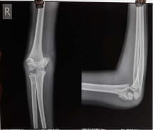
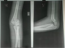
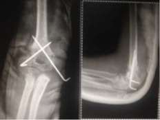
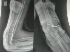
Introduction: Supracondylar fracture of the humerus is the most common elbow injury in children and makes up approximately 60% of all elbow injuries. The purpose of the present study is to evaluate the functional outcome of the displaced supracondylar humerus fracture treated with closed reduction and k-wire fixation by lateral and cross pinning technique. Material and Method: 50 children with fractures of the supracondylar humerus out of which 30 were boys and 20 were girls taken for prospective study at C.U. Shah Medical College from May 2017 to August 2019 was analyzed clinically and radiologically using Flynn’s criteria. Out of 50 cases 28 patients treated with lateral pinning and 22 patients treated with cross pinning technique based on the surgeon’s preference. Result: among patients treated with lateral pinning technique 19(68%) had an excellent result, 9(32%) had a good result. Similarly in patients treated with cross pinning technique, 9(41%), 7(32%), 2(9%), 4(18%) had excellent, good, fair, and poor outcomes respectively. 9 patients developed iatrogenic ulnar nerve palsy in cross pinning technique whereas 2 patients developed cubitus varus following cross pinning technique. Conclusion: Thus it can be concluded that closed reduction and K-wire fixation is an excellent method for the treatment of supracondylar fracture of the humerus with the significant difference in functional outcome between lateral pinning and cross pinning technique. The chances of ulnar nerve palsy increase following cross pinning technique which is not so in the case of lateral pinning. Thus suggesting the use of lateral pinning technique for the treatment of displaced supracondylar fracture of the humerus.

Keywords: Supracondylar fracture, Humerus, K-wire fixation

Corresponding Author How to Cite this Article To Browse
Baiju Patel, 3rd Year Resident Doctor, Department of Orthopaedics, C U Shah Medical College and Hospital, Surendranagar, Gujarat, India.
Email:
Vora J, Patel B, Vala G. A study to evaluate the functional outcome of displaced supracondylar humerus fracture in pediatrics treated with closed reduction and k-wire fixation. Surgical Review Int J Surg Trauma Orthoped. 2020;6(2):93-98.
Available From
https://surgical.medresearch.in/index.php/ijoso/article/view/158

Manuscript Received Review Round 1 Review Round 2 Review Round 3 Accepted
15-03-2020 25-03-2020 01-04-2020 06-04-2020
Conflict of Interest Funding Ethical Approval Plagiarism X-checker Note
No Nil Yes 14%

© 2020 by Jinesh Vora, Baiju Patel, Gaurav Vala and Published by Siddharth Health Research and Social Welfare Society. This is an Open Access article licensed under a Creative Commons Attribution 4.0 International License https://creativecommons.org/licenses/by/4.0/ unported [CC BY 4.0].

Introduction

Supracondylar fracture of the humerus is the most common elbow injury in children and makes up approximately 60% of all elbow injuries [1]. It becomes progressively more uncommon as the child approaches adolescence, the average age group of patients being 7⅟2 years. It is the fracture, which involves the lower end of the humerus just above both condyles and just proximal to the olecranon fossa, and does not directly involve the joint (extra-articular fracture) or distal humeral epiphysis.

Considering, the number of patients injured and the severity of the initial injury that occurs great diligence is required to secure an excellent result and to avoid or minimize the crippling complications, such as Volkmann’s ischaemic contracture, myositis ossification, stiffness, permanent nerve injuries, and malunion.

It is a general belief that accurate reduction in children is not essential for a good result, because growth may correct a deformity. It is true that functional end results of malalignment are generally very good but are also true the cosmetic end results are very poor. Stiffness of the elbow which sometimes follows relatively minor injuries, remarkable sensitivity of the injured joint, and too early passive movement add to the difficulties of treatment and prognosis. Recurrence of displacement occurs inspite of accurate closed reduction and immobilization in flexion.

These injuries of elbow demand respect because of their vascular damage and nerve injury they cause than any other injuries in the body [2]. There is no controversy regarding the management of undisplaced and partially displaced fracture but the treatment of a completely displaced fracture is not one but many.

Others have devised blind pinning after reduction or pinning under x-ray control. Some even advocate the extent to accept an unsatisfactory closed reduction, perform an osteotomy to correct the deformity at a later stage.

In 1800, Cooper and Hamilton use to treat these fractures with cuff and collar with the elbow in flexion, Liston in the 19th century and Robert in 1992 preferred immobilizing elbow in extension [3]. In 1898, Mouchet stressed the importance of accurate reduction to get satisfactory results. Cotton F.J. (1924) advised manipulation and maintenance of reduction with posterior splint with the elbow at

90⁰ flexion [4]. Bohler (1929) used percutaneous 'K' wire to hold a fracture fragment and a long arm slab. Attenbargh in 1995 showed that varus or valgus tilts are not corrected by remodeling. Karp in 1940 said that cause of varus is an epiphyseal injury to the lower end of humerus his views were shared by Lippilito in 1986. In 1945, Boyd and Altenherg and showed that radial, median and ulnar nerves were involved in that order of frequency [5]. The purpose of the present study is to evaluate the functional outcome of the displaced supracondylar humerus fracture treated with closed reduction and k-wire fixation by lateral and cross pinning technique.

Material and Method

50 children with fracture of the supracondylar humerus out of which 30 were boys and 20 were girls taken for prospective study at C.U. Shah Medical College from May 2017 to August 2019.The detailed history of each case was taken with a thorough clinical examination for the evaluation of the vascular and neurological status of extremity.

The radiograph anteroposterior and lateral view of the affected extremity was taken. The fractures are classified into flexion and extension type but the study contains extension type of fractures because of the rarity of flexion variety of fractures.

Extension type fracture is classified based on Gartland's classification i.e Type I- undisplaced, Type II- displaced with intact posterior cortex and Type III- displaced with no cortical contact. 15 were Gartland type 2 and 35 were Gartland type 3. Patients were analyzed clinically and radiologically using Flynn's criteria. Of the 50 cases, 28 patients were treated with lateral pinning and 22 with a cross pinning technique based on the surgeon’s preference.

Inclusion Criteria

  1. Age between 3 to 12 years.
  2. Closed fracture.

Exclusion Criteria

  1. Age less than 3 years and more than 12 years.
  2. Open fracture.
  3. Associated fracture in the same limb.
  4. Previous fracture in the same elbow.
  5. Fracture requiring open reduction.
  6. Floating elbow.

  1. Neurovascular injury.

Method

  • Palpate the bony landmarks, check for the direction of displacement.
  • Apply traction with the elbow flexed at 20 degrees and correct any lateral displacement.
  • Push the olecranon anteriorly to correct the posterior displacement and flex the elbow about 40 degrees.
  • Rotate the forearm externally to correct the usual internal rotation deformity.
  • Continue to flex the elbow above a right angle with maintaining pressure on the olecranon. The posterior displacement is reduced before this is done, otherwise, the brachial artery will be damaged between the fracture fragment. Flex the elbow until the olecranon lies anterior to the epicondyles.
  • Keep forearm in full pronation, to prevent varus deformity.
  • Check the position radiologically. Any angulation and rotational deformity should not be accepted and correct it.

For the lateral pinning technique, after reduction evaluation, two pins were inserted from the lateral aspect of the elbow. The pins were divergent and engaged in the medial cortex. The elbow was kept hyperflexion and in a position of pronation for inserting the lateral pins.

The elbow was then extended fully and fracture reduction and stability assessed clinically and radiologically under image intensifier.

For the cross pinning technique, after reduction evaluation, the lateral pin was inserted first, similar to the manner for the lateral pinning technique. The elbow was then extended to less than 90â?° position and a medial pin was inserted. For medial pin insertion, the surgeon palpated the ulnar nerve and pushed it posteriorly with the thumb. The fracture reduction and stability were assessed clinically and radiologically under the image intensifier.

The excess length of the pins was cut and then bent outside the skin to avoid migration. Betadine roller gauze dressing was applied to avoid pin track infection. An above elbow plaster slab was applied with the elbow in 90â?° flexion and full supination of the forearm.

All patients were discharged after 2 days. They were followed for clinical evaluation of carrying angle, elbow range of motion and neurological status, and radiologically evaluation of fracture displacement, Baumann angle, and the humor-capitellar angle at 3-4 weeks with final follow up at 4 months.Complication if any were noted. The slab and pins were removed after 4 weeks and an active elbow range of motion exercise was encouraged. At final follow Flynn’s criteria were used to grade the result as excellent, good, fair, and poor.

PRE-OP

surgery_158_01.jpg surgery_158_02.jpg

POST-OP

surgery_158_03.jpg


surgery_158_04.jpg

Results

Age Distribution

Table 1: Age Distribution

Age group Number of patients Percentage
0-5 15 30%
6-10 28 56%
11-15 7 14%
Total 50 100%

Table 1 shows that the commonest age group for supracondylar fracture of the humerus is between 6-10 years, followed by 0-5 years and 11-15 years respectively. The youngest patient in the study was 3 years old and the oldest being 12 years. The average age was 7.3 years.

Sex Distribution

Table-2: Sex Distribution

Male Female Total M:F Ratio
No. Percentage No. Percentage
30 60% 20 40% 50 1.5:1

Table no. 2 shows that male: female ratio is 1.5:1 indicating a higher incidence among males when compared to females.

Table-3: Classification Of Fracture

Type I Type II Type III Type IV
No. Percentage No. Percentage No. Percentage No. Percentage
- - 15 30% 35 70% - -

The table shows that most of the cases in the study group had Gartland type III supracondylar humerus fractures.

Table-4: Analysis of Result Based on Flynn’s Criteria

Result No. of patients Percentage
Excellent 28 56
Good 16 32
Fair 2 4
Poor 4 8

Result in the study are considered according to Flynn’s criteria for this cosmetic, functional, and neurological factor is taken into consideration. 56% of cases had excellent results followed by 32%,4%, and 8% of cases having good, fair, and poor results respectively.

Discussion

Supracondylar fractures of humerus are the most common elbow injury in children and makeup approximately 60% of all elbow injuries. It becomes progressively more uncommon as the child approaches adolescence, the average age group of patients being 71/2 years [6].

These injuries of elbow demand respect because of their vascular damage and nerve injuries they cause than any other injuries in the body. Unfortunately, it is the most difficult fracture to treat. Difficulty in treating this fracture lies in the fact that the fracture gets completed very often. Moreover, some of the complications are of nature, which threatens the use of extremity either temporarily or permanently.

Initial treatment and final management of these fractures are of utmost importance. This is quite often complicated due to a lack of awareness, poverty, and the presence of traditional bonesetter.

Fifty cases of displaced fracture supracondylar humerus in children between 3 to 12 years treated by closed reduction and K-wire fixation by cross pinning or lateral pinning technique at C.U. Shah Medical College and Hospital were taken in this study. The purpose of this study was to evaluate the functional outcome of displaced fracture supracondylar of the humerus in children treated by closed reduction and K-wire fixation using cross pinning or lateral pinning technique.

The age group of the patient was between 3 to 12 years. The peak incidence was in 6-10 years with an average age of 7.3 years.

In the present study incidence of supracondylar fracture was more in male children i.e. 60% and 40% in females. This male preponderance was noted in the study by Wilkins males 62.8% [7]

The left side was involved in 58% of cases, more common than the right forearm with a ratio of 1.38:1, which is comparable to that of Aronson and Prager with the ratio of the left side to the right side


of 1.8:1. [8] The common mechanism of injury in the present study was a fall on outstretched hand i.e. in 82% of cases which is comparable with Edward and Palmer study.

In the present study, 70% of cases were of Gartland Type III fracture whereas 30% of cases were of Gartland Type II fracture with all cases having extension variety of injury. The displacement of the distal fragment was Posteromedial in 42% of cases whereas it was Posterolateral and Posterior in 40% and 18% of cases. The result is comparable with other studies having predominately Posteromedial displacement Wilkins 75%, Aronson, and Prager 75%.9,10].

The average duration of hospital stay was 2.18 days whereas the average duration of hospital stay in other studies was 3.4 days by Aronson and Prager [11]. This rate is lower than other series due to better aseptic precaution, good antibiotics, and all surgeries were done with close reduction. There were no cases of post-infection in the present study.

In the present study out of 50 cases, 28 patients (56%) underwent lateral pinning and 22 patients (44%) underwent cross pinning. The choice of method of pin fixation was made according to the operating surgeon's personal preference [12,13,14].

There were 9 (18%) cases of iatrogenic nerve injury that occurred following medial pinning. In 5 cases there was only paraesthesia along with the ulnar nerve distribution which subsided spontaneously in one week. In the other 4 cases of nerve palsy, there were both motor and sensory deficits but complete neurological recovery occurred by the end of 4 months. The precautions were taken such as inserting the lateral pin first followed by extension of the elbow with the milking of ulnar nerve during medial pin placement.

The incidence of ulnar nerve injury with medial pinning in other series was 8% in study By Skaggs et al. there was no iatrogenic nerve injury following lateral pinning.

The correlation between the type of pinning and functional outcome was made on the basis of change in the carrying angle and range of motion as compared to the normal side. In the present study average change in carrying angle for cases treated with lateral pinning was 3.4 degree (range 0- 7 degree) with 4 patients having changes in carrying angle between 6-10 degrees. In the study by Aronson and Prager, this was 2.2 degrees (range 0

-8degree) [15,16]. The average change in carrying angle in cases treated by cross pinning was 4.2 with a range of 0-20 degrees. 2 patients had a loss of carrying angle above 10 degrees. There were 2 cases of cubitus varus deformity which occurred with cross pinning due to rotational instability. The deformity was associated with loss of flexion of 5 degrees and 10 degrees respectively. In the present study loss of movement occurred in 30% of cases. Loss of flexion was found in 14% of cases. Loss of extension was found in 16% of cases. The average loss of range of movement was 3.6 degrees for cases of cross pinning. For cases with lateral pinning the average loss of range of movement was 2.1 degree. The difference with regards to the loss of range of movement between the two groups was not statistically significant with both groups showing an excellent range of movement [17]. Functional outcome following two types of pinning was evaluated according to Flynn’s criteria. The functional outcome was excellent 56% and good in 32% of cases with the fair and poor result having 4% and 8% respectively. The cases treated with lateral pinning showed excellent results in 68% and good results in 32% of cases with no poor result. The cases treated with cross pinning showed excellent results in 41% and good, fair, and poor results in 32%, 9%, and 18% respectively.

Conclusion

In the present study, it was observed that closed reduction and percutaneous pinning is an excellent method for the treatment of displaced supracondylar fractureof humerus in children. Crossed pinning and lateral pinning is the treatment of choice in these fractures, careful technique during crossed pinning would reduce the chance of ulnar nerve injury.

What does the study add to the existing knowledge?

It was also observed that the lateral pinning is a good treatment of choice especially for the grossly swollen elbow in which the medial epicondyle is barely palpable with increased risk of ulnar nerve injury during placement of the medial pin.Both methods offered consistently satisfactory functional and cosmetic results. Iatrogenic ulnar nerve palsy was seen in cases of crossed pinning which was not so in case of lateral pinning. Cubitus varus is the commonest complication of this fracture in the present study.


Author's contribution

Dr. Jinesh Vora: Concept, study design

Dr. Baiju Patel: Manuscript preparation

Dr. Gaurav Vala: Statistical analysis

Reference

  1. Herring JA, editor. Fracture about the elbow, In- Tachdjian's Paediatric Orthopaedics. Philadelphia, WB Saunders. 3rd ed, 2002; Vol 3,2139-2221.
  2. David L, Skaggs and John M, Flynn. Supracondylar fracture of the distal humerus (Chapter 16), In Rockwood and Wilkins Fractures in Children. 8th edition, Lippincott Williams and Wilkins: Philadelphia. p 581-628.
  3. Weiland AJ, Meyer S, Tolo VT, Berg HL, Mueller J. Surgical treatment of displaced supracondylar fractures of the humerus in children, Analysis of fifty-two cases followed for five to fifteen years. J Bone Joint Surg Am. 1978;60(5)657-661.
  4. Zamzam MM, Bakarman KA. Treatment of displaced supracondylar humeral fractures among children- crossed versus lateral pinning. Injury. 2009;40(6)625-630.
  5. Vala GP, Chauhan R, Raiyani DM. AComparative Study of Crossed versus Lateral Pinning in theTreatment of Displaced Extension Type of SupracondylarFractures of the Humerus. GJRA. 2016;5(4).
  6. Rodriguez-Merchan EC. Supracondylar fractures of the humerus in children- treatment by overhead skeletal traction. J Pediatr Orthop. 1997;17(1);127.
  7. Celiker O, Pestilci FI, TuzunerM. Supracondylar fractures of the humerus in children- analysis of the result in 142 patients. J Orthop Trauma. 1990;4(3)265-269.
  1. Flynn C. Blind Pinning of Displaced of the Humerus Supracondylar in Children. J Bone Joint Surg Am. 1974;56(2)263-272.
  2. Gartland J. Management of Supracondylar fractures of the humerus in children. Surg gynecol obstet. 1959;109(2)145-154.
  3. Smith. Deformity following supracondylar fracture humerus. J Bone and Joint Surg. 1960;42A;235.
  4. Sharma A, Walia JSP, Brar BS, Sethi S. Early results of displaced supracondylar fractures of humerus in children treated by closed reduction and percutaneous pinning. Indian J Orthop. 2015;49(5)529-535.
  5. Boyd HB, Attenberg AR. Fracture about elbow in children. Arch Surg. 1944;49;231-234.
  6. Naik LG, Sharma GM, Badgire KS. Cross Pinning Versus Lateral Pinning in the Managementof Type III Supracondylar Humerus Fracture in Children. J Clin Diagnos Res. 2017;11(8)RC01-RC03.
  7. Henrikson B. Supracondylar fracture of humerus in children. ActaChirScand. 1956;369:1-72.
  8. Bharti SR, Magdum AK. A Comparative Study of Cross Pinning Versus Lateral Pinning by Percutaneous K-wire in Paediatric Supracondylar Fractures of Humerus. Int J Biomed Res. 2018;9(9)325-329.
  9. Aronson DD, Prager BI. Supracondylar fractures of humerus inchildren – A Modified technique of closed pinning. Clin Orthop Relat Res. 1987;218;174-184.
  10. Bhuyan BK. Close reduction and percutaneous pinning in displaced supracondylar humerus fractures in children. J Clin Orthop Trauma. 2012;3(2)89-93.