E-ISSN:2455-5436
P-ISSN:2456-9518
RNI:MPENG/2017/70870

Research Article

Pneumothorax

Surgical Review: International Journal of Surgery Trauma and Orthopedics

2022 Volume 8 Number 1 January February
Publisherwww.medresearch.in

Pneumothorax in Patients with Moderate to Severe Covid-19 Infection: A Retrospective Observational Study

George R.1, Paul Thomas V.2, Miriam Ipe S.3, Els Jojo J.4*
DOI: https://doi.org/10.17511/ijoso.2022.i01.03

1 Rahul George, Assistant Professor, Department of General Surgery, M.O.S.C Medical College & Hospital, Kolenchery, Ernakulam, Kerala, India.

2 Vijy Paul Thomas, Unit Chief and H.O.D, Department of General Surgery, M.O. S.C Medical College & Hospital, Kolenchery, Ernakulam, Kerala, India.

3 Shalini Miriam Ipe, Associate Professor, Department of Anesthesiology, M.O.S.C Medical College & Hospital, Kolenchery, Ernakulam, Kerala, India.

4* Joicy Els Jojo, Junior Resident, Department of General Surgery, M.O.S.C Medical College & Hospital, Kolenchery, Ernakulam, Kerala, India.

Background: Spontaneous pneumothorax as a complication of COVID-19 pneumonia is either uncommon or under-reported. The exact incidence and risk factors are still unknown. The objective of this study is to highlight an important complication, its incidence and explore the predisposing risk factors. Methods: We performed a retrospective review of COVID-19 pneumonia cases admitted to our hospital between February 2021 and June 2021. Data on their demographics, pre-existing risk factors, laboratory workup, imaging, treatment and clinical outcomes were gathered. Results: One thousand eight hundred and twenty-five patients have admitted to our institution between February 2021 and June 2021 with COVID-19 infection. 11 patients developed a pneumothorax (0.6%). 6 out of the 11 cases were patients who required mechanical ventilation (54.5%). All of these patients underwent chest tube insertion. Baseline imaging of these patients showed ground-glass opacities (GGO) and consolidation. 8 of the 11 patients succumbed to the disease (72.7%). Conclusion: In patients admitted with COVID-19 pneumonia and developing sudden respiratory compromise, pneumothorax is an important complication to be considered. Prompt identification of this complication and timely intervention is necessary to reduce morbidity and mortality. Low tidal volume lung-protective ventilation and the use of non-invasive ventilation for oxygenation remain the cornerstone in the management of COVID pneumonia. It significantly decreases iatrogenic complications like VILI (ventilator-induced lung injury) and P-SILI (patient self-inflicted lung injury.

Keywords: COVID-19, Pneumomediastinum, Pneumonia, Pneumothorax, Ventilation

Corresponding Author How to Cite this Article To Browse
Joicy Els Jojo, Junior Resident, Department of General Surgery, M.O.S.C Medical College & Hospital, Kolenchery, Ernakulam, Kerala, India.
Email:
Rahul George, Vijy Paul Thomas, Shalini Miriam Ipe, Joicy Els Jojo, Pneumothorax in Patients with Moderate to Severe Covid-19 Infection: A Retrospective Observational Study. Surgical Rev Int J Surg Trauma Orthoped. 2022;8(1):12-23.
Available From
https://surgical.medresearch.in/index.php/ijoso/article/view/256

Manuscript Received Review Round 1 Review Round 2 Review Round 3 Accepted
2022-01-03 2022-01-05 2022-01-12 2022-01-19 2022-01-26
Conflict of Interest Funding Ethical Approval Plagiarism X-checker Note
Nil Nil Yes 16%

© 2022by Rahul George, Vijy Paul Thomas, Shalini Miriam Ipe, Joicy Els Jojoand Published by Siddharth Health Research and Social Welfare Society. This is an Open Access article licensed under a Creative Commons Attribution 4.0 International License https://creativecommons.org/licenses/by/4.0/ unported [CC BY 4.0].

Introduction

Coronavirus is a family of an RNA virus and the disease is caused by SARS-CoV-2 capturing the world by its jugular. COVID-19 was declared a global health emergency by the WHO as of January 30, 2020. [1,2]. Pneumothorax is often a late manifestation and complication that presents during the treatment of COVID-19 pneumonia [3,4,5]. Pneumothorax increases the risk of mortality when undiagnosed or untreated. [6]. In a study published by Yang and colleagues in 92 deceased COVID-19 patients, one (1.1%) had a pneumothorax and died as a result of it 5 days after the initial presentation. [7]. Pneumothorax has also been linked to poor prognosis in patients infected with the acute Middle East respiratory syndrome coronavirus (MERS-CoV). [8]. Both pneumothorax and pneumomediastinum are known complications of mechanical ventilation. [3,9]. Nonetheless, even without barotrauma, pneumothorax,  pneumomediastinum, or more rarely both, can be present in COVID-19. [10,11]. In this study, we aim to explore the risk factors and understand the pathophysiology of pneumothorax in COVID-19 pneumonia.

Materials & Methods

A retrospective review of data of patients admitted to our institution with COVID-19 disease between February 2021 and June 2021 was performed. During this period one thousand eight hundred and twenty-five patients were treated in our institution. They were diagnosed via RT-PCR or antigen test. All patients were evaluated with a chest radiograph and CT thorax during the hospital stay. Pneumothorax was confirmed with a chest radiograph. Patients with pneumothorax were reviewed thoroughly. Details regarding risk factors (comorbidities, smoking, BMI, ventilation status and steroid use), laboratory parameters (CRP, LDH, Serum Ferritin, D-Dimer and total counts), imaging findings ( chest radiograph, CT severity score and CORADS), treatment modalities and outcome of disease were documented for each patient.

Sample Size: 1825

Sample size  equation- Estimation of proportion:

surgery_256_01.JPG

  • Sampling method: Convenient sampling.
  • Study design: Retrospective Observational study (from hospital records).
  • Study area: Department of General Surgery and Department of Anesthesiology, MOSC Medical College Hospital, Kolenchery, Ernakulam, Kerala.
  • Study population: Moderate to Severe COVID positive patients in Kerala.
  • Inclusion criteria: All patients who got admitted with Moderate to Severe COVID-19 infection.
  • Exclusion criteria: Patients who expired before appropriate evaluation or treatment.
  • Ethical Considerations: Approval was sought from the institutional ethics committee. Waiver of Informed consent was sought from the IEC.

Results

In our study, it was observed that the mean age of presentation was 51 years of age. The most common presenting symptoms were cough, fever and breathlessness. The average duration of symptoms was 4.9 days. It was noted that none of the patients was vaccinated at the time of presentation. 5 patients who developed pneumothorax were referred cases with severe COVID-19 pneumonia. (Table 1 & 2)

Patients who developed pneumothorax had comorbidities such as diabetes mellitus, hypertension, coronary artery disease, valvular heart disease, BPAD, nephrotic syndrome, COPD and pulmonary tuberculosis. Only one patient had a significant pulmonary disease (COPD, pulmonary tuberculosis) with a history of smoking. 2 patients had a history of intake of steroids. One patient had a BMI of less than 18.5kg/m2 (underweight), 2 patients had a BMI of more than 30kg/m2 (obese) and 8 patients had a BMI of 25-kg/m2 (overweight). (Table 1 & 2)


Table 1: Presenting Symptoms and Vaccination Status.

Sr no Symptoms Numbers & percentage Vaccination status
1 Cough 9(81.8%) Not vaccinated
2 Fever 7(63.6%) Not vaccinated
3 Breathlessness 8(72.7%) Not vaccinated
4 Diarrhoea 2(18.1%) Not vaccinated
5 Vomiting 1(9.09%) Not vaccinated
6 Swelling in the neck and chest 1(9.09%) Not vaccinated

Table 2: Comorbidities and Risk Factors.

Sr No. Comorbidities/ Risk factors Numbers and Percentages
1 Diabetes mellitus 7(63.6%)
2 Systemic hypertension 6(54.5%)
3 COPD & Pulmonary edema 1(9.09%)
4 CAD 2(18.18%)
5 Valvular heart disease 1(9.09%)
6 Nephrotic syndrome 1(9.09%)
7 Dyslipidemia 1(9.09%)

The mean serum LDH level was 825 U/L whereas the mean serum Ferritin level was 844.5 mcg/L. The D-Dimer, total counts and CRP levels were noted to be high in patients who developed pneumothorax. 9 patients who developed pneumothorax, had bilateral chest infiltrates on chest radiograph at presentation. CT findings included ground-glass opacities (GGO), consolidation and pneumomediastinum. (Figure 1 & 2).

surgery_256_02.JPG
Figure 1: Chest Xray Showing Left Pneumothorax and Right-Sided Consolidation.

Table 3: Salient Xray and Ct Findings of Patients with Pneumothorax. 

Sr No. Xray Findings Numbers and Percentages CT findings Numbers and Percentages
1 Chest infiltrates 9(81.8%) Ground glass opacities 8(72.72%)
2 Pneumothorax 2(18.18%) Consolidation 8(72.72%)
3     Pneumomediastinum 1(9.09%)
4     Pneumothorax 2(18.18%)

CT severity score was more than 12 in all patients with the mean score being 20.

surgery_256_03.JPG
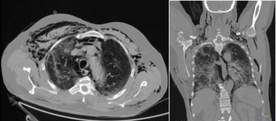
Figure 2: Axial and Coronal Sections of Ct Imaging Showing Ground Glass Opacities, Subcutaneous Emphysema, Pneumomediastinum and Pneumothorax.

The mean time of development of pneumothorax was noted to be 11 days after testing positive for COVID-19 infection. 6 patients who developed pneumothorax had subcutaneous emphysema. All patients underwent intercostal drainage and one patient required multiple same side intercostal drain (ICD) insertions for persistent non-resolving pneumothorax. One patient also underwent prophylactic ICD insertion on the opposite side to decrease the tracking of air from the affected lung. (Table 4).

Table 4: Clinical Features and Icd Details.

Sr no. Subcutaneous Emphysema Number and Percentages ICD position and number Number and Percentages
1 Present 6(54.54%) Unilateral 8(72.72%)
2 Absent                 5(45.45%) Bilateral 3(27.27%)
 3     Single ICD 8(72.72%)
4     Multiple ICD 3(27.27%)

In our study 11 out of 1825 patients developed a pneumothorax (0.6%). 5 out of them (45.4%) had a spontaneous pneumothorax. Four of the patients were on volume control ventilation, with a tidal volume (TV) of 6 to 8 ml/kg and positive end-expiratory pressure (PEEP) ranging from 7 to 12, while one was spontaneously breathing on a continuous positive airway pressure (CPAP) of 10mmHg. (Table 5).

Table 5: Mechanism of Pneumothorax and Mode of Ventilation.

Sr no. Mechanism of pneumothorax Numbers and Percentages Mode of non-invasive ventilation Numbers and percentages
1 Spontaneous 5(45.45%) BiPAP 2(18.18%)
2 Post ventilation 6(54.54%) SFM 2(18.18%)
3     HFNC 1(9.09%)

In our study, 5 patients (45.4%) who developed pneumothorax were on non-invasive ventilation (NIV). Out of these 5 patients, 2 patients (40%) were on SFM, 2 patients (40%) were on bilevel positive airway pressure (BiPAP) and 1 (20%) on high flow nasal cannula (HFNC). (Table 5).

All patients with pneumothorax were treated with steroids and antibiotics. 9 patients were started on anticoagulant therapy and 8 patients received anti-virals. 2 patients also received monoclonal antibodies as part of treatment. (Table 6).

Table 6: Treatment Modalities.

Sr no. Treatment administered Numbers and percentages
1 Antivirals 8(72.72%)
2 Antibiotics 11(100%)
3 Anticoagulants 9(81.81%)
4 Steroids 11(100%)
5 Monoclonal antibodies 2(18.18%)

3 of the patients survived while the remaining 8 succumbed to the fatal disease. (Table7).

Table 7: Outcomes in Patients with Pneumothorax.

Sr No. Outcome Numbers and Percentages
1 Survived 3(27.27%)
2 Succumbed 8(72.72%)

Discussion

Pneumothorax refers to air getting entrapped in the pleural space which can either be spontaneous or following trauma. [12]. Pneumothorax can also be classified as primary, secondary, iatrogenic (pacemaker insertion, thoracic surgeries) or following blunt/penetrating trauma. [12]. Diseased lung causes secondary pneumothorax whereas primary pneumothorax occurs in generally healthy people without an underlying lung pathology. [13]. In pneumothorax, loss of intrapleural negative pressure causes the lung to collapse partially or impair ventilation, oxygenation or both. The clinical presentation varies from being asymptomatic to life-threatening. [13]. Pneumothorax is a surgical emergency; prompt identification and management are important for a good prognosis. [12,13].

The primary risk factor for spontaneous pneumothorax (SP) is always an underlying lung pathology of which chronic obstructive pulmonary disease with emphysema outscores others such as tuberculosis, smoking, trauma, lung malignancy,

cystic fibrosis, alpha-1 antitrypsin deficiency, HIV associated Pneumocystis pneumonia and other lung diseases. [14-16]. Tall and thin built males in the age group of 10-30 years are known to be at risk for the development of primary spontaneous pneumothorax. [17]. Spontaneous pneumothorax and pneumomediastinum unrelated to intubation or positive pressure ventilation have been reported in various infections such as influenza, pneumocystis jirovecii pneumonia (PCP), herpes simplex, staphylococcus aureus pneumonia, cytomegalovirus, influenza bronchiolitis and severe acute respiratory syndrome (SARS). [18-23]. Other predisposing factors include asthma, corticosteroids, respiratory irritants and other anatomical abnormalities such as tracheomalacia. [24-27].

Development of bullae in otherwise normal lung parenchyma is seen in bullous lung disease. [28] Risk factors for the development of bullae include smoking, pulmonary sarcoidosis, alpha-1 antitrypsin, alpha-1 anti-chymotrypsin deficiency, connective tissue disorders (Marfan’s and Ehlers-Danlos syndrome), fibreglass exposure and addiction to drugs (marijuana). [29,30]. In COVID-19, sudden respiratory compromise in otherwise stable patients has been reported. The most prevalent cause for this sudden worsening was a pulmonary embolism, followed by pneumothorax as reported by Shaikha D Al-Shokri et al. [31, 32]. Cough, a common symptom of COVID-19 pneumonia poses an increased risk of spontaneous pneumothorax due to increased intrapulmonary pressure especially in the presence of underlying alveolar damage due to inflammation, ischemic parenchymal damage and other pleural abnormalities. [33-35]. Intense coughing can increase the intrathoracic pressure rapidly to up to 300mmHg. [36]

COVID-19 pneumonia is characterized by overwhelming inflammation and steroids are often recommended for reducing inflammatory responses and is thus a potential strategy for reversing progression in acute respiratory distress syndrome (ARDS). [37]. Steroid use in connective tissue disorders has been seen to weaken the pulmonary interstitial tissue causing an alveolar air leak. [38]. Corticosteroid therapy causes delayed wound healing in fragile lung tissue and leads to the formation of bullae and subsequent pneumothorax through the check valve mechanism. [39]. Even though pneumothorax is implicated in chronic


steroid use, a previous study describes a COVID-19 patient developing pneumothorax after just 6 days of steroid use from the time of hospitalization. [40]. In patients with SARS and pneumothorax, administration of methylprednisolone affected lung healing and the presence of a higher peak serum LDH and peripheral leukocyte count was postulated to depict a greater extent of lung injury, thus raising the risk of pneumothorax. [41].

COVID-19 data from Wuhan, China showed that obese patients had more severe illnesses and increased mortality. [42]. Studies have found obesity (BMI>=30kg/m2) to be an independent risk factor for pulmonary oedema, diffuse alveolar damage (DAD) and alveolar-capillary hemosiderosis. [43]. One can postulate that obese COVID-19 patients who are at risk for DAD are also at higher risk for developing SPM. Studies have demonstrated COVID-19 viral entry and cellular injury via angiotensin-converting enzyme -2 (ACE-2) receptor into target cells including surfactant-producing type II pneumocytes. [44]. The dysregulation of surfactant production contributes to the development of subcutaneous emphysema (SE) and pneumomediastinum from impaired lung compliance. The upregulation of ACE-2 expression found in chronic hypertension and diabetes mellitus may potentially explain the increased viral load in the population, leading to increased morbidity in COVID-19. [45]. COVID-19 infection can cause severe pneumonia leading to ARDS which is radiographically diagnosed by ground-glass opacities forming consolidative changes and at later stages to fibrotic changes. [7,4]. COVID 19 causes ischemic lung parenchymal damage by activation of inflammatory markers and fibroblasts precipitating fibro-myxoid exudates promoting pulmonary cyst formation. [46,29]. Development of severe intrapulmonary strain due to increased respiratory effort and persistent bouts of cough result in rupture of the alveolar cysts. [47]. Severe lung injury and diffuse alveolar damage contribute to the mechanism of spontaneous pneumothorax complicating severe acute respiratory distress syndrome. [41]. The rupture of alveoli secondary to diffuse alveolar damage as seen in COVID-19 may cause pulmonary interstitial emphysema (PIE), as is seen with neonates on ventilatory support. [48]. Hyperactive and dysregulated immune response known as cytokine storm syndrome leads to a hyperinflammatory form of ARDS which is

associated with critical illness and increased mortality in COVID-19 pneumonia. [49-51]. Hyperferritinemia, haemodynamic instability and multiorgan failure is now recognized as being the main cause of the severity of SARS-CoV-2 infection. [52]. This leads to weakening of bronchial walls causing edema, vascular congestion and microthrombi contributing to the rupture of preexisting bullae leading to pneumothorax. [29].

Laboratory evaluation has a predictive value to identify potential risk factors for spontaneous pneumomediastinum (SPM). Levels of cell death due to plasma membrane damage can be reflected by Lactate dehydrogenase (LDH). In the SARS outbreak, higher peak LDH was associated with SPM as compared with those without SPM (mean 863 U/L Vs 583 U/L). [53]. In the early phase of COVID-19 infection, leukopenia, lymphocytopenia, high level of CRP, high D-dimer, prolonged PT, and high levels of fibrinogen have been reported. [54,55]. A similar trend was noted in our study with the mean Serum LDH level being 825U/L. D-Dimer, total counts and CRP levels were also noted to be high in patients who developed pneumothorax. A study by Chen G et al reported that ferritin level was higher than 800mcg/L in 100% of patients with severe COVID-19 infection. [56]. It has also been reported that serum ferritin of more than 400mcg/L is a risk factor for progression to severe disease in a study by Ji D et al. [57]. The mean Serum ferritin level in our study was 844.5mcg/L

The hallmarks of the virus characterized by CT are bilateral interstitial infiltrates, ground-glass opacity (GGO) patterns, multiple lobar and subsegmental consolidations and air bronchograms. [58,10]. Features like pleural or pericardial effusions, cavitary lesions, bronchiectasis along uncommon features of pneumomediastinum and pneumothorax were also reported. [10,11,59,60]. CT remains the most effective method to detect lung abnormalities and estimate the evolution of the disease. Pneumothorax is a rare radiological finding seen in 1% of 99 patients was reported early in the pandemic by Chen et al. [61].


Severe COVID-19 patients are at high risk for coagulopathy like deep vein thrombosis (DVT), venous thromboembolism (VTE) and possible pulmonary embolism (PE) (25%). [62,63].


Increased D-Dimer, APLA, lactate dehydrogenase with mild to no changes in PT and PTT are seen in COVID-19 coagulopathy. [17,25,26]. Cytokine storm syndrome, DIC, immobilization, hypoxia secondary to excessive lung injury in COVID-19 infection can result in thromboembolic events. [64]. Mechanical ventilation causes overdistension of alveoli, causing the occurrence of a large pressure gradient between the marginal alveoli and lung interstitium, resulting in air leaks to the surrounding bronchovascular sheath (Macklin phenomenon). [65]. This explains how long term ventilation, results in accelerating cyst rupture and tracking air out of pulmonary tissue. [66]. This air traverses to the visceral pleura, mediastinum and cervical soft tissue causing pneumothorax, mediastinal emphysema, pneumomediastinum and cervicofacial subcutaneous emphysema. [34,67,68].

Invasive mechanical ventilation is often required in the treatment of COVID-19 and it appears to be a predominant risk factor for pneumothorax in COVID-19 pneumonia. The incidence of pneumothorax in mechanically ventilated patients is high and even higher in ARDS ranging from 14 to 87%. In our study 11 out of 1825 patients developed a pneumothorax (0.6%). 6 out of them (54.5%) had spontaneous pneumothorax while on mechanical ventilation. Four of the patients were on volume control ventilation, with a TV of 6 to 8 ml/kg and a PEEP ranging from 7 to 12, while one was spontaneously breathing on a CPAP of 10mmHg.The incidence of pneumothorax is seen to increase with the severity and duration of ARDS, duration of ventilation, comorbidities, barotrauma and volutrauma. Aiolfi et al, Yao W et al and Sun R et al reported cases of pneumothorax after mechanical ventilation. [69,3,5]. Sun R also suggested that ventilation using high PEEP may cause the development of pneumothorax, partly through the development of new bullae or rupture of existing bullae or cysts. [5]. Often manoeuvres performed to improve oxygenation such as high PEEP, high tidal volumes and high minute ventilation leads to high peak inspiratory pressures (greater than 40 to 50 cmH2O) alveolar overdistension and pneumothorax. In addition manipulation of the airway (intubation or tracheostomy) may cause alveolar rupture and pneumothorax. [70-72, 34]. Lung protective ventilation strategies such as low-volume and pressure-limited ventilation are essential to minimize barotrauma and volutrauma.


In a previous study, the rate of pneumothorax development was 5.9% in the first 24 hours following initiation of ventilation, and this rate increased to 10.4% after 1 day. [3]. Gattinoni et al, found that the longer the duration of mechanical ventilation in ARDS, the higher the incidence of pneumothorax (87% vs. 30% in those with more than 2 weeks of mechanical ventilation vs. less than 1 week). [73]. In addition, patients with bullae and low lung compliance had higher rates of pneumothorax.

It was interesting to note that patients on NIV (BIPAP, CPAP) and oxygen delivery devices (SFM, HFNC), who are thought to have a low risk of barotrauma and subsequent complications were seen to develop pneumothorax. In our study, 5 patients (45.4%) who developed pneumothorax were on NIV.Out of these 5 patients, 2 patients (40%) were on SFM, 2 patients (40%) were on BiPAP and 1 (20%) on HFNC. The proposed mechanism is that the end-expiratory pressure provided by BiPAP or CPAP increases the pressure gradient between alveoli and the interstitial space, causing alveolar rupture with the extension of air into the mediastinum, pleura, and subcutaneous tissues. That being said the vast majority of patients on NIV do not experience SE or SPM. It has been theorized that extensive airspace disease caused by COVID-19 makes normally low-risk patients susceptible to SE/SPM, perhaps related to the constellation of lung damage, supplemental oxygen, and decreased lung compliance. [74]. Wang et al and Zhou et al presented cases of pneumothorax without invasive ventilation. [40,11].

Wang et al reported a patient who presented with spontaneous pneumothorax, pneumomediastinum and subcutaneous emphysema without invasive ventilation. [40]. A case of pneumothorax in a spontaneously breathing patient was also reported by Zhou et al. [11]. High-flow nasal cannula (HFNC) therapy is an intermediate level of support between conventional oxygen delivery and non-invasive ventilation (NIV) and is capable of delivering up to 100% humidified and heated oxygen at a flow rate of 60L/min and a PEEP of up to 6 cm of water, meeting the increased flow demand and hence reduces the work of breathing in ARDS. [75].

HFNC helps mitigate (patient self-inflicted lung injury) P-SILI by delivering a variable pressure during the respiratory cycle with maximum pressure


in early expiration. This mechanism supplies a PEEP thus promoting alveolar recruitment but also avoids alveolar overdistension despite a high flow. Even though a few cases of possible barotrauma concerning HFNC have been reported in the pediatric population, the incidence of pneumothorax is reported to be as low as 1%. [76,77]. A Multicentric study from the UK comparing non-intubated patients and patients requiring positive pressure ventilation for the development of pneumothorax and pneumomediastinum concluded that both groups were susceptible for pneumothorax and barotrauma alone cannot explain this association. [78].

Conclusion

Having been over 2 years into the pandemic, we now realise that pneumonia is a common sequelae of COVID-19 infection. The mechanism of viral entry and cellular injury through the ACE-2 receptors into target cells as well as the cytokine storm leading to a hyperinflammatory form of ARDS have been delineated. Sudden deterioration of respiratory status in such patients should alert one to the possibility of pneumothorax as the precipitating factor, even in patients not on mechanical ventilation. Institution of lung protecting strategies and early intervention in cases of pneumothorax are thus important in ensuring better outcomes in these patients.

Authors Contribution: Conception and design of work: Rahul George, Vijy Paul Thomas, Data collection: Rahul George, Shalini Miriam Ipe, Data analysis and interpretation: Rahul George, Vijy Paul Thomas, Shalini Miriam Ipe, Joicy Els Jojo, Drafting the article: Rahul George, Shalini Miriam Ipe, Joicy Els Jojo, Critical review of the article: Vijy Paul Thomas, Shalini Miriam Ipe, Final approval of the version to be published: Rahul George, Vijy Paul Thomas, Shalini Miriam Ipe, Joicy Els Jojo

What does this study add to the existing knowledge?

Prompt identification of pneumothorax in COVID 19 pneumonia and timely intervention is necessary to reduce morbidity and mortality. Low tidal volume lung-protective ventilation and the use of non-invasive ventilation for oxygenation remain the cornerstone in the management of COVID pneumonia.  It significantly

decreases iatrogenic complications like VILI (ventilator-induced lung injury) and P-SILI (patient self-inflicted lung injury).

Reference

01. World Health Organization. Novel coronavirus (2019-nCoV) situation [report] [cited Jun 5, 2020]. Available from: https://www. who. int/emergencies/diseases/novel-coronavirus-2019/situation-reports. Vol. 11; 2020. Google Scholar [Crossref][PubMed][Google Scholar]

02. World Health Organization. Novel coronavirus (2019-nCoV) situation [report] [cited Jun 5, 2020]. Available from: https://www. who. int/emergencies/diseases/novel-coronavirus-2019/situation-reports. Vol. 1; 2020. Google Scholar [Crossref][PubMed][Google Scholar]

03. Yao W, Wang T, Jiang B, Gao F, Wang L, Zheng H, et al. Emergency tracheal intubation in 202 patients with COVID-19 in Wuhan, China: lessons learnt and international expert recommendations. Br J Anaesth. 2020 Jul;125(1):e28-e37. doi: 10.1016/j.bja.2020.03.026 [Crossref][PubMed][Google Scholar]

04. Hosseiny M, Kooraki S, Gholamrezanezhad A, Reddy S, Myers L. Radiology Perspective of Coronavirus Disease 2019 (COVID-19): Lessons From Severe Acute Respiratory Syndrome and Middle East Respiratory Syndrome. AJR Am J Roentgenol. 2020 May;214(5):1078-1082. doi: 10.2214/AJR.20.22969 [Crossref][PubMed][Google Scholar]

05. Sun, Ruihong, Hongyuan Liu, and Xiang Wang. Mediastinal emphysema, giant bulla, and pneumothorax developed during the course of COVID-19 pneumonia. Korean Journal of Radiology 21. 5 (2020): 541. [Crossref][PubMed][Google Scholar]

06. Xiang, Chunlin, and Gang Wu. SARS-CoV-2 pneumonia with subcutaneous emphysema, mediastinal emphysema, and pneumothorax: a case report. " Medicine 99. 20 (2020). [Crossref][PubMed][Google Scholar]

07. Yang F, Shi S, Zhu J, Shi J, Dai K, Chen X. Analysis of 92 deceased patients with COVID-19. J Med Virol. 2020 Nov;92(11):2511-2515. doi: 10.1002/jmv.25891 [Crossref][PubMed][Google Scholar]


08. Das KM, Lee EY, Al Jawder SE, Enani MA, Singh R, Skakni L, et al. Acute Middle East Respiratory Syndrome Coronavirus: Temporal Lung Changes Observed on the Chest Radiographs of 55 Patients. AJR Am J Roentgenol. 2015 Sep;205(3):W267-74. doi: 10.2214/AJR.15.14445 [Crossref][PubMed][Google Scholar]

09. Jacobi A, Chung M, Bernheim A, Eber C. Portable chest X-ray in coronavirus disease-19 (COVID-19): A pictorial review. Clin Imaging. 2020 Aug;64:35-42. doi: 10.1016/j.clinimag.2020.04.001 [Crossref][PubMed][Google Scholar]

10. Salehi S, Abedi A, Balakrishnan S, Gholamrezanezhad A. Coronavirus Disease 2019 (COVID-19): A Systematic Review of Imaging Findings in 919 Patients. AJR Am J Roentgenol. 2020 Jul;215(1):87-93. doi: 10.2214/AJR.20.23034 [Crossref][PubMed][Google Scholar]

11. Zhou, Changyu, et al. COVID-19 with spontaneous pneumomediastinum. " The Lancet Infectious Diseases 20. 4 (2020): 510. [Crossref][PubMed][Google Scholar]

12. Imran JB, Eastman AL. Pneumothorax. JAMA. 2017 Sep 12;318(10):974. doi: 10.1001/jama.2017.10476 [Crossref][PubMed][Google Scholar]

13. Zarogoulidis P, Kioumis I, Pitsiou G, Porpodis K, Lampaki S, Papaiwannou A, et al. Pneumothorax: from definition to diagnosis and treatment. J Thorac Dis. 2014 Oct;6(Suppl 4):S372-6. doi: 10.3978/j.issn.2072-1439.2014.09.24 [Crossref][PubMed][Google Scholar]

14. MacDuff A, Arnold A, Harvey J; BTS Pleural Disease Guideline Group. Management of spontaneous pneumothorax: British Thoracic Society Pleural Disease Guideline 2010. Thorax. 2010 Aug;65 Suppl 2:ii18-31. doi: 10.1136/thx.2010.136986 [Crossref][PubMed][Google Scholar]

15. Sahn SA, Heffner JE. Spontaneous pneumothorax. N Engl J Med. 2000 Mar 23;342(12):868-74. doi: 10.1056/NEJM200003233421207 [Crossref][PubMed][Google Scholar]

16. Noppen M. Spontaneous pneumothorax: epidemiology, pathophysiology and cause. Eur Respir Rev. 2010 Sep;19(117):217-9.

doi: 10.1183/09059180.00005310 [Crossref][PubMed][Google Scholar]

17. Gupta D, Hansell A, Nichols T, Duong T, Ayres JG, Strachan D. Epidemiology of pneumothorax in England. Thorax. 2000 Aug;55(8):666-71. doi: 10.1136/thorax.55.8.666 [Crossref][PubMed][Google Scholar]

18. López-Rivera F, Colón Rivera X, González Monroig HA, Garcia Puebla J. Pneumomediastinum and Pneumothorax Associated with Herpes Simplex Virus (HSV) Pneumonia. Am J Case Rep. 2018 Jan 30;19:109-113. doi: 10.12659/ajcr.906051 [Crossref][PubMed][Google Scholar]

19. She WH, Chok KSH, Li IWS, Ma KW, Sin SL, et al. Pneumocystis jirovecii-related spontaneous pneumothorax, pneumomediastinum and subcutaneous emphysema in a liver transplant recipient: a case report. BMC Infect Dis. 2019 Jan 18;19(1):66. doi: 10.1186/s12879-019-3723-y [Crossref][PubMed][Google Scholar]

20. Chu CM, Leung YY, Hui JY, Hung IF, Chan VL, Leung WS, et al. Spontaneous pneumomediastinum in patients with severe acute respiratory syndrome. Eur Respir J. 2004 Jun;23(6):802-4. doi: 10.1183/09031936.04.00096404 [Crossref][PubMed][Google Scholar]

21. Olliff JF, Williams MP. Radiological appearances of cytomegalovirus infections. Clin Radiol. 1989 Sep;40(5):463-7. doi: 10.1016/s0009-9260(89)80245-4 [Crossref][PubMed][Google Scholar]

22. Silva C, Almeida AF, Ferraz C, Nunes T, Guedes Vaz L. Spontaneous Pneumothorax With Subcutaneous Emphysema: A Rare Complication of Respiratory Syncytial Virus Infection. J Clin Med Res. 2016 Mar;8(3):260-2. doi: 10.14740/jocmr2353w [Crossref][PubMed][Google Scholar]

23. Murayama S, Gibo S. Spontaneous pneumomediastinum and Macklin effect: Overview and appearance on computed tomography. World J Radiol. 2014 Nov 28;6(11):850-4. doi: 10.4329/wjr.v6.i11.850 [Crossref][PubMed][Google Scholar]

24. Zylak CM, Standen JR, Barnes GR, Zylak CJ. Pneumomediastinum revisited. Radiographics. 2000 Jul-Aug;20(4):1043-57.


doi: 10.1148/radiographics.20.4.g00jl131043. Erratum in: Radiographics 2001 Nov-Dec;21(6):1616 [Crossref][PubMed][Google Scholar]

25. Lal A, Mishra AK, Sahu KK, Noreldin M. Spontaneous Pneumomediastinum: Rare Complication of Tracheomalacia. Arch Bronconeumol (Engl Ed). 2020 Mar;56(3):185-186. English, Spanish. doi: 10.1016/j.arbres.2019.09.017 [Crossref][PubMed][Google Scholar]

26. Meireles, José, et al. Spontaneous pneumomediastinum revisited. Respiratory Medicine CME 4. 4 (2011): 181-183. [Crossref][PubMed][Google Scholar]

27. Fishman, Alfred, et al. Fishman's pulmonary diseases and disorders. McGraw Hill Professional, 2008. [Crossref][PubMed][Google Scholar]

28. Goldberg C, Carey KE. Bullous lung disease. West J Emerg Med. 2013 Sep;14(5):450-1. doi: 10.5811/westjem.2013.3.16276 [Crossref][PubMed][Google Scholar]

29. Johnson MK, Smith RP, Morrison D, Laszlo G, White RJ. Large lung bullae in marijuana smokers. Thorax. 2000 Apr;55(4):340-2. doi: 10.1136/thorax.55.4.340 [Crossref][PubMed][Google Scholar]

30. Poissy J, Goutay J, Caplan M, Parmentier E, Duburcq T, Lassalle F, et al. Lille ICU Haemostasis COVID-19 Group. Pulmonary Embolism in Patients With COVID-19: Awareness of an Increased Prevalence. Circulation. 2020 Jul 14;142(2):184-186. doi: 10.1161/CIRCULATIONAHA.120.047430 [Crossref][PubMed][Google Scholar]

31. Al-Shokri SD, Ahmed AOE, Saleh AO, AbouKamar M, Ahmed K, Mohamed MFH. Case Report: COVID-19-Related Pneumothorax-Case Series Highlighting a Significant Complication. Am J Trop Med Hyg. 2020 Sep;103(3):1166-1169. doi: 10.4269/ajtmh.20-0713 [Crossref][PubMed][Google Scholar]

32. Joynt GM, Antonio GE, Lam P, Wong KT, Li T, Gomersall CD, et al. Late-stage adult respiratory distress syndrome caused by severe acute respiratory syndrome: abnormal findings at thin-section CT. Radiology. 2004 Feb;230(2):339-46. doi: 10.1148/radiol.2303030894 [Crossref][PubMed][Google Scholar]

33. Woodside, Kenneth J. , et al. Pneumothorax in patients with acute respiratory distress syndrome: pathophysiology, detection, and treatment. Journal of Intensive Care Medicine 18. 1 (2003): 9-20 [Crossref][PubMed][Google Scholar]

34. Akdogan, Raziye Ecem, et al. Pneumothorax in mechanically ventilated patients with COVID-19 infection. " Case Reports in Critical Care 2021 (2021). [Crossref][PubMed][Google Scholar]

35. McCool, F. Dennis. Global physiology and pathophysiology of cough: ACCP evidence-based clinical practice guidelines. " Chest 129. 1 (2006): 48S-53S [Crossref][PubMed][Google Scholar]

36. Russell B, Moss C, Rigg A, Van Hemelrijck M. COVID-19 and treatment with NSAIDs and corticosteroids: should we be limiting their use in the clinical setting? Ecancermedicalscience. 2020 Mar 30;14:1023. doi: 10. 3332/ecancer.2020.1023 [Crossref][PubMed][Google Scholar]

37. Korkmaz, C. , et al. Pneumomediastinum and subcutaneous emphysema associated with dermatomyositis. " Rheumatology 40. 4 (2001): 476-478 [Crossref][PubMed][Google Scholar]

38. Kadota T, Shimizu K, Tsurushige C, Kawaishi M, Araya J, Nakayama K, et al. Organizing pneumonia complicated by cyst and pneumothorax formation. Intern Med. 2012;51(22):3155-8. doi: 10.2169/internalmedicine.51.8319 [Crossref][PubMed][Google Scholar]

39. Wang W, Gao R, Zheng Y, Jiang L. COVID-19 with spontaneous pneumothorax, pneumomediastinum and subcutaneous emphysema. J Travel Med. 2020 Aug 20;27(5):taaa062. doi: 10.1093/jtm/taaa062 [Crossref][PubMed][Google Scholar]

40. Sihoe AD, Wong RH, Lee AT, Lau LS, Leung NY, Law KI, et al. Severe acute respiratory syndrome complicated by spontaneous pneumothorax. Chest. 2004 Jun;125(6):2345-51. doi: 10.1378/chest.125.6.2345 [Crossref][PubMed][Google Scholar]

41. Kass, David A. , Priya Duggal, and Oscar Cingolani. Obesity could shift severe COVID-19 disease to younger ages. The Lancet 395. 10236 (2020): 1544-1545 [Crossref][PubMed][Google Scholar]

42. Haque AK, Gadre S,


Taylor J, Haque SA, Freeman D, Duarte A. Pulmonary and cardiovascular complications of obesity: an autopsy study of 76 obese subjects. Arch Pathol Lab Med. 2008 Sep;132(9):1397-404. doi: 10.5858/2008-132-1397-PACCOO [Crossref][PubMed][Google Scholar]

43. Hoffmann, Markus, et al. SARS-CoV-2 cell entry depends on ACE2 and TMPRSS2 and is blocked by a clinically proven protease inhibitor. " cell 181. 2 (2020): 271-280. [Crossref][PubMed][Google Scholar]

44. Úri K, Fagyas M, Mányiné Siket I, Kertész A, Csanádi Z, Sándorfi G, et al. New perspectives in the renin-angiotensin-aldosterone system (RAAS) IV: circulating ACE2 as a biomarker of systolic dysfunction in human hypertension and heart failure. PLoS One. 2014 Apr 1;9(4):e87845. doi: 10.1371/journal.pone.0087845 [Crossref][PubMed][Google Scholar]

45. Do Lago VC, Cezare TJ, Fortaleza CMCB, Okoshi MP, Baldi BG, Tanni SE. Does COVID-19 Increase the Risk for Spontaneous Pneumothorax? Am J Med Sci. 2020 Dec;360(6):735-737. doi: 10. 1016/j.amjms.2020.07.024 [Crossref][PubMed][Google Scholar]

46. Ucpinar BA, Sahin C, Yanc U. Spontaneous pneumothorax and subcutaneous emphysema in COVID-19 patient: Case report. J Infect Public Health. 2020 Jun;13(6):887-889. doi: 10.1016/j.jiph.2020.05.012 [Crossref][PubMed][Google Scholar]

47. Morisot C, Kacet N, Bouchez MC, Rouland V, Dubos JP, Gremillet C, et al. Risk factors for fatal pulmonary interstitial emphysema in neonates. Eur J Pediatr. 1990 Apr;149(7):493-5. doi: 10.1007/BF01959402 [Crossref][PubMed][Google Scholar]

48. Qin C, Zhou L, Hu Z, Zhang S, Yang S, Tao Y, et al. Dysregulation of Immune Response in Patients With Coronavirus 2019 (COVID-19) in Wuhan, China. Clin Infect Dis. 2020 Jul 28;71(15):762-768. doi: 10.1093/cid/ciaa248 [Crossref][PubMed][Google Scholar]

49. Giamarellos-Bourboulis EJ, Netea MG, Rovina N, Akinosoglou K, Antoniadou A, Antonakos N, et al. Complex Immune Dysregulation in COVID-19 Patients with Severe Respiratory Failure. Cell Host Microbe. 2020 Jun 10;27(6):992-1000.

e3. doi: 10.1016/j.chom.2020.04.009 [Crossref][PubMed][Google Scholar]

50. Ye Q, Wang B, Mao J. The pathogenesis and treatment of the `Cytokine Storm' in COVID-19. J Infect. 2020 Jun;80(6):607-613. doi: 10.1016/j.jinf.2020.03.037 [Crossref][PubMed][Google Scholar]

51. Gao, Y‐M. , et al. Cytokine storm syndrome in coronavirus disease 2019: A narrative review. " Journal of internal medicine 289. 2 (2021): 147-161 [Crossref][PubMed][Google Scholar]

52. Chu CM, Leung YY, Hui JY, Hung IF, Chan VL, Leung WS, et al. Spontaneous pneumomediastinum in patients with severe acute respiratory syndrome. Eur Respir J. 2004 Jun;23(6):802-4. doi: 10.1183/09031936.04.00096404 [Crossref][PubMed][Google Scholar]

53. Tang N, Li D, Wang X, Sun Z. Abnormal coagulation parameters are associated with poor prognosis in patients with novel coronavirus pneumonia. J Thromb Haemost. 2020 Apr;18(4):844-847. doi: 10.1111/jth.14768 [Crossref][PubMed][Google Scholar]

54. Cui S, Chen S, Li X, Liu S, Wang F. Prevalence of venous thromboembolism in patients with severe novel coronavirus pneumonia. J Thromb Haemost. 2020 Jun;18(6):1421-1424. doi: 10.1111/jth.14830 [Crossref][PubMed][Google Scholar]

55. Chen G, Wu D, Guo W, Cao Y, Huang D, Wang H, et al. Clinical and immunological features of severe and moderate coronavirus disease 2019. J Clin Invest. 2020 May 1;130(5):2620-2629. doi: 10.1172/JCI137244 [Crossref][PubMed][Google Scholar]

56. Ji D, Zhang D, Chen Z, Xu Z, Zhao P, Zhang M, et al. Clinical characteristics predicting progression of COVID-19. (2020). [Crossref][PubMed][Google Scholar]

57. Simpson S, Kay FU, Abbara S, Bhalla S, Chung JH, Chung M, et al. Radiological Society of North America Expert Consensus Document on Reporting Chest CT Findings Related to COVID-19: Endorsed by the Society of Thoracic Radiology, the American College of Radiology, and RSNA. Radiol Cardiothorac Imaging. 2020 Mar 25;2(2):e200152. doi: 10.1148/ryct.2020200152 [Crossref][PubMed][Google Scholar]


58. Caruso D, Zerunian M, Polici M, Pucciarelli F, Polidori T, Rucci C, et al. Chest CT features of COVID-19 in Rome, Italy. " Radiology 296. 2 (2020): E79-E85. [Crossref][PubMed][Google Scholar]

59. Rohailla S, Ahmed N, Gough K. SARS-CoV-2 infection associated with spontaneous pneumothorax. CMAJ. 2020 May 11;192(19):E510. doi: 10.1503/cmaj.200609 [Crossref][PubMed][Google Scholar]

60. Chen N, Zhou M, Dong X, Qu J, Gong F, Han Y, et al. Epidemiological and clinical characteristics of 99 cases of 2019 novel coronavirus pneumonia in Wuhan, China: a descriptive study. Lancet. 2020 Feb 15;395(10223):507-513. doi: 10.1016/S0140-6736(20)30211-7 [Crossref][PubMed][Google Scholar]

61. Cui S, Chen S, Li X, Liu S, Wang F. Prevalence of venous thromboembolism in patients with severe novel coronavirus pneumonia. J Thromb Haemost. 2020 Jun;18(6):1421-1424. doi: 10.1111/jth.14830 [Crossref][PubMed][Google Scholar]

62. Zhang L, Feng X, Zhang D, Jiang C, Mei H, Wang J, et al. Deep Vein Thrombosis in Hospitalized Patients With COVID-19 in Wuhan, China: Prevalence, Risk Factors, and Outcome. Circulation. 2020 Jul 14;142(2):114-128. doi: 10.1161/CIRCULATIONAHA.120.046702 [Crossref][PubMed][Google Scholar]

63. Klok FA, Kruip MJHA, van der Meer NJM, Arbous MS, Gommers DAMPJ, Kant KM, et al. Incidence of thrombotic complications in critically ill ICU patients with COVID-19. Thromb Res. 2020 Jul;191:145-147. doi: 10.1016/j.thromres.2020.04.013 [Crossref][PubMed][Google Scholar]

64. Macklin, Madge Thurlow, and Charles C. Macklin. Malignant interstitial emphysema of the lungs and mediastinum as an important occult complication in many respiratory diseases and other conditions: an interpretation of the clinical literature in the light of laboratory experiment. " Medicine 23. 4 (1944): 281-358 [Crossref][PubMed][Google Scholar]

65. Fleming WH, Bowen JC. Early complications of long-term respiratory support. J Thorac Cardiovasc Surg. 1972 Nov;64(5):729-38. [Crossref][PubMed][Google Scholar]

66. Mason, R. , et al. Nadel’s textbook of respiratory medicine. (2005). [Crossref][PubMed][Google Scholar]

67. Vanzo V, Bugin S, Snijders D, Bottecchia L, Storer V, Barbato A. et al. Pneumomediastinum and pneumopericardium in an 11-year-old rugby player: a case report. " Journal of athletic training 48. 2 (2013): 277-281 [Crossref][PubMed][Google Scholar]

68. Aiolfi A, Biraghi T, Montisci A, Bonitta G, Micheletto G, Donatelli F, et al. Management of Persistent Pneumothorax With Thoracoscopy and Bleb Resection in COVID-19 Patients. Ann Thorac Surg. 2020 Nov;110(5):e413-e415. doi: 10.1016/j.athoracsur.2020.04.011 [Crossref][PubMed][Google Scholar]

69. Zantah, Massa, et al. Pneumothorax in COVID-19 disease-incidence and clinical characteristics. Respiratory Research 21. 1 (2020): 1-9. [Crossref][PubMed][Google Scholar]

70. Albelda SM, Gefter WB, Kelley MA, Epstein DM, Miller WT. Ventilator-induced subpleural air cysts: clinical, radiographic, and pathologic significance. Am Rev Respir Dis. 1983 Mar;127(3):360-5. doi: 10.1164/arrd.1983.127.3.360 [Crossref][PubMed][Google Scholar]

71. Gammon RB, Shin MS, Buchalter SE. Pulmonary barotrauma in mechanical ventilation. Patterns and risk factors. Chest. 1992 Aug;102(2):568-72. doi: 10.1378/chest.102.2.568 [Crossref][PubMed][Google Scholar]

72. Gattinoni L, Chiumello D, Caironi P, Busana M, Romitti F, Brazzi L, et al. COVID-19 pneumonia: different respiratory treatments for different phenotypes? Intensive Care Med. 2020 Jun;46(6):1099-1102. doi: 10. 1007/s00134-020-06033-2 [Crossref][PubMed][Google Scholar]

73. Carron M, Freo U, BaHammam AS, Dellweg D, Guarracino F, Cosentini R, et al. Complications of non-invasive ventilation techniques: a comprehensive qualitative review of randomized trials. Br J Anaesth. 2013 Jun;110(6):896-914. doi: 10.1093/bja/aet070 [Crossref][PubMed][Google Scholar]

74. Mauri, Tommaso, et al. Optimum support by high-flow nasal cannula in acute hypoxemic respiratory failure: effects of increasing


flow rates. Intensive care medicine 43. 10 (2017): 1453-1463. [Crossref][PubMed][Google Scholar]

75. Hegde S, Prodhan P. Serious air leak syndrome complicating high-flow nasal cannula therapy: a report of 3 cases. Pediatrics. 2013 Mar;131(3):e939-44. doi: 10.1542/peds.2011-3767 [Crossref][PubMed][Google Scholar]

76. Baudin F, Gagnon S, Crulli B, Proulx F, Jouvet P, Emeriaud G. Modalities and Complications Associated With the Use of High-Flow Nasal Cannula: Experience in a Pediatric ICU. Respir Care. 2016 Oct;61(10):1305-10. doi: 10.4187/respcare.04452 [Crossref][PubMed][Google Scholar]

77. Martinelli, Anthony W. , et al. COVID-19 and pneumothorax: a multicentre retrospective case series. " European Respiratory Journal 56. 5 (2020) [Crossref][PubMed][Google Scholar]