E-ISSN:2455-5436
P-ISSN:2456-9518
RNI:MPENG/2017/70870

Case Report

Large Bowel Lymphoma

Surgical Review: International Journal of Surgery Trauma and Orthopedics

2022 Volume 8 Number 1 January February
Publisherwww.medresearch.in

A Case Report on Primary Large Bowel Lymphoma

Shree Venkatesan B.1*, Senthil Kumar k.2, Thangamani P.3
DOI: https://doi.org/10.17511/ijoso.2022.i01.04

1* Bhaskaran Shree Venkatesan, Postgraduate, , MMC, Chennai, Tamil Nadu, India.

2 k Senthil Kumar, Assistant Professor, , MMC, Chennai, Tamil Nadu, India.

3 P Thangamani, Professor, Chief of General Surgery, MMC, Chennai, Tamil Nadu, India.

Large bowel (colorectal) lymphoma is a very rare tumor, accounting for <0.5% of primary colorectal malignancies, ~1.5% of all lymphomas, and ~15% of gastrointestinal lymphoma. The definition of primary GI lymphoma has differed among authors but typically refers to lymphoma that predominantly involves any section of the GI tract from the oropharynx to the rectum. The major site of extranodal non-Hodgkin lymphoma is the gastrointestinal tract(stomach>small intestine >large intestine). The disease appears later in life, predominantly in the male population. Complaints are nonspecific, requiring a high index of suspicion to establish the diagnosis Treatment includes surgery/chemotherapy/radiotherapy or a combination of all these three modalities. The aim of this paper is to report a rare case of primary colon lymphoma in a female patient.

Keywords: Large bowel lymphoma, Colorectal lymphoma, Primary

Corresponding Author How to Cite this Article To Browse
Bhaskaran Shree Venkatesan, Postgraduate, , MMC, Chennai, Tamil Nadu, India.
Email:
Bhaskaran Shree Venkatesan, k Senthil Kumar, P Thangamani, A Case Report on Primary Large Bowel Lymphoma. Surgical Rev Int J Surg Trauma Orthoped. 2022;8(1):24-28.
Available From
https://surgical.medresearch.in/index.php/ijoso/article/view/252

Manuscript Received Review Round 1 Review Round 2 Review Round 3 Accepted
2021-12-08 2021-12-10 2021-12-17 2021-12-24 2021-12-31
Conflict of Interest Funding Ethical Approval Plagiarism X-checker Note
Nil Nil Yes 17%

© 2022by Bhaskaran Shree Venkatesan, k Senthil Kumar, P Thangamaniand Published by Siddharth Health Research and Social Welfare Society. This is an Open Access article licensed under a Creative Commons Attribution 4.0 International License https://creativecommons.org/licenses/by/4.0/ unported [CC BY 4.0].

Introduction

Lymphoma (historical) lymphosarcoma was used for diffuse forms of the disease) is a malignancy arising from lymphocytes or lymphoblasts. Lymphoma can be restricted to the lymphatic system or can arise as an extranodal disease. This, along with variable aggressiveness results in a diverse imaging appearance. Thomas Hodgkin was the first to introduce lymphomas into medical science in 1832.

various classification systems have been used to differentiate lymphomas including the Rappaport Classification (used until the 70s) [1]. Kiel System, Working Formulation and Revised European-American Classification of Lymphoid Neoplasms (REAL) The latest WHO classification recognizes 5 histological subtypes of B cell lineage: Extranodal marginal lymphoma (MALT lymphoma), follicular lymphoma, mantle cell lymphoma, diffuse large B-cell lymphoma and Burkitt’s lymphoma [2].

All histological subtypes of nodal lymphomas may arise in the gastrointestinal tract out of which Diffuse large B-cell lymphoma is the most common subtype (~65%) of large bowel lymphoma. Other less common types include mucosa-associated lymphoid tissue (MALT) lymphoma and mantle cell lymphoma (MCL). 

The main idea of reporting this case is the rarity of presentation in the aforementioned age group and gender of the patient.

The mean age at diagnosis is 55 years. Men are affected twice as often as women.

Case Report

A 74-year-old lady presented with complaints of Altered bowel habits for the past 6 months, also with the loss of weight >6 kgs in the past 6 months, Pedal oedema for 1 month. The patient had fatigue and generalized myalgia as well.

History included T2DM on metformin 500mg bd well within control, Diastolic dysfunction on aspirin 150mg od, Lasix 20mg od and spironolactone 25mg od.

Menstrual history included pt had a normal menstrual cycle and attained menopause 30 years ago.

No significant family history.

The patient was a non-smoker, tobacco chewer, alcohol or betel nut chewer.

On examination, the mass of 3x4 cm was palpated at the right iliac fossa.

Initial blood works were normal, a CT scan revealed growth in ascending colon & caecum, and colonoscopic biopsies showed chronic active colitis.

Table 1: Blood investigation reports.

s.no wbc hb platelets urea creatinine Na K
1 5600 10.1 4.5 lakh        
2 4500 9.6 4.1 lakh 12 0.4 138 3.6
3 10000 11.4 4.7 lakh 18 0.6 138 4.5

Tumor marker: CEA 2.55 (normal)

CECT Abdomen suggested:

-Abnormal Circumferential wall thickening and luminal narrowing in the caecum, ic junction and terminal ileum.

-Multiple Lymphonodes in right ileocolic mesentery max dia 2x1 cm and aortocaval 1.7x1.4cm.

OGD- proliferative nodular growth at the cecum and edematous ileocecal valve noted, the biopsy was taken correspondingly.

Biopsy 1: chronic active colitis, no evidence of dysplasia or malignancy

Because of persistent symptoms proceeded with Right hemicolectomy

surgery_252_01.JPG
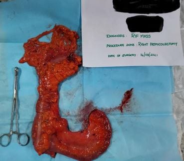
Fig 1: Right hemicolectomy specimen Final biopsy report.


surgery_252_02.JPG
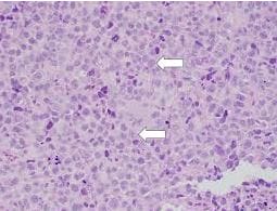
Fig 2: sheets of lymphocyte aggregates.

surgery_252_03.JPG
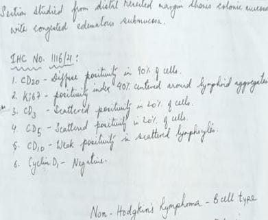
Fig 3: Final biopsy report.

Post-op follow up: The patient was monitored in the intensive care unit, recovered well and uneventful, started on the diet on the 5th postoperative day, and discharged on the 10th pod day. She was referred to the medical oncology and haematology department. She was asked to do a pet scan which showed.

a) Post right hemicolectomy status

ill-defined fat stranding and soft tissue thickening with increased metabolically active post-surgical sites and anterior abdominal wall.

b) Enlarged, homogeneously enhancing retroperitoneal and right external iliac nodes suggesting lymphomatous involvement. Planned for chemotherapy.

Discussion

The mean age at diagnosis

is 55 years. Men are affected twice as often as women. The most common symptoms of colonic lymphoma are Abdominal pain, Nausea, Vomiting, Weight loss, Abdominal mass, Change in bowel habits, Hematochezia, Obstruction, Intussusceptions, Acute peritonitis due to intestinal perforation. Lymphoma can often present with B symptoms (fever, night sweats and weight loss)

The infrequency of these complaints suggests that, despite the large size of the lymphoma, mucosal ulceration does not usually occur

The cecum is the most frequent location for primary colorectal lymphoma, probably due to the larger amount of lymphoid tissue in this region. In different series, the data on the frequency in the rectum varies from 8.3% to 35%.

The lack of specific complaints makes the diagnosis hard to establish. Unfortunately, for some patients, a surgical procedure is the only diagnostic tool. As a rare lesion, it requires a high index of suspicion. Due to a delay in diagnosis in 33%-65% of patients, the operative procedure is either urgent or emergent.

Dawson et al established criteria for the diagnosis of primary colorectal lymphomas in 1961(3)

These are:

(1) no enlarged superficial lymph nodes when the patient is first seen;

(2) chest radiographs without obvious enlargement of the mediastinal nodes;

(3) the white blood cell counts, both total and differential, are within normal range and bone marrow biopsy is also normal.

(4) at laparotomy only regional nodes are affected by the disease.

(5) the liver and spleen are free of tumors.

Table 2 : Published Studies in Large Bowel Lymphoma. Kindly refer Annexure 1

Anderson et al have shown that the distribution of NHL subtypes differs in various geographical areas and suggested that this could be a reflection of differences in etiological factors or the host response to these factors [4]. Unlike western countries, the frequency of T cell lymphoma in the East is reported to be 17.9%-42%.


Primary colorectal lymphoma differs from its gastric counterpart. The stomach is the most frequent site of origin of primary gastrointestinal lymphomas. The most common histological type found is MALT lymphoma Gastric MALT associated lymphoma can be successfully treated by Helicobacter pylori (H. Pylori) eradication. The same histological subtype localized in the large intestine does not have the same connection with H. pylori infection.

In more than half of patients, lymphoma is a bulky disease reaching over 5 cm in diameter. These bulky masses can usually be palpated by simple physical examination and can be viewed by ultrasonography.

This case is of particular interest as it has presented in an elderly female in her 7th decade with major symptom as weight loss and altered bowel habits.

Although the role of surgery in the treatment of gastrointestinal lymphoma is debatable, the majority of the reported patients were operated on irrespective of the disease stage. There are no controlled randomized trials due to the low incidence of the disease. According to some series, since the rate of spontaneous perforation is high (5 out of 17 patients), it is beneficial to perform a hemicolectomy to prevent this complication.

In the published studies, treatment varies from chemotherapy alone to multimodal therapies combining surgery, chemotherapy and radiotherapy. Chemotherapy remains the basis of treatment for the rapidly proliferating aggressive lymphomas because these malignancies almost always extend beyond local fields, encompassed by surgery or radiation.

The CHOP chemotherapeutic regimen (cyclophosphamide, doxorubicin, vincristine and prednisone) remains the first-line therapy for all moderate and high-grade B-cell lymphomas. Several prospective trials have shown that adding rituximab to a standard CHOP regimen (R-CHOP) resulted in higher response rates and better progression-free, event-free, disease-free and overall survival. Fan et al found significant improvement in the survival of patients with stage IIE who received adjuvant chemotherapy. However, the analysis of the subgroup of patients with high-grade lymphoma in stages I and II showed that adding chemotherapy did not significantly impact survival.

In 2005, Bilsel et al (16) reported a case of primary lymphoma of the rectum with a complete clinical response after a combination of radiation and chemotherapy. Similar results were published by other authors.

In case series published in 2002, Pricolo et al [17] treated patients with surgical resection and adjuvant chemo and radiotherapy.

Shimono et al (18) from Japan suggested preoperative radiotherapy.

Some authors proposed the so-called preventive chemotherapy in patients with stage IE, reaching five-year survival of 80%.

In cases of indolent lymphomas (mantle cell, follicular cell and T cell lymphomas), complete surgical resection and radiation are recommended because of decreased chemoresponsiveness.

Factors affecting survival differ among authors. The stage at the diagnosis has an impact on survival. According to Fan et al, only histological grade seems to be an adverse factor for prognosis. The urgency of the surgical procedure is also found to be an important factor affecting survival.

Conclusion

The vast majority of patients with primary colorectal lymphoma underwent some form of resection although the role of surgery and its extent is yet to be established. After resection, most patients undergo CHOP or other multi-agent therapy. The optimal management of primary lymphoma of the colon and rectum has never been determined by randomized trials. The small number of patients with various histological subtypes and different stages at presentation results in an unclear treatment protocol. Since all the published studies are retrospective, their scientific validity is partially diminished, imposing the need for conducting prospective multi-centric studies. Meanwhile, systematized clinical experiences contribute usefully to the knowledge of clinicians.

Reference

01. Rappaport, Henry. Tumors of the hematopoietic system. Atlas of tumor pathology (1966). [Crossref][PubMed][Google Scholar]

02. Harris NL, Jaffe ES, Diebold J, Flandrin G,


Muller-Hermelink HK, Vardiman J. Lymphoma classification--from controversy to consensus: the R. E. A. L. and WHO Classification of lymphoid neoplasms. Ann Oncol. 2000;11 Suppl 1:3-10 [Crossref][PubMed][Google Scholar]

03. Dawson Im, Cornes Js, Morson Bc. Primary malignant lymphoid tumours of the intestinal tract. Report of 37 cases with a study of factors influencing prognosis. Br J Surg. 1961 Jul;49:80-9. doi: 10.1002/bjs.18004921319 [Crossref][PubMed][Google Scholar]

04. Müller AM, Ihorst G, Mertelsmann R, Engelhardt M. Epidemiology of non-Hodgkin's lymphoma (NHL): trends, geographic distribution, and etiology. Ann Hematol. 2005 Jan;84(1):1-12. doi: 10.1007/s00277-004-0939-7 [Crossref][PubMed][Google Scholar]

05. Cai S, Cannizzo F Jr, Bullard Dunn KM, Gibbs JF, Czuczman M, Rajput A. The role of surgical intervention in non-Hodgkin's lymphoma of the colon and rectum. Am J Surg. 2007 Mar;193(3):409-12; discussion 412. doi: 10.1016/j.amjsurg.2006.12.007 [Crossref][PubMed][Google Scholar]

06. Bairey O, Ruchlemer R, Shpilberg O. Non-Hodgkin's lymphomas of the colon. Isr Med Assoc J. 2006 Dec;8(12):832-5. [Crossref][PubMed][Google Scholar]

07. Kim SJ, Choi CW, Mun YC, Oh SY, Kang HJ, Lee SI, et al. Multicenter retrospective analysis of 581 patients with primary intestinal non-hodgkin lymphoma from the Consortium for Improving Survival of Lymphoma (CISL). BMC Cancer. 2011 Jul 29;11:321. doi: 10.1186/1471-2407-11-321 [Crossref][PubMed][Google Scholar]

08. Doolabh N, Anthony T, Simmang C, Bieligk S, Lee E, Huber P, et al. Primary colonic lymphoma. J Surg Oncol. 2000 Aug;74(4):257-62. doi: 10.1002/1096-9098(200008)74:4<257::aid-jso3>3.0.co;2-u [Crossref][PubMed][Google Scholar]

09. Stanojević G, Stojanović M, Jovanović M, Stojanović M, Jeremić M, Branko B, et al. [Primary colorectal lymphomas]. Vojnosanit Pregl. 2009 Apr;66(4):295-301. Serbian. doi: 10.2298/vsp0904295s [Crossref][PubMed][Google Scholar]

10. Fan CW, Changchien CR, Wang JY, Chen JS, Hsu KC, Tang R, et al. Primary colorectal lymphoma. Dis Colon Rectum. 2000 Sep;43(9):1277-82. doi: 10.1007/BF02237436 [Crossref][PubMed][Google Scholar]

11. Cho MJ, Ha CS, Allen PK, Fuller LM, Cabanillas F, Cox JD. Primary non-Hodgkin lymphoma of the large bowel. Radiology. 1997 Nov;205(2):535-9. doi: 10.1148/radiology.205.2.9356641 [Crossref][PubMed][Google Scholar]

12. Gonzalez QH, Heslin MJ, Dávila-Cervantes A, Alvarez-Tostado J, de los Monteros AE, Shore G, et al. Primary colonic lymphoma. Am Surg. 2008 Mar;74(3):214-6. [Crossref][PubMed][Google Scholar]

13. Wong MT, Eu KW. Primary colorectal lymphomas. Colorectal Dis. 2006 Sep;8(7):586-91. doi: 10.1111/j.1463-1318.2006.01021.x [Crossref][PubMed][Google Scholar]

14. Busch E, Rodriguez-Bigas M, Mamounas E, Barcos M, Petrelli NJ. Primary colorectal non-Hodgkin's lymphoma. Ann Surg Oncol. 1994 May;1(3):222-8. doi: 10.1007/BF02303527 [Crossref][PubMed][Google Scholar]

15. Goran Z Stanojevic, Milica D Nestorovic, Branko R Brankovic, Miroslav P Stojanovic, Milan M Jovanovic, Milan D Radojkovic, Clinic for General Surgery, Clinical Center Nis, Bul Zorana Djindjica 48, 18000 Nis, Serbia. DOI: 10. 4251/wjgo. v3. i1.14 [Crossref][PubMed][Google Scholar]