E-ISSN:2455-5436
P-ISSN:2456-9518
RNI:MPENG/2017/70870

Research Article

Proximal Humerus Fracture

Surgical Review: International Journal of Surgery Trauma and Orthopedics

2021 Volume 7 Number 5 September October
Publisherwww.medresearch.in

A Clinical Study to Compare Functional Outcome of Proximal Humerus Fracture in Adults (18- 80yrs) Treated By Philos Platting and Conservative Modalities

Jetpariya D.1, Patel B.2*, Patel P.3, Patel H.4
DOI: https://doi.org/10.17511/ijoso.2021.i05.03

1 Divyesh Pragajibhai Jetpariya, Senior Resident, Department Of Orthopedics, M.P Shah Medical College, Jamnagar, Gujarat, India.

2* Baiju Mukeshbhai Patel, Senior Resident, Department Of Orthopedics, Smt. NHL Municipal Medical College, Ahmedabad, Gujarat, India.

3 Parth Pravinbhai Patel, Final Year MBBS, Department Of Orthopedics, C U Shah Medical College, Surendranagar, Gujarat, India.

4 Harsh Girishbhai Patel, Senior Resident, Department Of Orthopedics, Banas Medical College, Palanpur, Gujarat, India.

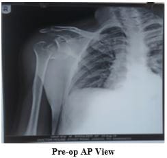
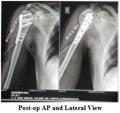
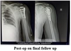
Introduction: It is the commonest fracture affecting the shoulder girdle in adults. Proximal humeral fractures account for almost 7% of all fractures and make up 80% of all humeral fractures. The purpose of this study is to carry out to enlighten functional outcome fracture proximal humerus treated with conservative and PHILOS platting and comparison of that. Material and Method: Twenty-six patients attended the Department Of Orthopaedics from May 2018 to August 2020 and were involved in this study prospectively, out of which 11 were male, and 15 were female. They were analysed clinically and radiologically using Constant and Murley shoulder scoring criteria. Out of 26 cases, 12 were treated conservatively, and 14 were treated by Pen reduction internal fixation (ORIF) Result: Out of 12 patients treated conservatively, three had excellent, seven had good, and 2 had fair functional outcome, whereas 14 patients treated by open reduction and internal fixation two had excellent, 4 had good, four had fair, and 4 had the poor functional outcome. Complications were seen in 9 patients. Out of that, five patients had shoulder stiffness, 3 had malunion, and 1 had impingement of implant. Conclusion: Proximal humerus fracture occurred most commonly in the 4th to 6th decade of life. For minimally displaced fracture, treatment of choice is nonoperative. For 2 part fracture treated with conservative modalities showed slightly better results than operated patients, but for 3 part fracture operated patients had somewhat better results than conservative.

Keywords: Proximal Humerus, Philos Plate, Shoulder Stiffness

Corresponding Author How to Cite this Article To Browse
Baiju Mukeshbhai Patel, Senior Resident, Department Of Orthopedics, Smt. NHL Municipal Medical College, Ahmedabad, Gujarat, India.
Email:
Divyesh Pragajibhai Jetpariya, Baiju Mukeshbhai Patel, Parth Pravinbhai Patel, Harsh Girishbhai Patel, A Clinical Study to Compare Functional Outcome of Proximal Humerus Fracture in Adults (18- 80yrs) Treated By Philos Platting and Conservative Modalities. Surgical Rev Int J Surg Trauma Orthoped. 2021;7(5):108-115.
Available From
https://surgical.medresearch.in/index.php/ijoso/article/view/245

Manuscript Received Review Round 1 Review Round 2 Review Round 3 Accepted
2021-10-07 2021-10-09 2021-10-16 2021-10-23 2021-10-30
Conflict of Interest Funding Ethical Approval Plagiarism X-checker Note
Nil Nil Yes 16%

© 2021by Divyesh Pragajibhai Jetpariya, Baiju Mukeshbhai Patel, Parth Pravinbhai Patel, Harsh Girishbhai Pateland Published by Siddharth Health Research and Social Welfare Society. This is an Open Access article licensed under a Creative Commons Attribution 4.0 International License https://creativecommons.org/licenses/by/4.0/ unported [CC BY 4.0].

Introduction

It is the commonest fracture affecting the shoulder girdle in adults [1]. Proximal humeral fractures account for almost 7% of all fractures and make up 80% of all humeral fractures. In patients above 65 years, proximal humeral fractures are the second most frequent upper extremity fracture and the third most common non-vertebral osteoporotic fracture after proximal femur and distal radius fractures, accounting for >10% of fractures in this patient population. [2,3].

These fractures challenge the treating orthopaedician because of their osteoporotic quality in older adults and the deforming forces of the muscles attached. Most proximal humerus fractures, in younger as well as in elderly patients, are stable & slightly or non-displaced, can be treated non operatively. Much controversy and confusion still exist, and no single treatment protocol or algorithm has been proved to be universally effective; areas still in question include radiographic diagnosis, operative or nonoperative treatment, consideration of patient age in treatment decision making, surgical approach, fracture fixation or hemiarthroplasty, type of internal fixation, and the rehabilitation protocol. Numerous authors have suggested that nonoperative treatment may be preferable for two-, three-, and four-part proximal humeral fractures in elderly patients, but pain and loss of function have been reported in high percentages of patients after this treatment approach. Several more recent reports, however, have indicated that the functional results of operative treatment are not significantly better than the results of nonoperative treatment in elderly patients, although radiograph results may be superior.[4].

The role of physiotherapy has emerged as a significant factor affecting overall prognosis. Therefore, this study is to carry out to enlighten functional outcome fracture proximal humerus treated with conservative and PHILOS platting and comparison of that.

Method and Material

Twenty-six patients were attended in the Department Of Orthopaedics from May 2018 to August 2020 and were involved in this study prospectively based on the following criteria.

Inclusion Criteria

  1. Closed fracture of proximal humerus without distal neurological and vascular deficit.
  2. Age: 18 years and above.
  3. Patients with a minimum of six months follow up. 

Exclusion Criteria:

  1. Compound fractures
  2. Age less than 18 years
  3. Pathological fractures
  4. Fracture with a distal neurological and vascular deficit
  5. Fractures that do not satisfy the inclusion criteria

After finding the suitability as per inclusion and exclusion criteria, patients were selected for the study and briefed about the nature of the study, the intervention, if any to be carried out and written, informed consent was obtained. History was obtained through verbal communication, clinical examination both local and systemic was done. All polytrauma patients were managed initially as per emergency care protocol. Once patients were vitally stable, X-rays were done, i.e. AP and Axillary views. If needed as per fracture type, CT scans were obtained. According to X rays, fractures were classified according to Neer's classification. Once the diagnosis was confirmed, the patient was given a shoulder immobiliser and analgesics. The further management of fracture was decided after senior consultants' opinion and treated accordingly.

All undisplaced or minimally displaced fractures were treated conservatively. In fractures with displacement more than 1 centimetre or angulation more than 45 degree of angulation was treated with open reduction and internal fixation with PHILOS (Proximal Humerus Internal Locking Osteosynthese) plate.

Those patients who were selected for operative treatment underwent routine preoperative investigation.

  • Blood and Urine investigation
  • HIV, HCV and HBsAg antibody detection and titers
  • ECG
  • X-ray Chest PA view

All patients were called for follow-up at one month, three months, six months, nine months and one year and evaluation using Constant Murley Scoring system along with appropriate radiological assessment was done. 

Non Operative Treatment: Non Operative treatment is preferable primarily for

  1. Elderly patients with osteoporosis.
  2. Severe comorbid conditions.
  3. Minimally displaced fractures.
  4. Impacted fractures.
  5. Surgical neck fractures with contact and no gross instability.
  6. Greater and lesser tuberosity fractures Displacement < 1 cm Overlap with the head < 20%.
  7. Varus posteromedial fractures Varus < 45 degrees or Minor tuberosity displacement.
  8. Valgus fractures where head not pointing superior or lateral Minor tuberosity displacement.

Closed reduction of highly comminuted or displaced fractures is challenging to reduce and manage often results in poor functional results. Conservative treatment is maintained with a triangular sling or U slab/cast for 4 to 6 weeks. Wrist & Elbow movement is encouraged immediately to minimise the risk of stiffness and edema. Passive mobilisation is allowed after two weeks when the pain is reduced and evidence of radiological union. The treatment of fracture of proximal humerus depends on various factors like age of the patient, occupation, displacement and angulation of fracture, the pattern of fracture and number of fracture fragments and others.

Operative Treatment

Indications for surgery:

  1. More than 1 cm displacement of a fracture fragment.
  2. Angulation of fracture fragments is 45° or greater.
  3. Greater tuberosity avulsion fracture if displacement is 5 mm or more.
  4. Two-part surgical neck fracture, displaced 3 or 4 part fractures.

However, other factors like quality of bone, the orientation of fracture, and soft tissue injuries, the age of the patient, comorbid condition and the surgeon's skill in treating these injuries also have a tremendous effect on indications of surgical treatment.

Surgical Approach: All the patients of operative management were treated with PHILOS Platting through Deltopectoral Approach. [2,4]

  • Make a 10- to 15-cm straight incision, following the line of the deltopectoral groove. The incision should begin just above the coracoid process deltopectoral track, with its cephalic vein retracting the pectoralis major medially with a cephalic vein.
  • The axillary artery is surrounded by the cords of the brachial plexus, which lie behind the pectoralis minor muscle. Abduction of the arm causes these neurovascular structures to become tight. It brings them close to the tip of the coracoid and the operative site.
  • Beneath the conjoined tendons lie the transversely running fibres of
  • the subscapularis muscle, which forms the only remaining anterior covering of the shoulder joint capsule.
  • Release the anterior portion of the deltoid to expose the fracture site.
  • If necessary, use a threaded pin as a joystick in the posterior humeral head to derogate the head into a reduced position. Sutures placed through the rotator cuff tendon (supraspinatus) also can be helpful for mobilisation.
  • For three-part or four-part fractures, place sutures into the rotator cuff tendons attached to the displaced tuberosity to aid in reduction.
  • For simpler fracture patterns, reduce the fracture and provisionally fix it with Kirschner wires; confirm reduction with fluoroscopy. If medial comminution is present, check to ensure that a varus malreduction has not occurred.
  • Place the plate onto the greater tuberosity, posterior to the biceps tendon, and provisionally fix it in place with Kirschner wires; confirm correct plate position with fluoroscopy. A plate placed too far proximally may cause impingement, and a plate placed too

  • close to the biceps tendon may damage the anterior humeral circumflex artery.
  • Place two locking screws through the plate holes into the humeral head segment and one or two screws into the shaft. Confirm subchondral placement of the proximal screws and the quality of the reduction with fluoroscopy; this is easier with the fluoroscopy unit on the opposite side of the table from the surgeon.
  • When accurate reduction is confirmed, insert remaining screws under direct fluoroscopic guidance.
  • For fractures with medial comminution, fix the plate to the proximal segment with screws and reduce the shaft segment to the plate. This helps avoid varus malposition, which is associated with higher failure rates. Screw fixation into the inferomedial humeral head also adds stability for fractures with medial comminution.
  • In three-part or four-part fractures, sutures inserted into the supraspinatus and subscapularis tendons aid in controlling the fracture fragments.
  • Reduce the tuberosities to the articular surface and each other with pins or sutures or both; Observation or palpation through the rotator interval may aid in the reduction of the lesser tuberosity to the humeral head. Often there is a small segment of the articular surface with the lesser tuberosity that is a key to reduction. Fluoroscopy is helpful during complex proximal humeral reconstruction.
  • Fix the plate in the same manner as for a two-part fracture. Rotator cuff sutures can be incorporated into the plate for added stability.
  • Confirm reduction and screw placement on anteroposterior and lateral fluoroscopy images.
  • Through wash with normal saline is given and closer done in successive layers.[1, 2]

Post Operative Protocol

  • Postoperatively, the arm was immobilised in a sling. The dressing was done on post-op days 2nd, and 6th and stitch were removed by 10 to 14th postoperative day
  • The time for commencement of shoulder rehabilitation was determined by the
  • stability of fixation, quality of bone, and patient compliance. Passive ROM exercises (i.e., pendulums, passive forward elevation, and external rotation) generally were begun on the 2nd postoperative day, provided that a stable reduction was achieved.
  • Active ROM of the Elbow, wrist, and hand was also started immediately after surgery.
  • The patient progressed through a three-phase rehabilitation program, consisting of passive assisted exercises early, active exercises beginning at approximately six weeks postoperatively, and strengthening or resisted exercises beginning 10 to 12 weeks after surgery.

Result

  • A sample size of 26 (11 male, 15 female) patients was selected to evaluate for proximal humerus fractures treated by the nonoperative or operative method. The majority (10) of the patients were in the age group 51-60 years (Range 18-80 years).
  • The predominant cause of trauma was a domestic fall (76.9%) followed by a road traffic accident.
  • Neer classification of proximal humerus fractures showed 3 of 1 part, 6 of 2 part surgical neck, 2 of 2 part greater tuberosity, 10 of 3 part and 4 of 4 part fracture.
  • Three patients (11.53%) with minimally displaced fractures were treated conservatively by shoulder immobiliser application. Out of six patients with 2 part surgical neck fracture, 3 (11.53%) had conservative treatment, 3(11.53%) had Open reduction and PHILOS as treatment. 2(7.69%) patients of 2 part isolated Greater tuberosity were treated conservatively. Out of 10 patients with 3 part surgical neck + greater tuberosity fracture, 4 (15.38%) had conservative treatment in the form of shoulder immobiliser application, 6 (23.8%) had Open reduction and PHILOS Platting as treatment. All 4 part fracture 5(19.23) were treated with open reduction and PHILOS platting.
  • The average time is taken for radiological union 14.1 weeks (12 to 16 weeks).
  • Complications were seen in 9 patients. Out of that, five patients had shoulder stiffness, 3 had malunion, and 1 had impingement of implant.

  • According to Constant and Murley, shoulder scoring criteria out of 12 patients treated conservatively 3 had excellent, seven had good, and 2 had fair functional outcome whereas 14 patients treated by open reduction and internal fixation 2 had excellent, four had good, four had fair, and 4 had a poor functional effect.
  • A good result was seen in 2 part fractures treated conservatively than operated patients. In contrast, excellent results were seen in 3 part fractures treated by open reduction and internal fixation than treated conservatively. 


Table 1: Sex Incidence

Sex No. of patients
Male 11 (42.3%)
Female 15 (57.7%)
Total 26 (100%)

Table 2: Mode of Injury

Mode of injury No. of patients
Domestic fall 20 (76.9%)
Fall from height 1 (3.9%)
Road traffic accident 5 (19.2%)
Total 26(100%)

Table 3: Type of Fracture 

Type of fracture (Based on Neer's) No. of patients
1 part 3 (11.52%)
2 part Surgical Neck 6 (23.07%)
2 part Greater Tuberosity 2 (7.69%)
3 part (Greater tuberosity + Surgical neck) 10 (38.46%)
4 part 4 (19.26%)
Total 26 (100%)

Table 4: Analysis of Result Based On Constant Score 

Results Conservative OR+IF (PHILOS) Total
Excellent 3 2 5 (19.2%)
Good 7 4 11 (42.31%)
Fair 2 4 6 (23.08%)
Poor 0 4 4 (15.39%)
Total 12 (46.15%) 14 (53.85%) 26 (100%)


Case 1: 52 years of female H/o fall at home diagnosed with Fracture Proximal Humerus (3 part fracture) treated conservatively

surgery_245_01.JPG
surgery_245_02.JPG

Case 2: 43 year of male H/o RTA diagnosed with fracture Proximal Humerus with anterior dislocation (2 part fracture surgical neck), treated with open reduction and PHILOS platting
surgery_245_03.JPG


surgery_245_04.JPG
surgery_245_05.JPG

Discussion

Proximal humerus fractures, defined as fractures occurring at or proximal to the surgical neck of the humerus, are prevalent. These fractures are the second most frequent shoulder girdle injury in adults and comprise 4% of all fractures and approximately one-half of all humerus fractures. These fractures have a dual age distribution, occurring either in young people following high energy trauma or in those older than 65 years with low-velocity injuries like simple fall.[2, 4].

The vast majority of proximal humerus fractures are treated nonoperatively. According to Neer's classification, one part and minimal displaced two-part fractures are generally treated conservatively, suitable for prognosis. [5]. However, surgical treatment is becoming more frequent because recent advances in the understanding of anatomy, good surgical skills and better instrumentation

have led to various modalities for the treatment of displaced fractures like percutaneous pinning, intramedullary nailing, plate fixation or Prosthetic replacement.[6].

High-quality radiographic projections are complemented by the use of CT with three-dimensional reconstruction. An effort is made to understand the fracture pattern, measure displacement, and suggest treatment recommendations taking into account the patient's age and comorbidities. 

In the present study of 26 patients with proximal humerus fractures, we managed 14 patients by open reduction and internal fixation (PHILOS) and 12 conservative treatments.

Indications for surgery in our series were –

  1. Failure of closed reduction in two-part fractures.
  2. All displaced fractures three and four-part

(>1 cm displacement and > 45° angulation).

The purpose of the study was to evaluate the functional outcome of proximal humerus fractures in adults treated by the Philos platting and conservative method.

In our study, the average age of the patients included was over 60 years, with fractures occurring more commonly in females than males, which was following an epidemiological study performed by Charles et al. [7]. In our study, five patients had a complication of shoulder stiffness comparable with the Rajinder et al. study. [8] One patient had the complication of implant impingement, as seen in Aggarwal et al. [9]. Three patients had the complication of malunion, which is comparable with the Resch et al. study. [10]. In our research, Out of 12 patients treated conservatively, 3 (25%) had excellent outcomes, 7 (58.33%) had a good outcome, 2 (16.67%) patients had fair outcomes, and none had a poor outcome. A similar result has been noted in literature by Young and colleagues [11]. Gaebler et al. [12], Canbora et al. [13] and others. Most fractures of proximal humerus are found to be undisplaced and conservative modality is the treatment of choice in such patients. In our study, Out of 14 patients treated by Open Reduction and Internal Fixation (PHILOS), 2 (14.28%) had excellent outcomes, 4 (28.57%) had a good outcome, 4 (28.57%) patients had fair outcomes, and 4 (28.57%) had a poor outcome. The Constant


mean score at the final follow up was 66.71. These results are comparable to results available in the literature. And implies that in displaced 2,3 and 4 part fracture, this modality provides a good outcome if anatomical reduction and stable fixation are achieved, along with appropriate post-op rehabilitation. In normal conventional plates, the chance of backing out or cutting out of screws is more. It is difficult to hold the bones as they are highly fragile due to osteoporosis, thereby affecting proper reduction. The normal screws are highly prone to soft tissue dissection, and all these accounts for the high rate of failure in procedures using conventional plates in an osteoporotic bone.

With the advent of locking plates, the fraction of backing out or cutting out of screws is reduced due to the locking head and fixed angle present in fixed angle screws. The multidirectional nature of screws in the locking plate, which spans through the sphericity of the head and not the centre alone, reduces the failure in fixation and collapse of the head of the humerus.

Suturing of tendons with eyelets of the plate is possible in locking plates which reduces the risk in fixation of small fragments of osteoporotic bone, which was otherwise hard, and also reduces the possibility of collapse. Soft-tissue dissection rates are similar in both conventional and interlocking plates, but with the surgeon's skills and meticulous surgical procedures, this negativity can be overcome. In the bone plate interface, the reduced compression effect of locking plates, when compared to conventional containers, play a high role in reducing avascularity of the bony fragments and head of the humerus. The literature says anatomical neck fractures of proximal humerus account for only 0.54% of proximal humeral fractures. Displaced anatomical neck fractures cause complete disruption of blood supply to the articular segment. The success rate of closed pinning and headless screw fixation is significantly less. The chance of avascular necrosis of the humeral head increases by five times in these types of fractures. Still, the overall functional outcome is good even after avascular necrosis in many cases. The preferred treatment for displaced anatomical neck fracture is primary arthroplasty. The Neer's four-part fractures and 4part fracture-dislocation are rare compared to other proximal humerus fractures. The chances of avascular necrosis are very high. The Neer's primary hemiarthroplasty

is the preferred treatment. [14]. The treatment of complex humeral 3- or 4-part fractures represents a challenge. The surgeon must obtain an exact anatomical reduction and stable fixation, and at the same time, minimise the iatrogenic risk of screw penetration and avascular necrosis of the humeral head by maximal protection of the periarticular soft tissues.

Poor results in these complex fractures are due to the following causes:

  1. Inadequate fracture reduction, especially the medial cortex.
  2. Unstable fixation.
  3. Incorrect positioning of the fixation devices.

Conclusion

  • An increase in the incidence of proximal humerus fracture is probably due to increasing RTA and osteoporosis in the geriatric population
  • Proximal humerus fracture occurred most commonly in the 4th to 6th decade of life
  • There is no significant difference in sex in the occurrence of proximal humerus fracture but in our study average age of male is 47 years and female is 59 years that suggest incidence in young male due to increasing RTA and Accidental injury at work while in geriatric female incidence is due to trivial trauma due to osteoporosis
  • For minimally displaced fracture at any age group, treatment of choice is nonoperative
  • For 2 part fracture treated with conservative modalities showed slightly better results than operated patients, but for 3 part fracture operated patients had somewhat better results than conservative, but these results are not that of significance, so for 2 and 3 part fracture there is no significant difference in the outcome
  • All 4 part fracture treated with operative management
  • In 3 part fracture, operative management had a slight edge over conservative, probably due to immediate mobilisation after rigid fixation with PHILOS plate
  • Prophylactic antibiotics before surgery helped in the reduction of the rate of infection.

  • Under certain circumstances where the patient is not fit for surgery or doesn't give consent or with high comminution, advanced age, severe osteoporosis is treated conservatively.
  • The options as to the surgical approach or the type of implant used to depend on the pattern of the fracture, the quality of the bone encountered, the patient's goals and the surgeon's familiarity with the techniques. The learning curve with the implants chosen certainly also plays a role. An adequate surgical procedure will minimise complications, and an aggressive rehabilitation regime will ensure the best possible result.

Author’s contribution: Divyesh Jetpariya: study design, Baiju Patel: manuscript preparation, Harsh Patel & Parth Patel: statistical analysis.

Reference

01. Tarantino U, Cerocchi I, Scialdoni A, Saturnino L, Feola M, Celi M, Liuni FM, Iolascon G, Gasbarra E. Bone healing and osteoporosis. Aging Clin Exp Res. 2011 Apr;23(2 Suppl):62-4. [Crossref][PubMed][Google Scholar]

02. Philipp N. Streubel, Joaquin Sanchez-Sotelo, and Scott P. Steinmann: Rockwood and Greens Fractures in Adults, 9th edition. [Crossref][PubMed][Google Scholar]

03. Bell MJ, Beauchamp CG, Kellam JK, McMurtry RY. The results of plating humeral shaft fractures in patients with multiple injuries. The Sunnybrook experience. J Bone Joint Surg Br. 1985 Mar;67(2):293-6. doi: 10.1302/0301-620X.67B2.3980544 [Crossref][PubMed][Google Scholar]

04. R. H. Cofield (1988): Campbell's operative orthopedics 13th edition. [Crossref][PubMed][Google Scholar]

05. Neer CS 2nd. Displaced proximal humeral fractures. II. Treatment of three-part and four-part displacement. J Bone Joint Surg Am. 1970 Sep;52(6):1090-103 [Crossref][PubMed][Google Scholar]

06. Jost B, Spross C, Grehn H, Gerber C. Locking plate fixation of fractures of the proximal humerus: analysis of complications, revision strategies and outcome. J Shoulder Elbow Surg. 2013 Apr;22(4):542-9. doi: 10.1016/j.jse.2012.06.008 [Crossref][PubMed][Google Scholar]

07. Court-Brown CM, Garg A, McQueen MM. The epidemiology of proximal humeral fractures. Acta Orthop Scand. 2001 Aug;72(4):365-71. doi: 10.1080/000164701753542023 [Crossref][PubMed][Google Scholar]

08. Gaheer RS, Hawkins A. Fixation of 3- and 4-part proximal humerus fractures using the PHILOS plate: mid-term results. Orthopedics. 2010 Sep 7;33(9):671. doi: 10.3928/01477447-20100722-11 [Crossref][PubMed][Google Scholar]

09. Aggarwal S, Bali K, Dhillon MS, Kumar V, Mootha AK. Displaced proximal humeral fractures: an Indian experience with locking plates. J Orthop Surg Res. 2010 Aug 23;5:60. doi: 10.1186/1749-799X-5-60 [Crossref][PubMed][Google Scholar]

10. Resch H, Povacz P, Fröhlich R, Wambacher M. Percutaneous fixation of three- and four-part fractures of the proximal humerus. J Bone Joint Surg Br. 1997 Mar;79(2):295-300. doi: 10.1302/0301-620x.79b2.6958 [Crossref][PubMed][Google Scholar]

11. Rhee, Yong Girl, et al. "Indirect reduction technique in proximal humeral fractures stabilised by locking plates. " Clinics in Shoulder and Elbow 17. 1 (2014): 2-9. [Crossref][PubMed][Google Scholar]

12. Joseph D. Zuckerman, James P. Ward :Disorder Of The Shoulder , Diagnosis and Management : Shoulder trauma ,3rd edition. [Crossref][PubMed][Google Scholar]

13. Canbora MK, Kose O, Polat A, Konukoglu L, Gorgec M. Relationship between the functional outcomes and radiological results of conservatively treated displaced proximal humerus fractures in the elderly: A prospective study. Int J Shoulder Surg. 2013 Jul;7(3):105-9. doi: 10.4103/0973-6042.118911 [Crossref][PubMed][Google Scholar]

14. Kristiansen B, Barfod G, Bredesen J, Erin-Madsen J, Grum B, Horsnaes MW, et al. Epidemiology of proximal humeral fractures. Acta Orthop Scand. 1987 Feb;58(1):75-7. doi: 10.3109/17453678709146347 [Crossref][PubMed][Google Scholar]