E-ISSN:2455-5436
P-ISSN:2456-9518
RNI:MPENG/2017/70870

Research Article

Population

Surgical Review: International Journal of Surgery Trauma and Orthopedics

2021 Volume 7 Number 3 May-June
Publisherwww.medresearch.in

Comparison of functional outcome and complications in Gartland’s Type III fracture Supracondylar Humerus in paediatric population by different pinning technique

Upadhyay S.1, Kumar Kirar S.2*, Singh S.3, Varshney A.4
DOI: https://doi.org/10.17511/ijoso.2021.i03.03

1 Sanjay Upadhyay, Assistant Professor, Department of Orthopaedics, ABV Government Medical College, Vidisha, Madhya Pradesh, India.

2* Sunil Kumar Kirar, Senior Resident, Department of Orthopaedics, ABV Government Medical College, Vidisha, Madhya Pradesh, India.

3 Sanat Singh, Associate Professor, Department of Orthopaedics, ABV Government Medical College, Vidisha, Madhya Pradesh, India.

4 Atul Varshney, Professor & HOD, Department of Orthopaedics, ABV Government Medical College, Vidisha, Madhya Pradesh, India.

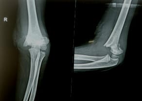
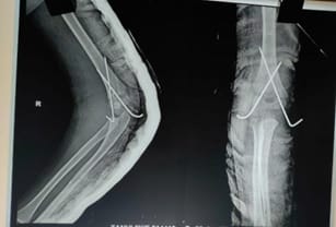
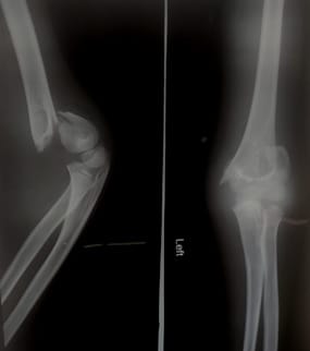
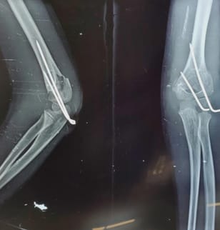
Background: Fracture Supracondylar humerus is one among common fracture in children between age 5-7 years. Boys are frequently affected than girls. Extension variety is more common. The conventional approach to treat fracture supracondylar humerus (Type III) is a close reduction with percutaneous fixation. There have been controversies as to which surgical technique should be used, cross pinning or two-wire lateral pinning. This study aims to find which method of pinning is most appropriate to fix fracture supracondylar humerus. Method and material: A Retrospective comparative study was designed to analyze the outcome of the cross pinning and lateral pinning method. A total of 60 patients were included in the study. They were divided into two groups of 30 each. Group A comprised of fixation by cross pinning method. Group B comprised of fixation by two-wire lateral pinning method. Results of both groups were analysed about Flynn’s criteria and complications. Result: The mean age in Group A was 5.1 years and in Group B was 4.8 years. One patient was lost to follow up in Group A. On the final follow up, there was statistically no difference in terms of outcome according to Flynn’s criteria in both groups. According to Flynn’s criteria>95% of patients had a satisfactory outcome in both groups. Among Group A, there were 2 cases of iatrogenic ulnar nerve praxis whereas in group B there was one case of pin loosening. Conclusion: On comparing both techniques there was no significant difference in the outcome. However, there is a slight increase in the chances of iatrogenic ulnar nerve injury in the cross pinning method.

Keywords: Fracture supracondylar humerus, Cross pinning, Lateral pinning, Flynn’s criteria, Iatrogenic nerve injury

Corresponding Author How to Cite this Article To Browse
Sunil Kumar Kirar, Senior Resident, Department of Orthopaedics, ABV Government Medical College, Vidisha, Madhya Pradesh, India.
Email:
Upadhyay S, Kirar SK, Singh S, Varshney A. Comparison of functional outcome and complications in Gartland’s Type III fracture Supracondylar Humerus in paediatric population by different pinning technique. Surgical Rev Int J Surg Trauma Orthoped. 2021;7(3):51-56.
Available From
https://surgical.medresearch.in/index.php/ijoso/article/view/231

Manuscript Received Review Round 1 Review Round 2 Review Round 3 Accepted
2021-03-28 2021-04-08 2021-04-15 2021-04-20
Conflict of Interest Funding Ethical Approval Plagiarism X-checker Note
No Nil Yes 9%

© 2021 by Sanjay Upadhyay, Sunil Kumar Kirar, Sanat Singh, Atul Varshney and Published by Siddharth Health Research and Social Welfare Society. This is an Open Access article licensed under a Creative Commons Attribution 4.0 International License https://creativecommons.org/licenses/by/4.0/ unported [CC BY 4.0].

Introduction

Approximately 60% of all elbow injury in the first decade of life are constituted by Fracture supracondylar humerus. [1]. Children are susceptible to this type of fracture because of weak metaphyseal sclerotin of the distal humerus and thin ridge of metaphyseal bone between the coronoid fossa and olecranon fossa.[2]. Such injuries are challenging to orthopaedician due to associated immediate and late complications.[3]. These complications are malunion, volksman ischemic contracture, neurovascular damage, compartment syndrome to name a few. Fracture supracondylar humerus is divided into extension and flexion variety. Extension variety is further subcategorized into

Type 1- Undisplaced fracture

Type 2 –Partially displaced fracture with undisplaced posterior hinge

Type 3- Completely displaced fracture.

Wilkins [4] further categorized type 3 fracture in

Type 3A- Posteromedial

Type 3B - Posterolateral

Type 3 fracture requires close reduction and pinning. Pinning is commonly done by two methods – either a medial pin and lateral pin in a cross manner or two lateral pins [5,6]. Cross pinning was preferred to lateral pinning due to more stability. [7,8]. However surgeons believe that cross approach leads to increased chances of iatrogenic ulnar nerve injury. [9,10,11,12]. The present study aims to compare the outcome of lateral and cross pinning technique in fracture supracondylar humerus.

Material and method

A Retrospective study comprising of 60 cases of fracture supracondylar humerus type III was conducted from the period of August 2018 to January 2021 at a tertiary care centre in ABVGMC and associated hospital, Vidisha. All patients were randomly divided into two groups. The process of randomization was done by the odd and even number technique. The patient with an odd number was allotted group A (n=30) and with even number was allotted group B (n=30). Group A patients underwent cross entry K wire technique (Figure – 1) and Group B patients underwent lateral entry K wire

technique (Figure – 2). A thorough pre-operative clinical and radiological workup was done. Fracture supracondylar humerus was classified according to Gartland’s classification system. Written and informed consent was taken from the guardians of all participants. Regular follow up was done at the end of 1 week, 3 weeks, 6 weeks. Later on patients were followed at the end of one and two years. Flynn’s criteria were used to assess the outcome.[13]. The criteria have two components a) Functional and b) Cosmetic. Each component is further subdivided into excellent, good, moderate and poor at the interval of five degrees. The functional component consists of measuring the arc of motion in a sagittal plane which includes flexion and extension. The cosmetic component consists of measuring of carrying angle which indicates the coronal movement at the elbow joint.

Operative technique: Patient kept in supine position with an affected arm on the armrest and under general anesthesia. Under aseptic condition scrubbing, painting and draping have done. Using traction-counter traction technique fracture supracondylar humerus reduction achieved under IITV guidance and keeping elbow in hyperflexion, K wire (size-1.8mm) pinning done in a divergent manner in lateral entry K wire technique. In the cross pinning technique, lateral K wire insertion done in elbow hyperflexion and medial K wire insertion done in extension to avoid ulnar nerve injury under IITV guidance. After satisfactory reduction K wire was bent and cut ends are kept outside the skin. Above elbow slab is given in 90-degree elbow flexion in all cases. Postoperative limb elevation and monitoring done to assess the neurovascular status and wound dressing done on a postoperative day 2 following which patients were discharged.

surgery_231_01.jpg

Figure-1: Preoperative X-ray


surgery_231_02.jpg

Figure-2: Postoperative X-ray (cross pinning)

surgery_231_03.jpg

Figure-3: Preoperative X-ray

surgery_231_04.jpg

Figure-4: Postoperative X-ray (lateral pinning)

Results

The cross entry Group A comprised of 30 patients. The average age of Group A was 5.1 years. The total numbers of male patients in Group A were22 and female patients were 08 in number. The right elbow was involved in 17 patients whereas the left elbow was involved in 13 patients. One patient was lost to follow-up (Table-1). The lateral entry Group B also comprised of 30 patients. The average age of Group B was 4.8 years. Male patients constituted 20 cases whereas female cases were 10 in number in Group B. The right elbow was involved in 14 patients and the left elbow was involved in 16 patients (Table -1). Flynn’s criteria were applied at the end of the final follow up (02 years). Among Group A(n=29) excellent outcome was seen in 22 cases, Good in 04 cases, fair in 02 cases and poor outcome in 01 cases. The overall satisfactory outcome was in 96.55% of cases and unsatisfactory outcome in 03.45% of cases (Table-2). In Group B (n=30) excellent outcome was seen in 24 cases, Good in 04 cases, Fair in 01case and poor in 01 cases. 96.67% of patients had satisfactory outcome whereas 03.33% had unsatisfactory outcome (Table- 2). There is no statistically significant difference in outcome among both groups (student ‘t’ test,p=0.93). The complication seen in Group A was iatrogenic ulnar nerve praxia in two patients. While in group B pin loosening was seen in one case.

Table 1- Showing patient wise distribution.

  Group A (n=30) Group B(n=30)
Mean Age 5.1 4.8
Male 22 20
Female 08 10
Right Elbow 17 14
Left Elbow 13 16

Table 2- Showing outcome according to Flynn’s criteria

  Group A (n=29) Group B(n=30)
Excellent 22 24
Good 04 04
Fair 02 01
Poor 01 01

Discussion

Fracture supracondylar humerus is a common childhood injury that usually results from a fall on an outstretched hand.


Such type of fractures is challenging to treating surgeons. The main aim of Gartland’s type III fractures is an anatomical reduction with stable internal fixation, so the gold standard of treatment is a close reduction with K wire fixation. K wire has many added advantages like easy to use, low cost and decreases hospitalization stay [14,15].

Although the consensus is the use of percutaneous pinning controversies exist in optimal pin configuration [16,17,18]. Cross pinning is supposed to provide superior biomechanical stability to lateral pinning but chances of injury to the ulnar nerve are high.[19]. Some author claimed that the use of three lateral pins or two lateral entry pins that are divergent and are located in both lateral and the central column provide torsional rigidity similar to cross pinning. [11,20,21,22]. Some researchers have highlighted the reason for less fixation stability with the lateral pinning technique.

They have emphasized faulty techniques such as failure to engage both fragments with at least two pins, failure to achieve bicortical fixation with at least two pins and failure to achieve more than or equal to 2mm of pin separation at the fracture site. They suggested checking the stability of fixation by stressing the fracture under fluoroscopy after the procedure and when instability is found, it should be rectified with a third lateral pin or a medial pin.[23]. The mean age of patients in our study was 5.1 years in Group A and 4.8 years in Group B respectively.

Similar results were obtained by Babal JC et al [24]. where the mean age of the patients was 7.20 years and 6.28 in Group A and B respectively. Khademolhosseini M et al [25]. also obtained similar findings. The male patients in group A were 22 in number and Group B were 20 in number. The female patients were 8 and 10 in Group A and B respectively. Another study showing more male patients as compared to female patients was done by Lokesh Naik et al [26]. There were 21 (36.8%) females and 36 (63.2%) males in their study.

In our study we found that among Group A 22 cases achieved excellent outcome, 04 cases have a good outcome,02 cases have a fair outcome and 01 cases have a poor outcome. While in Group B 24 cases have an excellent outcome,04 cases have a good outcome, 01 cases have a fair outcome and 1 case have a poor outcome. In Group A96.55% cases had satisfactory results and 03.45% cases had unsatisfactory results.

In Group B 96.67% of cases had satisfactory results and 03.33% cases had unsatisfactory results. There was no significant difference between the functional outcomes of both groups. These results were comparable to the studies done by Yen YM [27].

who found both lateral entry pin fixation (n=28) and medial and lateral entry pin fixation (n=24) are effective in the treatment of completely displaced (type-III) extension supracondylar fractures of the humerus in children. The study conducted by Lokesh Naik et al [26]. showed the functional factor in Group A was satisfactory in 27(96.4%) patients and the functional factor was satisfactory in all the patients of Group B. Another study by Rajesh Govindaswamy[28] et al they found in both techniques, there were excellent functional results with less than 5-degree loss of range of motion in most children (96%).

Rizk et al [29] conducted a study on 50 patients. They found that according to the modified Flynn’s criteria, the functional outcome was excellent in 25 patients in group I, whereas the outcome was excellent in 24 patients and good in one patient in group II. No fair or poor functional or cosmetic outcome was obtained in both groups. There was no significant statistical difference between both groups regarding both the functional (P=0.276).

Ulnar nerve praxia is usually a major concern in patients of cross pinning. In our study we found 2 cases of ulnar nerve praxia in group A. In another study done by Lyons JP et al [21]. they found 6% of patients had nerve injury. Lokesh et al [26]. found that 6.8% of cases in the cross pinning group had ulnar nerve neuropraxia postoperatively and who recovered completely within three weeks of surgery.

However Yen YM does not found any case of ulnar nerve injury in either group.[27]. In another study by Holsam M et al [19]. the incidence of ulnar nerve neuropraxia ranged from 1.4 to 15.6%. The reason suggested by them is that the ulnar nerve becomes vulnerable to injury with elbow flexion because it subluxate over the medial epicondyle.

They have suggested precautions to eliminate the risk of ulnar nerve injury such as fixation from the lateral side only, a small incision over medial epicondyle for direct visualization, insertion of the medial pin with an elbow in some extension. We found a single case of pin loosening in group B. Similar finding were found by Lokesh Gudda et al [26].


Conclusion

We found that both techniques have similar outcome so either of them can be used to treat fracture supracondylar humerus. Lateral pinning also provides good stability as compared to cross pinning. However cross pinning method has slightly increased the chances of ulnar nerve injury.

What does this study add to present knowledge?

Both cross pinning and two-wire lateral pinning have a similar outcome in the treatment of Gartland’s Type III fracture Supracondylar Humerus in the paediatric population.

Author contribution

SKK, SU: conceptual framework, data collection. SU, SS, AV: a review of literature, methodology review. SKK, SU: manuscript writing and editing.

Reference

01. Herring JA. Tachdjian’s Pediatric Orthopaedics. 3rd ed, Vol 3, Philadelphia- WB Saunders, Fracture about the elbow. 2002;2139–221. [Crossref][PubMed][Google Scholar]

02. Na Y, Bai R, Zhao Z, Han C, Kong L, Ren Y, Liu W. Comparison of lateral entry with crossed entry pinning for pediatric supracondylar humeral fractures- a meta-analysis. J Orthop Surg Res. 2018 Apr 3;13(1)68. doi: 10.1186/s13018-018-0768-3 [Crossref][PubMed][Google Scholar]

03. Millis MB, Singer IJ, Hall JE. Supracondylar fracture of the humerus in children, Further experience with a study in orthopaedic decision-making. Clin Orthop Relat Res. 1984 Sep;(188)90-7. [Crossref][PubMed][Google Scholar]

04. Wilkins KE. The operative management of supracondylar fractures. Orthop Clin North Am. 1990 Apr;21(2)269-89. [Crossref][PubMed][Google Scholar]

05. Kocher MS, Kasser JR, Waters PM, Bae D, Snyder BD, Hresko MT, et al. Lateral entry compared with medial and lateral entry pin fixation for completely displaced supracondylar humeral fractures in children- A randomized clinical trial. J Bone Joint Surg Am. 2007;89(4)706-12. doi: 10.2106/JBJS.F.00379 [Crossref][PubMed][Google Scholar]

06. Ramachandran M, Skaggs DL, Crawford HA, Eastwood DM, Lalonde FD, Vitale MG, Do TT, Kay RM. Delaying treatment of supracondylar fractures in children- has the pendulum swung too far?. J Bone Joint Surg Br. 2008 Sep;90(9)1228-33. doi: 10.1302/0301-620X.90B9.20728 [Crossref][PubMed][Google Scholar]

07. Paul D Choi, Rojeh Melikian, David L Skaggs. Risk factors for vascular repair and compartment syndrome in the pulseless supracondylar humerus fracture in children. J Pediatr Orthop. 2010;30(1)50-6. doi: 10.1097/BPO.0b013e3181c6b3a8 [Crossref][PubMed][Google Scholar]

08. De Pellegrin M, et al. "Frattura sovracondiloidea di omero in età infantile, Osteosintesi percutanea in posizione prona. " Giornale Italiano di Ortopedia e Traumatologia. 34(2008). [Crossref][PubMed][Google Scholar]

09. Dua A, Eachempati KK, Malhotra R, Sharma L, Gidaganti M. Closed reduction and percutaneous pinning of displaced supracondylar fractures of humerus in children with delayed presentation. Chin J Traumatol. 2011 Feb 1;14(1)14-9. [Crossref][PubMed][Google Scholar]

10. Kim WY, Chandru R, Bonshahi A, Paton RW. Displaced supracondylar humeral fractures in children- results of a national survey of paediatric orthopaedic consultants. Injury. 2003 May;34(4)274-7. doi: 10.1016/s0020-1383(02)00321-2 [Crossref][PubMed][Google Scholar]

11. Lee SS, Mahar AT, Miesen D, Newton PO. Displaced pediatric supracondylar humerus fractures- biomechanical analysis of percutaneous pinning techniques. J Pediatr Orthop. 2002 Jul-Aug;22(4)440-3. [Crossref][PubMed][Google Scholar]

12. Woratanarat P, Angsanuntsukh C, Rattanasiri S, Attia J, Woratanarat T, Thakkinstian A. Meta-analysis of pinning in supracondylar fracture of the humerus in children. J Orthop Trauma. 2012 Jan;26(1)48-53. doi: 10.1097/BOT.0b013e3182143de0 [Crossref][PubMed][Google Scholar]

13. Flynn JC, Matthews JG, Benoit RL. Blind pinning of displaced supracondylar fractures of the humerus in children- Sixteen years' experience with long-term follow-up. J Bone Joint Surg Am. 1974 Mar;56(2)263-72. [Crossref][PubMed][Google Scholar]


14. Sahu RL. Percutaneous K-wire fixation in paediatric Supracondylar fractures of humerus- A retrospective study. Niger Med J. 2013 Sep;54(5)329-34. doi: 10.4103/0300-1652.122355 [Crossref][PubMed][Google Scholar]

15. Slobogean BL, Jackman H, Tennant S, Slobogean GP, Mulpuri K. Iatrogenic ulnar nerve injury after the surgical treatment of displaced supracondylar fractures of the humerus- number needed to harm, a systematic review. J Pediatr Orthop. 2010;30(5)430-6. doi: 10.1097/BPO.0b013 e3181e00c0d [Crossref][PubMed][Google Scholar]

16. Ariño VL, Lluch EE, Ramirez AM, Ferrer J, Rodriguez L, Baixauli F. Percutaneous fixation of supracondylar fractures of the humerus in children. J Bone Joint Surg Am. 1977 Oct;59(7)914-6. [Crossref][PubMed][Google Scholar]

17. Eidelman M, Hos N, Katzman A, Bialik V. Prevention of ulnar nerve injury during fixation of supracondylar fractures in children by 'flexion-extension cross-pinning' technique. J Pediatr Orthop B. 2007;16(3)221-4. doi: 10.1097/BPB.0b013 e328010b684 [Crossref][PubMed][Google Scholar]

18. Wind WM, Schwend RM, Armstrong DG. Predicting ulnar nerve location in pinning of supracondylar humerus fractures. J Pediatr Orthop. 2002 Jul-Aug;22(4)444-7. [Crossref][PubMed][Google Scholar]

19. Abubeih, Hossam MA, et al. "Percutaneous cross-pinning versus two lateral entry pinning in Gartland type III pediatric supracondylar humerus fractures". The Egyptian Orthopaedic Journal. 2019;54(1)52. [Crossref][PubMed][Google Scholar]

20. Skaggs DL, Hale JM, Bassett J, Kaminsky C, Kay RM, Tolo VT. Operative treatment of supracondylar fractures of the humerus in children- The consequences of pin placement. J Bone Joint Surg Am. 2001;83(5)735-40. [Crossref][PubMed][Google Scholar]

21. Lyons JP, Ashley E, Hoffer MM. Ulnar nerve palsies after percutaneous cross-pinning of supracondylar fractures in children's elbows. J Pediatr Orthop. 1998 Jan-Feb;18(1)43-5. [Crossref][PubMed][Google Scholar]

22. Gordon JE, Patton CM, Luhmann SJ, Bassett GS, Schoenecker PL. Fracture stability after pinning of displaced supracondylar distal humerus fractures in children. J Pediatr Orthop. 2001 May-Jun;21(3)313-8. [Crossref][PubMed][Google Scholar]

23. Sankar WN, Hebela NM, Skaggs DL, Flynn JM. Loss of pin fixation in displaced supracondylar humeral fractures in children- causes and prevention. J Bone Joint Surg Am. 2007 Apr;89(4)713-7. doi: 10.2106/JBJS.F.00076 [Crossref][PubMed][Google Scholar]

24. Babal JC, Mehlman CT, Klein G. Nerve injuries associated with pediatric supracondylar humeral fractures- a meta-analysis. J Pediatr Orthop. 2010 Apr-May;30(3)253-63. doi: 10.1097/BPO.0b013e3181d213a6 [Crossref][PubMed][Google Scholar]

25. Khademolhosseini M, Abd Rashid AH, Ibrahim S. Nerve injuries in supracondylar fractures of the humerus in children- is nerve exploration indicated?. J Pediatr Orthop B. 2013 Mar;22(2)123-6. doi: 10.1097/BPB.0b013e32835b2e14 [Crossref][PubMed][Google Scholar]

26. Naik LG, Sharma GM, Badgire KS, Qureshi F, Waghchoure C, Jain V. Cross Pinning Versus Lateral Pinning in the Management of Type III Supracondylar Humerus Fractures in Children. J Clin Diagn Res. 2017 Aug;11(8)RC01-RC03. doi: 10.7860/JCDR/2017/28481.10351 [Crossref][PubMed][Google Scholar]

27. Yen YM, Kocher MS. Lateral entry compared with medial and lateral entry pin fixation for completely displaced supracondylar humeral fractures in children, Surgical technique. J Bone Joint Surg Am. 2008 Mar;90 Suppl 2 Pt 1;20-30. doi: 10.2106/JBJS.G.01337 [Crossref][PubMed][Google Scholar]

28. Rajesh Govindasamy, Ramkumar Gnanasundaram, SaravananKasirajan, Fawas Thonikadavath, Rajeev K. Tiwari, Cross pinning versus lateral pinning in type III supracondylar fracture- a retrospective analysis. Int J Res Orthop. 2016 Sep;2(3)138-142. [Crossref][PubMed][Google Scholar]

29. Rizk A S, Kandil M I. Conventional versus lateral cross-pinning (Dorgan’s technique) for fixation of displaced pediatric supracondylar humeral fractures- a randomized comparative study. The Egyptian Orthopaedic Journal. 2018;53(4)348. [Crossref][PubMed][Google Scholar]