E-ISSN:2455-5436
P-ISSN:2456-9518
RNI:MPENG/2017/70870

Research Article

Analysis

Surgical Review: International Journal of Surgery Trauma and Orthopedics

2021 Volume 7 Number 2 March-April
Publisherwww.medresearch.in

A Retrospective Analysis of Plate Fixation of Humerus Fracture

Kumar Kirar S.1, Upadhyay S.2*, Singh S.3, Varshney A.4
DOI: https://doi.org/10.17511/ijoso.2021.i02.06

1 Sunil Kumar Kirar, Senior Resident, Department of Orthopaedics, ABV Government Medical College, Vidisha, Madhya Pradesh, India.

2* Sanjay Upadhyay, Assistant Professor, Department of Orthopaedics, ABV Government Medical College, Vidisha, Madhya Pradesh, India.

3 Sanat Singh, Associate Professor, Department of Orthopaedics, ABV Government Medical College, Vidisha, Madhya Pradesh, India.

4 Atul Varshney, Professor & HOD, Department of Orthopaedics, ABV Government Medical College, Vidisha, Madhya Pradesh, India.

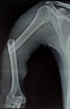
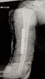
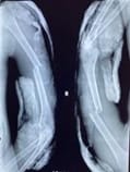
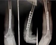
Introduction: Humeral shaft fractures represent 1–3% of all the fractures coming in our OPD. These fractures are mostly treated conservatively. Both the plate fixation and nailing techniques are being used but plate fixation has the advantage of lesser rates of malunion and non-union. Our study aimed to evaluate the results of plate fixation in our hospital that employs plate fixation as the golden standard. Materials and Methods: This was a retrospective cohort study of all patients treated for a humeral shaft fracture in our hospital (District Hospital Vidisha associated with ABV Medical College, Vidisha between July 2018 and June 2020 with a mean follow-up of 6 months. Results: Plate fixation was performed in 40 patients with a humeral shaft fracture. The mean age was 50 (SD 20) years with 60 % (n = 24) being male. There were 55 % (n = 22) fractures in the right and 45 % (n =18) fractures in the left. None of the patients develops superficial surgical site infection. Complications like Radial Nerve palsy, Deep surgical site infections and Non-union occurred in 2.5 % (n = 1), 2.5 % (n = 1) and 5 % (n = 2) of patients, respectively. The median duration of radiological fracture healing was 18 (range 10–42) weeks. Conclusion: Plate fixation for humeral shaft fractures has low risks of complications. The complications can be further minimized with greater surgical expertise.

Keywords: Plate fixation, Humerus shaft fracture, Complication

Corresponding Author How to Cite this Article To Browse
Sanjay Upadhyay, Assistant Professor, Department of Orthopaedics, ABV Government Medical College, Vidisha, Madhya Pradesh, India.
Email:
Kirar SK, Upadhyay S, Singh S, Varshney A, A Retrospective Analysis of Plate Fixation of Humerus Fracture. Surgical Rev Int J Surg Trauma Orthoped. 2021;7(2):28-35.
Available From
https://surgical.medresearch.in/index.php/ijoso/article/view/229

Manuscript Received Review Round 1 Review Round 2 Review Round 3 Accepted
2021-04-06 2021-04-15 2021-04-19 2021-04-25
Conflict of Interest Funding Ethical Approval Plagiarism X-checker Note
No Nil Yes 7%

© 2021 by Sunil Kumar Kirar, Sanjay Upadhyay, Sanat Singh, Atul Varshney and Published by Siddharth Health Research and Social Welfare Society. This is an Open Access article licensed under a Creative Commons Attribution 4.0 International License https://creativecommons.org/licenses/by/4.0/ unported [CC BY 4.0].

Introduction

Humeral shaft fractures represent 1–3% of all the fractures coming in our OPD [1]. Currently, there are no defined gold standards for the treatment of humeral shaft fractures [2,3]. Most of these fractures are mostly treated conservatively. Although nonoperative treatment has a long and successful history in certain cases [4]. It requires good patient compliance for a successful outcome. Therefore, new surgical treatment methods have been developed to reduce soft tissue damage, improve early training and prevent long uncomfortable periods of immobilization, which can be associated with nonoperative treatment [3,5,8].

Internal fixation may be indicated in open fractures with nerve injury, fractures associated with vascular injury, fractures extending into neighbouring joints, bilateral fractures and inpatient with multiple injuries. [9, 10]. The internal fixation methods for humerus shaft fractures can be broadly grouped into plating or intramedullary techniques. Plate osteosynthesis remains the gold standard of fixation of humeral shaft fractures compared to other methods [11]. In our centre we mainly used plate fixation for treating humerus fractures, mainly two-part fractures [12]. Nailing was rarely used when plating was not possible due to fracture pattern.

Although plate fixation has the advantage of lesser rates of malunion and non-union, it causes soft tissue disruption, more blood loss and there are higher chances of infection and radial nerve damage with an open plating procedure [13,14]. Union was defined as a radiologically detectable callus bridge or at least three visible cortices on the radiographs [15]. Depending on the type of callus formation, fracture healing was denoted as direct fracture healing (absolute stability: no callus formation) or indirect fracture healing (relative stability: callus formation) [16].

Nonunion was defined as failed fracture healing six months after the initial trauma. It was assessed clinically by the presence of pain and radiographically by the absence of a callus bridge or the persistence of visible fracture lines. Infection was defined by positive clinical signs of an infection, such as local pain, erythema, warmth, swelling and draining wounds in the affected limb leading to revision surgery [17]. Our study aimed to evaluate the results of plate fixation in our hospital that employs plate fixation as the golden standard.

Materials And Methods

This was a retrospective cohort study of all the patients treated for a humeral shaft fracture in our hospital (District Hospital Vidisha associated with ABV Medical College, Vidisha) between July 2018 and June 2020 with a mean follow-up of 6 months.

Inclusion criteria:

  • Age 18-70 years
  • Fracture pattern: Two part , > 20° angulations anteriorly, > 30° varus deformation, > 3 cm dislocation and > 20° rotation.

Both close and open fractures were included in the study

  • Failed conservative treatment
  • Concomitant vascular injury

Exclusion criteria:

  • Age : <18 years and > 70 years
  • Shaft fractures extending into the metaphysic
  • Pathological fractures
  • Follow up less than 6 months

Both the lateral and AP view radiographs were obtained and based on X-ray; fracture was classified on basis of AO classification ( Fig 1 & 3). Humerus diaphyseal fracture was classified as type A (Simple), type B (wedge) and type C (Complex) [18]. The Gustilo-Anderson classification was used in cases of open fractures [19]. In our study, there were 3 such cases, two types 1 and one types 2 open wounds. Patients were operated under regional/general anesthesia in the supine or lateral position. All patients received a single dose of ceftriaxone 1 gram injection half an hour before surgery as antibiotic prophylaxis.

Tourniquet was not used during the surgery. 4.5 mm Locking Compression Plate (LCP) was used to fix the fracture (Fig 2& 4). Both the anterolateral, posterior, and minimally invasive approaches were used depending on the fracture pattern and soft tissue conditions. Postoperatively, the patient stayed in the hospital till the first check dressing which was done on the second postoperative day (2nd POD). X-ray was taken on the first operative day (1st POD) and the patient was discharged on prophylactic oral antibiotics for 10 days.

Follow up was done on 8th POD (second check dressing),12th POD (stitch removal), one month (2nd check x-ray ), 3 months (3rd check x-ray) and


6 months (4th check x-ray). Clinical evaluation in form of a range of movement of shoulder and elbow joint was also done in every follow-up. In each follow up under the supervision of the operating surgeon and physiotherapist, early non-weight- bearing range of motion exercises was taught to the patient to do at home. Superficial surgical site infections were classified as either superficial or deep according to the definition of the Centres for Disease Control and Prevention [20]. Non-union was defined as the absence of bone healing after 6 months. Patients with impairment of shoulder and elbow function were classified as having either no impairment of range of motion (full recovery) or suffering from impairment in a range of motion regardless of severity (shoulder/elbow impairment).

Results

The median hospital stay was 6 days (range 2–18). Patients were followed up for 6 months. Patients with follow up less than 6 months were not included in the study

Plate fixation (4.5mm Locking Compression Plate, LCP) was performed in 40 patients with a humeral shaft fracture (Fig 2 & 4). The mean age was 50 (SD 20) years with 60 % (n = 24) being male. There were 55 % (n = 22) fractures in the right and 45 % (n = 18) fractures in the left. Out of 40 patients, 21 patients (52.5%) had an AO/OTA type-A fracture, 13 patients (32.5%) had type-B and 6 patients (15 %) had type-C fracture. Anterolateral approach (in supine position) was done in 24 patients (60 %) of cases and posterior approach (in lateral position) was done in the remaining 16 (40%) cases. All fractures were fixed by a 4.5 mm narrow LCP plate (Fig 2 & 4). None of the patients develops superficial surgical site infection. Complications like radial nerve palsy, deep surgical site infections and non-union occurred in 2.5 % (n = 1), 2.5 % (n = 1) and 5 % (n = 2) of patients respectively.

In one patient who developed radial nerve palsy postoperatively, the palsy recovered spontaneously in 4 months. In our study, we found no radial nerve palsy before surgery. In one case of deep surgical infection, culture sensitivity of wound was done and accordingly intra-venous antibiotic injections were given for 5 days postoperatively followed by oral antibiotics for next 9 days and the patient stayed in the hospital during this period for dressings. The patient was discharged when the wound healed and stitch removal was done.

Both the cases of non-union were reoperated (six months after primary surgery). Cancellous autologous iliac bone grafts were used along with a longer LCP plate in both the revision surgeries. The fracture healed in both cases after the second surgery (mean period was 18 weeks after second surgery). Radiological fracture healing and patients’ ability to fully weight bear was seen after an average of 18 weeks. Full recovery of range of movement of the shoulder joint was found in 34 (85%) cases and in the case of the elbow joint, full recovery was seen in 36 (90%) cases.

Table 1: Distribution of patient based on Gender

Sex Cases
Male 24
Female 16
Total 40

Table 2: Distribution of patient based on side involved:

Side Total
Right 22
Left 18
Total 40

Table 3: Distribution of patient based on AO classification:

AO classification Cases
Type A 21
Type B 13
Type C 06
Total 40

Table 4: Distribution of patient based on etiology:

Etiology Cases
RTA (road trafiic accident) 16
Assault 8
Fall of a heavy object 5
Other causes 11
Total 40

Case 1:

surgery_229_01.jpg


Fig 1:Pre-op X-ray

surgery_229_02.jpg

Fig 2: Post-op X-ray

Case 2:

surgery_229_03.jpg

Fig 3: Pre-op X-ray

surgery_229_04.jpg

Fig 4:  Post-op X-ray

Discussion

Most of the humerus fractures are treated conservatively. Non-operative treatment requires good patient compliance for a successful outcome. It is generally accepted that conservative management is best for isolated closed fractures of the shaft of the humerus [9,21-24].

In a study by Vichare (1972) he found that standard conservative methods of treatment in patients with multiple injuries can lead to a high incidence of malunion and non-union and so he devised a traction system to manage such fractures with multiple injuries [25]. However, an important aim in the management of these patients is to be able to sit them upright with pain-free extremities [26]. Failing this, prolonged recumbency may lead to considerable morbidity.

Early mobilisation of the limb helps to prevent the “fracture disease” [27]. In two recent reviews by Clement and Gosler et al., they demonstrated a deficiency in the current literature of level one evidence for the treatment of humeral shaft fractures (3,5). In a study by Papasoulis et al., they found that the union rate ranged from 77 to 100% and good functional results were achieved after the nonsurgical treatment of humeral shaft fractures(28,29).

Also in a study by Matsunaga et al., comparing functional bracing and bridge plating for humeral shaft fractures, it was found that nonsurgical treatment was associated with a significantly higher rate of nonunion and angular displacement (anteroposterior) than bridge plating (8). Xuqi Hu et al. presented the results of a systematic review and meta-analysis of eight studies, including four randomized controlled trials (RCTs), two prospective cohort trials and two retrospective cohort trials [30].

Of these eight studies, four compared ORIF to MIPO, and none of the four studies showed a significant difference in terms of the postoperative radial injury incidence, union rate or functional outcome between the two groups [31-34]. ORIF offers the opportunity for the exact reduction and anatomical fixation of the fracture and can enable primary or secondary fracture healing depending on the type of osteosynthesis and fracture pattern (15).

Boschi et al. in their study find out the outcomes of the treatment of 280 humeral shaft fractures with ORIF in terms of the approach and plate location. The overall healing rate was 98.2%, without a significant difference in the approach or plate location [35]. Humeral shaft fractures are commonly associated with lesions of the radial nerve. The anatomical proximity and association of the bone and nerves in the humeral shaft explain the incidence between 8 % and 12 % [36,37].


The best treatment for humeral shaft fractures complicated with radial nerve injury is highly controversial [38,39]. While concomitant nerve injury has been used as an argument for the immediate surgical treatment of fractures in the past (using a posterior approach and visualizing the radial nerve) (40), recent investigations have shown no significant difference in radial nerve palsy recovery between initial operative and nonoperative management strategies [36,41].

Most radial nerve injuries in cases of humeral shaft fracture are caused by traction or compression of the nerve, which is known as neuropraxia. Much fewer nerve injuries are identified as discontinuity of the nerve (axonotmesis or neurotmesis) [42]. Neuropraxia is a reversible injury, resulting in spontaneous reversibility in a large portion of traumatic radial nerve palsy cases (36). Plate fixation is the golden standard in our hospital.

Our study included only those cases of humerus fractures that were managed surgically by plate fixation It is a well-known fact that a higher level of training generally yields better results [43,44)]. This greater expertise is likely reflected in the results of this study. With greater experience, complication rates can be minimized in patients undergoing plate fixation for humeral shaft fractures. The objections which have been made to humeral shaft plating are that it may lead to non-union [45]. or to radial nerve injury [21,46]. or to infection, or that the fixation may fail with refracture through the screw holes (9).

Although no long-term complications occurred in our study, one limitation is the lesser duration of follow-up time (six months). Due to limited sample size and lesser duration of follow-up and therefore studies with big sample size and longer duration of follow up will be required to prove the advantage of plating in humerus fractures We only calculated the proportion of patients with full recovery of the shoulder in 34 cases (85%) and elbow function in 36 cases (90%) following surgery.

These proportions are comparable to those found in similar studies [47,48,49]. Nevertheless, functional results are equally important to take into consideration when determining the optimal treatment for patients with humeral shaft fractures. There were several limitations in our study that should be considered when interpreting the results. It was a retrospective study and will have certain amounts of selection bias.

Also, the ability to fully weight bear is a subjective outcome that ideally should be measured on a day to day basis using but it was determined at fixed intervals when the patient was called for follow up. Consequently, this outcome is also determined by the length of the intervals. This applies to a lesser extent for radiological fracture union as changes in radiographs require several weeks to become detectable.

Conclusion

Plate fixation for humeral shaft fractures has low risks of complications. The complications can be further minimized with greater surgical expertise.

What does this study add to present knowledge?

Plate fixation for humeral shaft fractures has low risks of complications.

Author contribution

SKK, SU: conceptual framework, data collection.

SU, SS, AV: a review of literature, methodology review.

SKK, SU: manuscript writing and editing.

Reference

01. Ekholm R, Adami J, Tidermark J, Hansson K, Törnkvist H, Ponzer S. Fractures of the shaft of the humerus- An epidemiological study of 401 fractures. J Bone Joint Surg Br. 2006 Nov;88(11)1469-73. doi: 10.1302/0301-620X.88B11.17634 [Crossref][PubMed][Google Scholar]

02. Ouyang H, Xiong J, Xiang P, Cui Z, Chen L, Yu B. Plate versus intramedullary nail fixation in the treatment of humeral shaft fractures- an updated meta-analysis. J Shoulder Elbow Surg. 2013 Mar;22(3)387-95. doi: 10.1016/j.jse.2012.06.007 [Crossref][PubMed][Google Scholar]

03. Gosler MW, Testroote M, Morrenhof JW, Janzing HM. Surgical versus non-surgical interventions for treating humeral shaft fractures in adults. Cochrane Database Syst Rev. 2012 Jan;1;CD008832. doi: 10.1002/14651858.CD008832 [Crossref][PubMed][Google Scholar]


04. Sarmiento A, Kinman PB, Galvin EG, Schmitt RH, Phillips JG. Functional bracing of fractures of the shaft of the humerus. J Bone Joint Surg Am. 1977 Jul;59(5)596-601. [Crossref][PubMed][Google Scholar]

05. Clement ND. Management of Humeral Shaft Fractures- Non-Operative Versus Operative. Arch Trauma Res. 2015 Jun 20;4(2)e28013. doi: 10.5812/atr.28013v2 [Crossref][PubMed][Google Scholar]

06. Zhao JG, Wang J, Meng XH, Zeng XT, Kan SL. Surgical interventions to treat humerus shaft fractures- A network meta-analysis of randomized controlled trials. PLoS One. 2017;12(3)e0173634. doi: 10.1371/journal.pone.0173634 [Crossref][PubMed][Google Scholar]

07. Schoch BS, Padegimas EM, Maltenfort M, Krieg J, Namdari S. Humeral shaft fractures: national trends in management. J Orthop Traumatol. 2017 Sep;18(3)259-263. doi: 10.1007/s10195-017-0459-6 [Crossref][PubMed][Google Scholar]

08. Matsunaga FT, Tamaoki MJ, Matsumoto MH, Netto NA, Faloppa F, Belloti JC. Minimally Invasive Osteosynthesis with a Bridge Plate Versus a Functional Brace for Humeral Shaft Fractures- A Randomized Controlled Trial. J Bone Joint Surg Am. 2017;99(7)583-592. doi: 10.2106/JBJS.16.00628 [Crossref][PubMed][Google Scholar]

09. Rüedi T, Moshfegh A, Pfeiffer KM, Allgöwer M. Fresh fractures of the shaft of the humerus-conservative or operative treatment?. Reconstr Surg Traumatol. 1974;14(0)65-74. [Crossref][PubMed][Google Scholar]

10. Mast JW, Spiegel PG, Harvey JP Jr, Harrison C. Fractures of the humeral shaft- a retrospective study of 240 adult fractures. Clin Orthop Relat Res. 1975 Oct;(112)254-62. [Crossref][PubMed][Google Scholar]

11. Schemitsch E H, M Bhandari. "Fractures of the diaphyseal humerus". Skeletal trauma. 2001(1481) 1514. [Crossref][PubMed][Google Scholar]

12. Kim YG, Park KH, Kim JW, Oh JK, Yoon JP, Kim HJ, Oh CW. Is minimally invasive plate osteosynthesis superior to open plating for fixation of two-part fracture of the proximal humerus?. J Orthop Surg (Hong Kong). 2019 May-Aug;27(2)2309499019836156. doi: 10.1177/2309499019836156 [Crossref][PubMed][Google Scholar]

13. Hohmann E, Glatt V, Tetsworth K. Minimally invasive plating versus either open reduction and plate fixation or intramedullary nailing of humeral shaft fractures- a systematic review and meta-analysis of randomized controlled trials. Journal of shoulder and elbow surgery. 2016;25(10)1634-1642. [Crossref][PubMed][Google Scholar]

14. Zarkadis NJ, Eisenstein ED, Kusnezov NA, Dunn JC, Blair JA. Open reduction-internal fixation versus intramedullary nailing for humeral shaft fractures- an expected value decision analysis. J Shoulder Elbow Surg. 2018 Feb;27(2)204-210. doi: 10.1016/j.jse.2017.08.004 [Crossref][PubMed][Google Scholar]

15. Lotzien S, Hoberg C, Rausch V, Rosteius T, Schildhauer TA, Gessmann J. Open reduction and internal fixation of humeral midshaft fractures- anterior versus posterior plate fixation. BMC Musculoskelet Disord. 2019 Nov 10;20(1)527. doi: 10.1186/s12891-019-2888-2 [Crossref][PubMed][Google Scholar]

16. Megas P. Classification of non-union. Injury. 2005 Nov;36 Suppl 4;S30-7. doi: 10.1016/j.injury.2005.10.008 [Crossref][PubMed][Google Scholar]

17. Trampuz A, Zimmerli W. Diagnosis and treatment of infections associated with fracture-fixation devices. Injury. 2006 May;37 Suppl 2;S59-66. doi: 10.1016/j.injury.2006.04.010 [Crossref][PubMed][Google Scholar]

18. Meinberg EG, Agel J, Roberts CS, Karam MD, Kellam JF. Fracture and Dislocation Classification Compendium-2018. J Orthop Trauma. 2018 Jan;32 Suppl 1;S1-S170. doi: 10.1097/BOT.000000000000 1063 [Crossref][PubMed][Google Scholar]

19. Gustilo RB, Mendoza RM, Williams DN. Problems in the management of type III (severe) open fractures- a new classification of type III open fractures. J Trauma. 1984 Aug;24(8)742-6. doi: 10.1097/00005373-198408000-00009 [Crossref][PubMed][Google Scholar]

20. Health Protection Agency. Surveillance of surgical site infection in England- October 1997–September 2005. Health Protection Agency, London. 2006. [Crossref][PubMed][Google Scholar]

21. Boehler L. Conservative Treatment Of Fresh Closed Fractures Of The Shaft Of The Humerus. J Trauma. 1965 Jul;5;464-8. [Crossref][PubMed][Google Scholar]


22. Klenerman L. Fractures of the shaft of the humerus. J Bone Joint Surg Br. 1966 Feb;48(1)105-11. [Crossref][PubMed][Google Scholar]

23. Christensen S. Humeral shaft fractures, operative and conservative treatment. Acta Chir Scand. 1967;133(6)455-60. [Crossref][PubMed][Google Scholar]

24. Sarmiento A, Kinman PB, Galvin EG, Schmitt RH, Phillips JG. Functional bracing of fractures of the shaft of the humerus. J Bone Joint Surg Am. 1977 Jul;59(5)596-601. [Crossref][PubMed][Google Scholar]

25. Vichare NA. Fractures of the humeral shaft associated with multiple injuries. Injury. 1974 May;5(4)279-82. [Crossref][PubMed][Google Scholar]

26. McMurtry R Y, J Pickard. "The multiply injured patient- a challenging orthopaedic nursing problem. " Can Orthop Nurs Assoc J. 1980(3)2-5. [Crossref][PubMed][Google Scholar]

27. Miller ME, Allg6wer A, Schneider R, Wlllnegger H. Manual of internal fixation. - technique. s recommended hv the AO group. 2nd ed. Berlin, Heidelberg, New York- Springer-Yerlag. 1979 [Crossref][PubMed][Google Scholar]

28. Papasoulis E, Drosos GI, Ververidis AN, Verettas DA. Functional bracing of humeral shaft fractures- A review of clinical studies. Injury. 2010 Jul;41(7)e21-27. doi: 10.1016/j.injury.2009.05.004 [Crossref][PubMed][Google Scholar]

29. Leung KS, Kwan M, Wong J, Shen WY, Tsang A. Therapeutic functional bracing in upper limb fracture-dislocations. J Orthop Trauma. 1988;2(4) 308-13. doi: 10.1097/00005131-198802040-00007 [Crossref][PubMed][Google Scholar]

30. Hu X, Xu S, Lu H, Chen B, Zhou X, He X, Dai J, Zhang Z, Gong S. Minimally invasive plate osteosynthesis vs conventional fixation techniques for surgically treated humeral shaft fractures- a meta-analysis. J Orthop Surg Res. 2016;11(1)59. doi: 10.1186/s13018-016-0394-x [Crossref][PubMed][Google Scholar]

31. Esmailiejah AA, Abbasian MR, Safdari F, Ashoori K. Treatment of Humeral Shaft Fractures- Minimally Invasive Plate Osteosynthesis Versus Open Reduction and Internal Fixation. Trauma Mon. 2015 Aug;20(3)e26271. doi: 10.5812/traumamon. 26271v2 [Crossref][PubMed][Google Scholar]

32. Kim JW, Oh CW, Byun YS, Kim JJ, Park KC. A prospective randomized study of operative treatment for noncomminuted humeral shaft fractures- conventional open plating versus minimal invasive plate osteosynthesis. J Orthop Trauma. 2015 Apr;29(4)189-94. doi: 10.1097/BOT.0000000000000232 [Crossref][PubMed][Google Scholar]

33. Oh CW, Byun YS, Oh JK, Kim JJ, Jeon IH, Lee JH, Park KH. Plating of humeral shaft fractures: comparison of standard conventional plating versus minimally invasive plating. Orthop Traumatol Surg Res. 2012 Feb;98(1)54-60. doi: 10.1016/j.otsr.2011.09.016 [Crossref][PubMed][Google Scholar]

34. Wang C, Li J, Li Y, Dai G, Wang M. Is minimally invasive plating osteosynthesis for humeral shaft fracture advantageous compared with the conventional open technique?. J Shoulder Elbow Surg. 2015 Nov;24(11)1741-8. doi: 10.1016/j.jse.2015.07.032 [Crossref][PubMed][Google Scholar]

35. Boschi V, Pogorelic Z, Gulan G, Vilovic K, Stalekar H, Bilan K, Grandic L. Subbrachial approachto humeral shaft fractures- new surgical technique and retrospective case series study. Can J Surg. 2013;56(1)27–34. doi: 10.1503/cjs.011911 [Crossref][PubMed][Google Scholar]

36. Shao YC, Harwood P, Grotz MR, Limb D, Giannoudis PV. Radial nerve palsy associated with fractures of the shaft of the humerus- a systematic review. J Bone Joint Surg Br. 2005 Dec;87(12)1647-52. doi: 10.1302/0301-620X.87B12.16132 [Crossref][PubMed][Google Scholar]

37. Bono CM, Grossman MG, Hochwald N, Tornetta P 3rd. Radial and axillary nerves- Anatomic considerations for humeral fixation. Clin Orthop Relat Res. 2000 Apr;(373)259-64. [Crossref][PubMed][Google Scholar]

38. Rocchi M, Tarallo L, Mugnai R, Adani R. Humerus shaft fracture complicated by radial nerve palsy- Is surgical exploration necessary?. Musculoskelet Surg. 2016 Dec;100(Suppl 1)53-60. doi: 10.1007/s12306-016-0414-3 [Crossref][PubMed][Google Scholar]

39. Schacher B, Schnick U, Böttcher R, Casper D, Eisenschenk A, Ekkernkamp A. Paresen des N radialis bei Humerusschaftfrakturen. Obere Extremität. 2015;10(3)137-143. [Crossref][PubMed][Google Scholar]


40. Packer JW, Foster RR, Garcia A, Grantham SA. The humeral fracture with radial nerve palsy- is exploration warranted?. Clin Orthop Relat Res. 1972;88;34-8. doi: 10.1097/00003086-197210000-00005 [Crossref][PubMed][Google Scholar]

41. Liu GY, Zhang CY, Wu HW. Comparison of initial nonoperative and operative management of radial nerve palsy associated with acute humeral shaft fractures. Orthopedics. 2012 Aug 1;35(8)702-8. doi: 10.3928/01477447-20120725-10 [Crossref][PubMed][Google Scholar]

42. Grassmann J P, Jungbluth P, Bullermann L, Hakimi M, Gehrmann S V, Thelen S, Wild M. Radial nerve palsy associated with humeral shaft fractures-early exploration or expectant procedure?- An analysis concerning current strategies of treatment. Zeitschrift fur Orthopadie und Unfallchirurgie. 2010;148(6)691-696. [Crossref][PubMed][Google Scholar]

43. Birkmeyer JD, Stukel TA, Siewers AE, Goodney PP, Wennberg DE, Lucas FL. Surgeon volume and operative mortality in the United States. N Engl J Med. 2003 Nov 27;349(22)2117-27. doi: 10.1056/NEJMsa035205 [Crossref][PubMed][Google Scholar]

44. Chowdhury MM, Dagash H, Pierro A. A systematic review of the impact of volume of surgery and specialization on patient outcome. Br J Surg. 2007 Feb;94(2)145-61. doi: 10.1002/bjs.5714 [Crossref][PubMed][Google Scholar]

45. Gartland JJ, Ferguson AB Jr, Goodman MC, Rodriguez EE, Skovrou M. Fresh midshaft fractures of the humerus in adults- evaluation of treatment in Pennsylvania during 1952-1956, made by the Scientific Research Committee, Pennsylvania Orthopedic Society. Penn MeJ J. 1939;52;848. [Crossref][PubMed][Google Scholar]

46. Titze A. The operative treatment of fractures of the shaft of the humerus. Reconstr Surg Traumatol. 1974;14(0)75-83. [Crossref][PubMed][Google Scholar]

47. Gausden EB, Christ AB, Warner SJ, Levack A, Nellestein A, Lorich DG. The triceps-sparing posterior approach to plating humeral shaft fractures results in a high rate of union and low incidence of complications. Arch Orthop Trauma Surg. 2016 Dec;136(12)1683-1689. doi: 10.1007/s00402-016-2578-6 [Crossref][PubMed][Google Scholar]

48. Kumar BS, Soraganvi P, Satyarup D. Treatment of Middle Third Humeral Shaft Fractures with Anteromedial Plate Osteosynthesis through an Anterolateral Approach. Malays Orthop J. 2016 Mar;10(1)38-43. doi: 10.5704/MOJ.1603.007 [Crossref][PubMed][Google Scholar]

49. van de Wall BJM, Ganzert C, Theus C, van Leeuwen RJH, Link BC, Babst R, Beeres FJP. Results of plate fixation for humerus fractures in a large single-center cohort. Arch Orthop Trauma Surg. 2020 Oct;140(10)1311-1318. doi: 10.1007/s00402-019-03319-z [Crossref][PubMed][Google Scholar]