E-ISSN:2455-5436
P-ISSN:2456-9518
RNI:MPENG/2017/70870

Research Article

Fracture

Surgical Review: International Journal of Surgery Trauma and Orthopedics

2021 Volume 7 Number 3 May-June
Publisherwww.medresearch.in

Open versus closed reduction and K-wire fixation for supracondylar fracture of the humerus (Gartland type 3) in children

Kumar Kirar S.1, Upadhyay S.2*, Singh S.3, Varshney A.4
DOI: https://doi.org/10.17511/ijoso.2021.i03.02

1 Sunil Kumar Kirar, Senior Resident, Department of Orthopaedics, ABV Government Medical College, Vidisha, Madhya Pradesh, India.

2* Sanjay Upadhyay, Assistant Professor, Department of Orthopaedics, ABV Government Medical College, Vidisha, Madhya Prdaesh, India.

3 Sanat Singh, Associate Professor, Department of Orthopaedics, ABV Government Medical College, Vidisha, Madhya Pradesh, India.

4 Atul Varshney, Professor & HOD, Department of Orthopaedics, ABV Government Medical College, Vidisha, Madhya Pradesh, India.

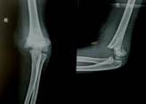
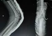
Background: The purpose of the study was to compare the presentation and postoperative results of children treated by open reduction and closed reduction for completely displaced Gartland type III supracondylar humerus fractures (SCFs). Method: Supracondylar fracture of the humerus is a common paediatric fracture seen in our OPD. Among them Type III fractures are displaced with no cortical contact, and reduction is difficult, and maintaining reduction is almost impossible without some form of internal fixation. Therefore during surgery of type 3 fractures, fixation is done by two methods. 1 open reduction and fixation with 2 cross k-wire 2. closed reduction and fixation with 2 cross k-wire fixation. Following pinning, the elbow was immobilized in an above elbow slab in pronation with the elbow at 75 degrees of flexion. Result: The average age of patients was 5 years (age range, 3 to 10 years). The test population consisted of 18female (36%) and 32 male (64%) patients. There were 31 fractures (62%) in the right elbow and 19 fractures (38%) in the left. Group 1 patients stayed in the hospital for 5 days while Group 2 stayed for only 2 days in the hospital. Also group 1 patient required follow-up at eight postoperative days (for check dressing) and 11 postoperative days for stitch removal while group 2 patients were directly called for k-wire removal at 3 weeks postoperatively. Both groups of patients were called after three weeks for k-wire removal. Mean clinical follow-up for both groups was 6 months. Conclusion: The closed reduction technique was preferred because it required less hospitalization time, less number followup, and resulted in almost no visible surgical scars.

Keywords: Closed reduction, Displaced fracture, Supracondylar fracture, Trauma

Corresponding Author How to Cite this Article To Browse
Sanjay Upadhyay, Assistant Professor, Department of Orthopaedics, ABV Government Medical College, Vidisha, Madhya Prdaesh, India.
Email:
Kirar SK, Upadhyay S, Singh S, Varshney A. Open versus closed reduction and K-wire fixation for supracondylar fracture of the humerus (Gartland type 3) in children. Surgical Rev Int J Surg Trauma Orthoped. 2021;7(3):44-50.
Available From
https://surgical.medresearch.in/index.php/ijoso/article/view/225

Manuscript Received Review Round 1 Review Round 2 Review Round 3 Accepted
2021-03-25 2021-04-05 2021-04-14 2021-04-16
Conflict of Interest Funding Ethical Approval Plagiarism X-checker Note
No Nil Yes 6%

© 2021 by Sunil Kumar Kirar, Sanjay Upadhyay, Sanat Singh, Atul Varshney and Published by Siddharth Health Research and Social Welfare Society. This is an Open Access article licensed under a Creative Commons Attribution 4.0 International License https://creativecommons.org/licenses/by/4.0/ unported [CC BY 4.0].

Background

Supracondylar fracture of the humerus is one of the most common fracture (only after clavicle and both bone forearm fracture) in the paediatric age group with a male predominance accounting for 16% of all paediatric fractures and 60% of all paediatric elbow fractures, classically occurring as a result of fall on an outstretched hand [1,3]. It is of two types- extension type most common (95-98%) and flexion type less common (<5%). Extension type injury is more common than flexion type [4]. It is frequently found in the non-dominant extremity. The flexion type is common in elderly children [5]. The Gartland classification is a commonly used system for the evaluation and treatment of supracondylar humerus fractures in children. [6]. Type I injuries are immobilized with an above elbow slab for 3 to 4 weeks, with radiographic alignment checked at 1 week. Type IIA injuries can be treated with closed reduction and slab or percutaneous pinning, whereas type IIB injuries should have closed reduction and percutaneous pinning to prevent coronal and/or rotational malalignment. Types III injuries also are treated with closed reduction and percutaneous pinning, as are flexion-type injuries, with possible open reduction and internal fixation if closed reduction is unsuccessful [6].

Table 1: Gartland Classification of the supracondylar humerus:

TYPE DESCRIPTION OF FRACTURE
I Non displaced
II A Intact posterior cortex, hinged in extension, no rotation or translation
B Intact posterior cortex, hinged in extension with some degree of rotational displacement or translation
III Complete displacement

The purpose of the study was to compare the presentation and postoperative results of children treated by open reduction and closed reduction for completely displaced Gartland type III supracondylar humerus fractures (SCFs). Criteria taken into consideration were final (functional and cosmetic) outcomes, period of hospitalization, the time needed for healing and the expected common complications.

Material and Method

A retrospective comparative study of 50 paediatric patients with type III supracondylar fracture of the humerus was done in our hospital (District hospital

associated with ABV Govt Medical College, Vidisha).

Patients who attended our hospital between January 2019 –September 2020 were included in the study. The diagnosis was based on clinical and X-ray findings. Earlier our hospital didn’t have the facility of C-arm intensifier (from January 2019-November 2019) so all the patients were treated with open reduction (Group-1). Later when our hospital received C-arm in December 2019, thereafter from December 2019 to September 2020, all patients were treated with close reduction (Group-2) We divided the patients into two groups.

Group 1 consist of patients who were treated with open reduction and 2 cross k-wire fixation (ORIF, open reduction and internal fixation), and Group 2 consist of patients who were treated with close reduction and 2 cross k-wire fixation (CRIF, close reduction and internal fixation) [7,8]. There are various studies in which the main difference in opinion is mainly to whether using closed reduction and percutaneous k-wire fixation [7, 8]. or perform open reduction and fixation under direct vision. Some studies preferred closed treatment [9]. but others found it did not yield optimal results when they applied it to their patients [10]. Functional and cosmetic assessments were conducted utilizing the Flynn et al. outcome criteria [7].

Table 2: Flynn Criteria

Results RATING LOSS OF CARRYING ANGLE LOSS OF MOTION
Satisfactory Excellent 0º-5º 0º-5º
Good 5º-10º 5º-10º
Fair 10º-15º 10º-15º
Unsatisfactory Poor >15º >15º

Data records regarding name, age, sex, left or right elbow, time of arrival and time of surgical treatment for each patient were collected. Patients with open fractures and /or associated neurovascular injuries were excluded from the studies. The surgical technique for Group 1(open reduction) was as follows. Patients were prepared on an ordinary operative table, in a supine position. After anaesthesia (general/regional), painting and draping were done.

A pneumatic tourniquet was inflated. Using a poster-lateral approach, the triceps was split, and the ulnar nerve was identified and protected. The fracture was reduced and fixed by two crossed k-wires, medially and laterally. Haemostasis was secured, and the wound was closed without leaving a drain.


The surgical technique for Group 2 (close reduction) was as follows.

An elbow support device was placed at the lateral edge of the table to keep the joint stable during the k-wire fixation. After anesthesia (general/regional), painting and draping were done and the fracture was reduced closely under C-arm intensifier. Two crossed k-wires were introduced laterally and medially without using a tourniquet.

In both groups, a P.O.P. above-elbow slab was applied. Group 1 patients stayed in the hospital for 5 days while Group 2 stayed for only 2 days in the hospital. One check dressing on the second postoperative day of group 1 patient was done during their stay at the hospital. Postoperative check X-ray was done of all patients of both groups 1 & 2 and they were discharged from the hospital on oral medication.

Group 1 patients received prophylactic antibiotics (Amoxicillin syrup) till stitch removal in addition to painkillers. Also group 1 patient required two additional follow-ups at eight postoperative days (for check dressing) and 11 postoperative days for stitch removal. Both groups’ patients were called at four weeks for k-wire removal. Active range of motion and early physiotherapy started on the day of wire removal (after 4 weeks)

Schedule of follow-up of Group1: 8th POD,11th POD,2nd week, 4th week, 6th week,3rd month,6th month. Schedule of follow-up of Group2: 2nd week, 4th week, 6th week, 3rd month, 6th month.

During the postoperative period in hospital and after discharge from the hospital, during follow-up we assessed the patient based on the accuracy of the reduction and looking for possible early complications such as nerve injury, compartment syndrome, range of motion, presence of any malunion or cubitus varus and presence of infection

We depended on the Flynn et al. (7) criteria of evaluation to assess the results regarding any deformity and range of motion at the elbow joint Statistical data were presented by mean ± SD (standard deviation) and percentage. Analysis of data was done using SPSS version 22, utilizing independent sample t-test and one-way ANOVA according to p-value (P ≤ 0.05).

Results

Fig 1: Preop X-ray

surgery_225_01.jpg

surgery_225_02.jpg

Fig 2 :Post-op X-ray

During the defined period for our study (January 2019 –September 2020), we registered a total of 50 patients in both groups for our study. Group 1 (ORIF group) during January 2019 to November 2019 when the C-arm facility was not available in our hospital, we registered 33 patients. Group 2 (Close reduction group) from December 2019 to September 2020, we registered 17 patients The average age of patients was 5 years (age range, 3 to 10 years). The test population consisted of 18 females (36%) and 32 males (64%) patients. There were 31 fractures (62%) in the left elbow and 19 fractures (38%) in the right (right hand was dominant in all cases).

Table 3: Distribution of cases in each group:

  Group 1 (Open reduction) Group 2 (Close Reduction)
No. of cases 33 17

Table 4: Comparison of age distribution in each group:

Age: Group 1 (Open reduction) Group 2 (Close Reduction)
Frequency % Frequency %
<3 years 7 21.2 2 11.8
3-6 16 48.5 9 52.9

7-10 10 30.3 6 35.3
Total 33   17  

Table 5: Comparison of sex distribution in each group:

Sex Group 1 (Open reduction) Group 2 (Close Reduction) Total
Frequency % Frequency %
Male 20 60.6 12 70.6 32
Female 13 39.4 5 29.4 18
Total 33   17   50

Table 6: Comparison of side distribution in each group:

Side Group 1 (Open Reduction) Group 2 (Close Reduction) Total
Frequency % Frequency %
Left 18 54.5 13 76.5 31
Right 15 45.5 4 23.5 19
Total 33   17   50

Table 7: Comparison of both groups using Flynn Criteria:

Result (grade) Group 1 (Open reduction) Group 2 (Close Reduction) Total
Frequency % Frequency %
Excellent 22 66.7 11 64.7 33
Good 8 24.2 5 29.4 13
Fair 2 6 1 5.9 3
Poor 1 3 0 0 1
Total 33   17   50

As per the Flynn et al. criteria of evaluation, we obtained the following results.

In group 1: Out of a total of 33 patients, we reported 22 patients (66.7%) scored an excellent result, 8 patients (24.2%) scored a good result, 2 patients (6%) scored a fair result, and only one patient (3%) scored a poor result (Table 7).

In group 2: Out of a total of 17 patients, we reported 11 patients (64.7 %) scored an excellent result, 5 patients (29.4%) scored a good result, 1 patient (5.9%) had a fair result, and no patient had a poor result (Table 7).

Complications like pin tract infection occurred in 3 patients (1 from group 1 and 2 from group 2) and ulnar nerve palsy was seen in two patients of group 2. Pin tract infection recovered after daily dressing and a short course of antibiotics.

Ulnar nerve palsy also recovered in both the patients after 6 weeks. Statistically, there were no significant differences (P > 0.05) between patients of both groups regarding the Flynn et al. criteria.

Discussion

In this study, our main aim was to achieve the functionality as early as possible, a stable elbow joint and no obvious deformity at the end. Surgery is required in all cases of Gartland type 3 supracondylar humerus fractures. In the closed method of reduction with k-wire pinning the infection rate is very low. Also it requires a shorter stay in hospital [9-12]. But sometimes it requires multiple attempts to restore perfect anatomical reduction which may later lead to elbow joint stiffness and myositis ossificans [13,14].

Also, there is more risk of ulnar nerve injury while passing medial k-wire in close procedure than in open procedure as in open procedure k-wire is passed under direct vision after isolating the nerve In this study, the sample size was relatively small for the duration of the study and therefore further studies with bigger sample size and longer follow up may be required to prove the advantage of one technique over the other. We had noted that in most of the published articles regarding the results of ORIF, there was not great precision or strict randomization.

Furthermore, in previous studies, open reduction was performed after trials of closed reduction in which perfect reduction was not achieved. In our study, there was the same surgery team (all the authors of this study) who did both types of surgeries (open reduction and close reduction). this may be the reason that we found no significant statistical differences between the results of both groups.

In both treatment groups, these results are very similar to most of the published articles. This is true especially when considering the method of closed reduction as an option of choice for a displaced Gartland type III supracondylar fracture of the humerus [9,15,20]. Furthermore, as a comparison between the two methods, the ratio of excellent, good results, fair and poor results are almost identical in both treatment groups.

That is almost shown in other studies, as well, as mentioned by Ababneh et al. [10]. in which even better results were shown using CRIF than ORIF. This occurred, as well, in other studies [9,11,12]. In a study by Mazda et al, a prospective study of 116 cases, they first tried closed reduction under general anesthesia with fluoroscopic control and lateral percutaneous pinning using two parallel pins and


when closed reduction failed, open reduction and internal fixation by cross-pinning were done [21]. In a study by D'Ambrosia normal elbow motion resulted following each method of closed treatment but open reduction caused some loss in extension [22]. In a study conducted by Devkota et al, they found closed reduction and percutaneous K– wire pinning in the management of supracondylar fractures of the humerus in children is safe as regards avoidance of vascular complications, effective in obtaining good results and relatively economical regarding hospitalization [23].

The disadvantage is the need for proficiency and the availability of C– arm fluoroscopy. In a study by Flynn et al, percutaneous pinning after closed reduction of supracondylar fractures has got several advantages (7). Immediate fixation of these fractures reduces the duration of hospital stay.

There were two cases of ulnar nerve neurapraxia (11.7%) in the closed treatment group which was probably due to a bad trial of pin entry medially. This result is comparable to other studies [24,27]. In studies by (Cemal Kazim Oglu), there was a high percentage of ulnar nerve insult in both types of treatment post-operatively, where it was 9.7% in the closed reduction group versus 5.4% in the ORIF group 928).

Both cases of ulnar nerve injury recovered spontaneously within 6 weeks. This is consistent with most cases [28, 29]. In this study, only the crossed-pin method of fixation was used, as we think it is sufficient to provide good stability, and it is mentioned in many articles [9,29,33]. As a comparison, although both techniques do not carry a significant rate of complication, in group 1, there was a longer post-operative hospitalization time.

Additionally, there is the cosmetically permanent surgical scar, which is a special concern for female patients.

Conclusion

In our study, we found no significant difference in the outcome for patients with Gartland type III supracondylar humerus fractures treated either by open reduction (Group1) or by close reduction (Group2) as per the Flynn et al. evaluation criteria. Although the sample size was relatively small, we recommend the closed reduction technique as the first choice of treatment, if C-arm facilities are available.

This conclusion was drawn because it required less hospitalization time, less number follow up, and resulted in almost no visible surgical scars.

What does this study add to present knowledge?

The closed reduction technique is the first choice of treatment, if C-arm facilities are available.

Author contribution

SKK, SU: conceptual framework, data collection. SU, SS, AV: a review of literature, methodology review. SKK, SU: manuscript writing and editing.

Reference

01. Cheng JC, Shen WY. Limb fracture pattern in different pediatric age groups- a study of 3,350 children. J Orthop Trauma. 1993;7(1)15-22. doi: 10.1097/00005131-199302000-00004 [Crossref][PubMed][Google Scholar]

02. Wilkins KE. Fractures in children. 3rd ed, Philadelphia- JB Lippincott. 1991. [Crossref][PubMed][Google Scholar]

03. Skaggs D, Pershad J. Pediatric elbow trauma. Pediatr Emerg Care. 1997 Dec;13(6)425-34. doi: 10.1097/00006565-199712000-00021 [Crossref][PubMed][Google Scholar]

04. Mahan ST, May CD, Kocher MS. Operative management of displaced flexion supracondylar humerus fractures in children. J Pediatr Orthop. 2007;27(5)551-6. doi: 10.1097/01.bpb.0000279 032.04892.6c [Crossref][PubMed][Google Scholar]

05. Skaggs DL. Elbow Fractures in Children- Diagnosis and Management. J Am Acad Orthop Surg. 1997;5(6)303-312. doi: 10.5435/00124635- 199711000-00002 [Crossref][PubMed][Google Scholar]

06. Gartland Jj. Management of supracondylar fractures of the humerus in children. Surg Gynecol Obstet. 1959 Aug;109(2)145-54. [Crossref][PubMed][Google Scholar]

07. Flynn JC, Matthews JG, Benoit RL. Blind pinning of displaced supracondylar fractures of the humerus in children, Sixteen years' experience with long-term follow-up. J Bone Joint Surg Am. 1974 Mar;56(2)263-72. [Crossref][PubMed][Google Scholar]


08. Paradis G, Lavallee P, Gagnon N, Lemire L. Supracondylar fractures of the humerus in children, Technique and results of crossed percutaneous K-wire fixation. Clin Orthop Relat Res. 1993 Dec;(297)231-7. [Crossref][PubMed][Google Scholar]

09. Hadlow AT, Devane P, Nicol RO. A selective treatment approach to supracondylar fracture of the humerus in children. J Pediatr Orthop. 1996 Jan-Feb;16(1)104-6. doi: 10.1097/00004694-199601000-00021 [Crossref][PubMed][Google Scholar]

10. Ababneh M, Shannak A, Agabi S, Hadidi S. The treatment of displaced supracondylar fractures of the humerus in children- A comparison of three methods. Int Orthop. 1998;22(4)263-5. doi: 10.1007/s002640050255 [Crossref][PubMed][Google Scholar]

11. Haddad RJ Jr, Saer JK, Riordan DC. Percutaneous pinning of displaced supracondylar fractures of the elbow in children. Clin Orthop Relat Res. 1970;71;112-7. [Crossref][PubMed][Google Scholar]

12. Nacht JL, Ecker ML, Chung SM, Lotke PA, Das M. Supracondylar fractures of the humerus in children treated by closed reduction and percutaneous pinning. Clin Orthop Relat Res. 1983 Jul-Aug;(177)203-9. [Crossref][PubMed][Google Scholar]

13. Pirone AM, Graham HK, Krajbich JI. Management of displaced extension-type supracondylar fractures of the humerus in children. J Bone Joint Surg Am. 1988 Jun;70(5)641-50. Erratum in: J Bone Joint Surg [Am] 1988 Aug;70(7):1114 [Crossref][PubMed][Google Scholar]

14. Chen RS, Liu CB, Lin XS, Feng XM, Zhu JM, Ye FQ. Supracondylar extension fracture of the humerus in children, Manipulative reduction, immobilisation and fixation using a U-shaped plaster slab with the elbow in full extension. J Bone Joint Surg Br. 2001 Aug;83(6)883-7. doi: 10.1302/0301-620x.83b6.11705 [Crossref][PubMed][Google Scholar]

15. Battaglia TC, Armstrong DG, Schwend RM. Factors affecting forearm compartment pressures in children with supracondylar fractures of the humerus. J Pediatr Orthop. 2002 Jul-Aug;22(4)431-9. [Crossref][PubMed][Google Scholar]

16. Ozkoc G, Gonc U, Kayaalp A, Teker K, Peker TT. Displaced supracondylar humeral fractures in children- open reduction vs closed reduction and pinning. Arch Orthop Trauma Surg. 2004 Oct;124(8)547-51. doi: 10.1007/s00402-004-0730-1 [Crossref][PubMed][Google Scholar]

17. Gartland J. Management of supracondylar fractures of the humerus in children. Surg Gynecol Obstet. 1959 Aug;109(2)145-54. [Crossref][PubMed][Google Scholar]

18. Rockwood CA Jr, Wilkins KE, Beaty JH. Fractures in children, vol 3. Lippincott-Raven, Philadelphia. 1996. [Crossref][PubMed][Google Scholar]

19. Mehlman CT, Crawford AH, McMillion TL, Roy DR. Operative treatment of supracondylar fractures of the humerus in children- the Cincinnati experience. Acta Orthop Belg. 1996;62 Suppl 1;41-50. [Crossref][PubMed][Google Scholar]

20. Paradis G, Lavallee P, Gagnon N, Lemire L. Supracondylar fractures of the humerus in children, Technique and results of crossed percutaneous K-wire fixation. Clin Orthop Relat Res. 1993 Dec;(297)231-7. [Crossref][PubMed][Google Scholar]

21. Mazda K, Boggione C, Fitoussi F, Penneçot GF. Systematic pinning of displaced extension-type supracondylar fractures of the humerus in children- A prospective study of 116 consecutive patients. J Bone Joint Surg Br. 2001 Aug;83(6)888-93. doi: 10.1302/0301-620x.83b6.11544 [Crossref][PubMed][Google Scholar]

22. D'Ambrosia RD. Supracondylar fractures of humerus-prevention of cubitus varus. J Bone Joint Surg Am. 1972 Jan;54(1)60-6. [Crossref][PubMed][Google Scholar]

23. Devkota P, Khan JA, Acharya BM, Pradhan NM, Mainali LP, Singh M, Shrestha SK, Rajbhandari AP. Outcome of supracondylar fractures of the humerus in children treated by closed reduction and percutaneous pinning. JNMA J Nepal Med Assoc. 2008 Apr-Jun;47(170)66-70. [Crossref][PubMed][Google Scholar]

24. Mubarak S J. Ischemia from fractures and injuries about the elbow- The elbow. Saunders, Philadelphia. 1985;289-301. [Crossref][PubMed][Google Scholar]

25. Weber BG, Brunner C, Freuler F, Casey PA. Treatment of fractures in children and adolescents. J Pediatr Orthop. 1981;1;231–232. [Crossref][PubMed][Google Scholar]


26. Kalenderer O, Reisoglu A, Surer L, Agus H. How should one treat iatrogenic ulnar injury after closed reduction and percutaneous pinning of paediatric supracondylar humeral fractures?. Injury. 2008 Apr;39(4)463-6. doi: 10.1016/j.injury.2007.07.016 [Crossref][PubMed][Google Scholar]

27. Lyons JP, Ashley E, Hoffer MM. Ulnar nerve palsies after percutaneous cross-pinning of supracondylar fractures in children's elbows. J Pediatr Orthop. 1998 Jan-Feb;18(1)43-5. [Crossref][PubMed][Google Scholar]

28. Kazimoglu C, Cetin M, Sener M, Aguş H, Kalanderer O. Operative management of type III extension supracondylar fractures in children. Int Orthop. 2009 Aug;33(4)1089-94. doi: 10.1007/s00264-008-0605-0 [Crossref][PubMed][Google Scholar]

29. Cramer KE, Devito DP, Green NE. Comparison of closed reduction and percutaneous pinning versus open reduction and percutaneous pinning in displaced supracondylar fractures of the humerus in children. J Orthop Trauma. 1992;6(4)407-12. doi: 10.1097/00005131-199212000-00002 [Crossref][PubMed][Google Scholar]

30. Oh CW, Park BC, Kim PT, Park IH, Kyung HS, Ihn JC. Completely displaced supracondylar humerus fractures in children- results of open reduction versus closed reduction. J Orthop Sci. 2003;8(2)137-41. doi: 10.1007/s007760300023 [Crossref][PubMed][Google Scholar]

31. Sankar WN, Hebela NM, Skaggs DL, Flynn JM. Loss of pin fixation in displaced supracondylar humeral fractures in children- causes and prevention. J Bone Joint Surg Am. 2007 Apr;89(4)713-7. doi: 10.2106/JBJS.F.00076 [Crossref][PubMed][Google Scholar]

32. Skaggs DL, Cluck MW, Mostofi A, Flynn JM, Kay RM. Lateral-entry pin fixation in the management of supracondylar fractures in children. J Bone Joint Surg Am. 2004 Apr;86(4)702-7. doi: 10.2106/00004623-200404000-00006 [Crossref][PubMed][Google Scholar]

33. Zionts LE, McKellop HA, Hathaway R. Torsional strength of pin configurations used to fix supracondylar fractures of the humerus in children. J Bone Joint Surg Am. 1994 Feb;76(2)253-6. doi: 10.2106/00004623-199402000-00013 [Crossref][PubMed][Google Scholar]