E-ISSN:2455-5436
P-ISSN:2456-9518
RNI:MPENG/2017/70870

Case Report

Cancer

Surgical Review: International Journal of Surgery Trauma and Orthopedics

2021 Volume 7 Number 2 March-April
Publisherwww.medresearch.in

A rare case of osseous metaplasia in anaplastic thyroid cancer

N Wagh A.1, Samprathi D.2*, R Bhagvat S.3, Tandur A.4, N Dhimole N.5
DOI: https://doi.org/10.17511/ijoso.2021.i02.04

1 Amol N Wagh, Associate Professor, Department of General Surgery, Grant Government Medical College and JJ Group of Hospitals, Mumbai, Maharashtra, India.

2* Samprathi D, Resident, Department of General Surgery, Grant Government Medical College and JJ Group of Hospitals, Mumbai, Maharashtra, India.

3 Shirish R Bhagvat, Professor, Department of General Surgery, Grant Government Medical College and JJ Group of Hospitals, Mumbai, Maharashtra, India.

4 Amarjeet Tandur, Assistant Professor, Department of General Surgery, Grant Government Medical College and JJ Group of Hospitals, Mumbai, Maharashtra, India.

5 Nikhil N Dhimole, Resident, Department of General Surgery, Grant Government Medical College and JJ Group of Hospitals, Mumbai, Maharashtra, India.

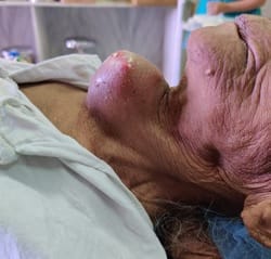
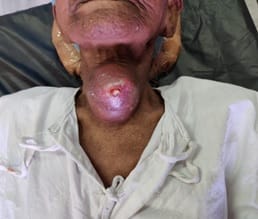
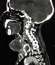
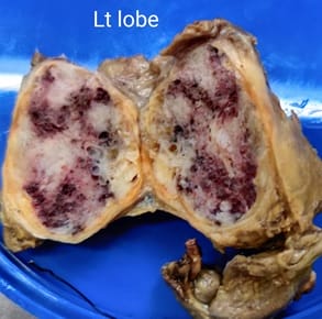
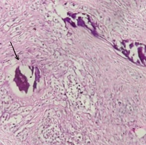
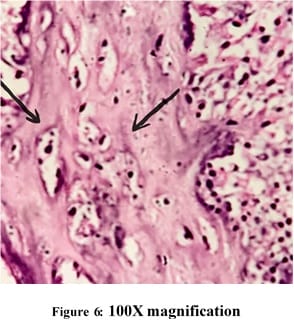
Anaplastic thyroid cancer is the most advanced and aggressive thyroid cancer. It is very rare and is found in less than 2% of patients with thyroid cancer. It most commonly occurs in people over the age of 60 years. It is usually diagnosed at later stages when the disease has already spread to surrounding structures or has grown large enough to compress the trachea or esophagus causing pressure symptoms. The patient here, a 75-year-old female, presented with a large swelling in the front of the neck in euthyroid status which was diagnosed as anaplastic thyroid cancer and was managed surgically and the surgical specimen was sent for histopathological examination. The presence of osseous metaplasia with mature bone formation in anaplastic thyroid carcinoma is a rare entity and has been discussed in this case report.

Keywords: Anaplastic, Thyroid, Neoplasm, Osseous, Metaplasia

Corresponding Author How to Cite this Article To Browse
Samprathi D, Resident, Department of General Surgery, Grant Government Medical College and JJ Group of Hospitals, Mumbai, Maharashtra, India.
Email:
Wagh AN, Samprathi D, Bhagvat SR, Tandur A, Dhimole NN. A rare case of osseous metaplasia in anaplastic thyroid cancer. Surgical Rev Int J Surg Trauma Orthoped. 2021;7(2):17-21.
Available From
https://surgical.medresearch.in/index.php/ijoso/article/view/222

Manuscript Received Review Round 1 Review Round 2 Review Round 3 Accepted
2021-03-23 2021-03-24 2021-04-03 2021-04-13
Conflict of Interest Funding Ethical Approval Plagiarism X-checker Note
No Nil Yes 9%

© 2021 by Amol N Wagh, Samprathi D, Shirish R Bhagvat, Amarjeet Tandur, Nikhil N Dhimole and Published by Siddharth Health Research and Social Welfare Society. This is an Open Access article licensed under a Creative Commons Attribution 4.0 International License https://creativecommons.org/licenses/by/4.0/ unported [CC BY 4.0].

Introduction

Thyroid cancer, a malignant tumor of the thyroid gland is relatively uncommon compared to other cancers. Papillary thyroid cancer is the most common type of all thyroid cancers. Anaplastic thyroid cancer is the most advanced and aggressive thyroid cancer. Anaplastic thyroid cancer is very rare and is found in less than 2% of patients. It most commonly occurs in people over the age of 60 years [1]. Most cases at the time of diagnosis are associated with extensive local disease spread and distant metastasis in 20%–50% of cases accounting for high mortality rates [2]. Patients with anaplastic thyroid cancer (ATC) face a uniformly dismal prognosis, with average 5-year survival rates of around 7% and a median survival time of 6 months [3]. The major cause of death of this high-grade malignancy is either distant metastasis or is due to the involvement of the vital structures of the neck [4] [5].

Anaplastic thyroid cancer may present with various symptoms. Most often it presents as a lump or nodule in the neck. These tumors grow very quickly and often the growth can be seen by the patient or their family/friends. In some cases, anaplastic thyroid cancer presents as a neck mass with difficulty in swallowing, difficulty in breathing, or hoarseness of voice if one of the vocal cords is paralyzed by the tumor. About 10% of patients have anaplastic thyroid cancer that is present only in the thyroid, and approximately 40% of those diagnosed have cancer that is localized in the neck and/or lymph nodes. The remaining patients have anaplastic thyroid cancer that has metastasized to other parts of the body at the time of diagnosis [1]. Anaplastic thyroid carcinoma has shown histopathological variants in the past in the form of chondrosarcomatous differentiation or osteoclastic variants [6] [7]. Osseous metaplasia with mature bone formation has been reported on various occasions in follicular adenoma of the thyroid. Here we present a case of the rare occurrence of osseous metaplasia with mature bone formation in a patient with anaplastic thyroid carcinoma which has been documented only once in the past.

Case Report

A 75-year-old female presented with complaints of swelling in the anterior aspect of the neck for 2 months and pain at the site of swelling for 1 month.

The patient who was apparently alright 2 months back noticed the swelling, which showed sudden progression in size in the past 1 month associated with pain. She had occasional dysphagia to solids with no respiratory complaints. There were no endocrinological manifestations. On examination, a tender swelling palpable in the neck with the local rise of temperature and redness was seen. The anterior-most part of the swelling was ulcerated showing an unhealthy floor (figure 1,2). The patient had symptoms of fever and pain during the course of her stay in the hospital for evaluation.

surgery_222_01.jpg

Figure 1: Swelling in front of the neck showing ulceration at its tip as seen in lateral view

surgery_222_02.jpg

Figure 2: Swelling in front of the neck showing ulceration at its tip as seen from anteriorly

Plain radiograph of the neck revealed globular soft tissue shadow in the anterior part of the lower neck with sub-centimetre densities in the posterior part.


Ultrasonography of the neck showed a heterogenous lesion of 5.5 x 4 x 5.5cm in the right lobe of the thyroid with a hypo-echoic lesion of 2.5x2cm with punctate calcifications in the inner aspect of the lesion with no evidence of significant lymphadenopathy. Fine Needle Aspiration Cytology (FNAC) of the swelling revealed a Bethesda category- V lesion in the right lobe of the thyroid gland.

High Resolution Computed Tomography (HRCT) of the neck was done (figure 3) which showed a bulky thyroid gland with multiple heterogeneously enhancing nodules within. Few nodules in both lobes of the thyroid show thick peripheral calcifications. The laryngeal architecture is maintained. No evidence of significant cervical lymphadenopathy. Findings are suggestive of aggressive neoplasm of the thyroid gland.

surgery_222_03.jpg

Figure 3: HRCT of the neck showing bulky thyroid gland with multiple heterogeneously enhancing nodules within.

Her blood investigation revealed hemoglobin of 9.1gm%, total leukocyte count of 12,680, TSH of 0.48, total T3, T4 of 57.84 and 10.71 respectively. She was taken up for total thyroidectomy after proper counselling & consent of the patient and her relatives. Total thyroidectomy was done without neck lymph nodes dissection. During the procedure, both the RLN were visualized and no surrounding structures involvement was seen.

The tissue was sent for histopathological examination in formalin solution. Post-operative period was uneventful with no voice changes. On follow up after 7 days, suture line was healthy with no complications. Due to poor performance status of the patient, she wasn’t given chemo or radiotherapy.

surgery_222_04.jpg

Figure 4: Gross specimen, on cut section showing greyish white areas of necrosis with brownish compressed thyroid parenchyma.

On gross examination, both the lobes are hard and gritty to cut. The cut section shows greyish white friable tumor with areas of necro- sis along with compressed surrounding brownish colored thyroid parenchyma. (Figure 4).

Histopathology revealed Anaplastic carcinoma tumor cells with osseous metaplasia (mature bone tissue as marked by black arrows) (figure 5 & 6).

surgery_222_05.jpg


Figure 5: Histopathology showing anaplastic carcinoma tumor cells with osseous metaplasia (mature bone tissue as marked by black arrow)

surgery_222_06.jpg

Figure 6: Histopathology showing anaplastic carcinoma tumor cells with osseous metaplasia (mature bone tissue as marked by black arrow)

Discussion

Anaplastic thyroid carcinoma often originates in an abnormal thyroid gland; a history of goitre is reported in >80% of cases [8]. It is more common in women than in men [9]. Most patients present with a neck mass and symptoms of dyspnea, dysphagia and dysphonia [10]. Anaplastic Thyroid carcinoma is one of the rare thyroid cancers seen in about 2% of the cases of thyroid malignancies [11].

In this case, the patient is a 75year old female with no previous history of goitre, no symptoms of tracheal or esophageal compression. The highly variable microscopic appearance of Anaplastic thyroid carcinoma can be broadly categorized into three patterns that can occur alone or in any combination: sarcomatoid, giant cell, and epithelial.

The secondary features of ATC include acute inflammation, macrophage infiltration, and osteoclast-like multi-nucleated giant cells. The other reported rare variants of ATC to include paucicellular variant [12]. angiomatoid [13].

lymphoepithelioma-like, and small-cell variant. Osseous metaplasia in follicular adenoma and papillary carcinoma of the thyroid has been reported frequently. Cases of ectopic bone formation and extramedullary hematopoiesis in thyroid nodule (goitre) have been reported too. But, osseous metaplasia in anaplastic thyroid carcinoma has been reported only once before in a study of 59 thyroid malignancies of which 7 were diagnosed with ATC, where the histopathological examination of one patient showed spindle cells with osseous metaplasia [14].

In our case, a differential diagnosis of metastatic deposits of bone tissue in the thyroid could be considered but the histopathology showed benign bone tissue in the background of malignant ATC cells hence confirming osseous metaplasia. The median survival time of patients with ATC following diagnosis is approximately 5 months [8]. The cause of death is attributable to upper airway obstruction and suffocation, which often develop despite tracheostomy in 50% of patients with ATC; in the remaining patients, the cause of death includes complications of local and distant disease or therapy [15].

If the tumor appears resectable, an attempt should be made for total thyroidectomy with complete gross tumor resection together with selective resection of all involved local or regional structures and lymph nodes [16].

Conclusion

Surgical excision is the mainstay of treatment in anaplastic thyroid cancer. It is due to its aggressive nature and early metastasis. Histopathological confirmation of the clear margins and type of cancer cells plays role in the prediction of the post-operative course and prognosis of the patient. Variants of anaplastic thyroid cancer such as osseous metaplasia with mature bone formation are rare. There is a paucity of literature about the implications of such variants on the management and prognosis of patients. Further studies are needed for a better understanding of the disease.

Reference

  1. Anaplastic Thyroid Cancer. American Thyroid Association. [cited 2021 Apr 12].

  1. Giuffrida D, Gharib H. Anaplastic thyroid carcinoma: current diagnosis and treatment. Ann Oncol. 2000 Sep;11(9)1083-9.
  2. Venkatesh YS, Ordonez NG, Schultz PN, Hickey RC, Goepfert H, Samaan NA. Anaplastic carcinoma of the thyroid- A clinicopathologic study of 121 cases. Cancer. 1990 Jul 15;66(2)321-30.
  3. Fletcher CDM. Diagnostic histopathology of tumors [Internet]. 2013 [cited 2021 Apr 12].
  4. Carcangiu ML, Steeper T, Zampi G, Rosai J. Anaplastic thyroid carcinoma- A study of 70 cases. Am J Clin Pathol. 1985 Feb;83(2)135-58.
  5. Liu J, Cui N, Ding W, Dong X, Chen X, Jiang J, Liu Y. Anaplastic thyroid carcinoma with chondrosarcomatous differentiation- a case report. Diagn Pathol. 2020 Jul 21;15(1)89.
  6. Sharma Mahima. "Anaplastic thyroid carcinoma and its osteoclastic variant." Natl J Med Res. 2014;4;98-100.
  7. American Thyroid Association Guidelines for Management of Patients with Anaplastic Thyroid Cancer. Thyroid [Internet]. [cited 2021 Apr 12].
  8. McIver B, Hay ID, Giuffrida DF, Dvorak CE, Grant CS, Thompson GB, van Heerden JA, Goellner JR. Anaplastic thyroid carcinoma: a 50-year experience at a single institution. Surgery. 2001 Dec;130(6):1028-34.
  9. Chiacchio S, Lorenzoni A, Boni G, Rubello D, Elisei R, Mariani G. Anaplastic thyroid cancer: prevalence, diagnosis and treatment. Minerva Endocrinol. 2008 Dec;33(4):341-57.
  1. Thyroid Cancer [Internet]. American Thyroid Association. [cited 2021 Apr 12].
  2. Wan SK, Chan JK, Tang SK. Paucicellular variant of anaplastic thyroid carcinoma- A mimic of Reidel's thyroiditis. Am J Clin Pathol. 1996 Apr;105(4)388-93.
  3. Njim L, Moussa A, Hadhri R, Gassab I, Ben Yahia N, Mahmoudi H, et al. Tumeur angiomatoïde de la thyroïde- angiosarcome primitif ou variante du carcinome anaplasique? [Angiomatoid tumor of the thyroid gland- primitive angiosarcoma or variant of anaplastic carcinoma?]. Ann Pathol. 2008 Jun;28(3)221-4.
  4. Ghosh A, Nepal N, Gharti M D, Basnet S, Baxi M, Talwar O P. Anaplastic carcinoma of the thyroid-clinicomorphological spectrum and review of literature. Journal of pathology of Nepal. 2011;1(1)45-48.
  5. Shaha AR. Airway management in anaplastic thyroid carcinoma. Laryngoscope. 2008 Jul;118(7)1195-8.
  6. Haddad RI, Lydiatt WM, Ball DW, Busaidy NL, Byrd D, Callender G, et al. Anaplastic Thyroid Carcinoma, Version 2, 2015. J Natl Compr Canc Netw. 2015 Sep;13(9)1140-50.