E-ISSN:2455-5436
P-ISSN:2456-9518
RNI:MPENG/2017/70870

Research Article

Transcutaneous

Surgical Review: International Journal of Surgery Trauma and Orthopedics

2021 Volume 7 Number 2 March-April
Publisherwww.medresearch.in

Nitroglycerin transcutaneous patch: boon to salvaging post-operative partial flap necrosis, simple and effective method

K. Malviya M.1, Soni S.2*, Gupta D.3
DOI: https://doi.org/10.17511/ijoso.2021.i02.03

1 Manohar K. Malviya, Assistant Professor, Department of surgery, Chirayu Medical College and Hospital, Bhopal, Madhya Pradesh, India.

2* Sameer Soni, Assistant Professor, Department of surgery, Chirayu Medical College and Hospital, Bhopal, Madhya Prdaesh, India.

3 Dipangi Gupta, Junior Resident, Department of surgery, Chirayu Medical College and Hospital, Bhopal, Madhya Prdaesh, India.

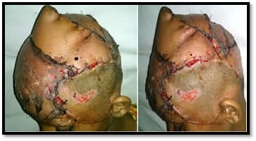
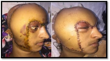
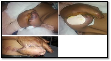
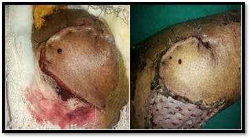
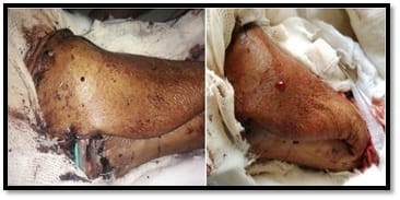
Introduction: The majority of surgical complications after tissue transfer surgery (Local transposition of the fasciocutaneous flap, Pedicle flap, Free flap or musculocutaneous flap) are related to vascular thrombosis, which usually occurs within 3 days of surgery. Venous congestion usually results in oedema and darkening of the skin colour. During early venous obstruction, a needle stick will cause rapid bleeding of dark blood and arterial obstruction or spasm will cause delayed bleeding. Patients and methods: This is prospective study was carried out during the period from January 2018 to February 2021 at the Plastic surgery unit-Chirayu Medical College And Hospital Bhopal, India. This study included patients aged 13 to 70 years undergoing reconstructive surgery with flaps (Fasciocutaneous Pedicle flap, Free flap, local transposition flap or musculocutaneous flap) for the wounds at any part of the body. The NTG patch was applied over the cutaneous surface of the compromised flap and then flap insufficiency was observed. Results: In this study total of 50 patients with flaps reconstruction were included. Among which 34 %( 17 patients) had skin changes and 66 % (33 patients) had congested bleed on needle prick. NTG patches were applied on the flap surface at regular intervals. After 1 week follows up, the changes in 82% (41) flaps were reversed back and the flap remained healthy. 18% (nine) flaps had partial and or complete necrosis. Conclusion: There was a marked reduction in partial flap necrosis in patients who received nitroglycerin patch. The flap survival was significantly improved and prevents the re-exploration of flaps. Their application is a simple, safe, and effective way to help salvage the flaps.

Keywords: Flap salvage, Flap necrosis, Post-operative flap congestion, Flap ischemia

Corresponding Author How to Cite this Article To Browse
Sameer Soni, Assistant Professor, Department of surgery, Chirayu Medical College and Hospital, Bhopal, Madhya Prdaesh, India.
Email:
Malviya MK, Soni S, Gupta D. Nitroglycerin transcutaneous patch: boon to salvaging post-operative partial flap necrosis, simple and effective method. Surgical Rev Int J Surg Trauma Orthoped. 2021;7(2):10-16.
Available From
https://surgical.medresearch.in/index.php/ijoso/article/view/218

Manuscript Received Review Round 1 Review Round 2 Review Round 3 Accepted
2021-03-05 2021-03-15 2021-04-12 2021-04-30
Conflict of Interest Funding Ethical Approval Plagiarism X-checker Note
No Nil Yes 8%

© 2021 by Manohar K. Malviya, Sameer Soni, Dipangi Gupta and Published by Siddharth Health Research and Social Welfare Society. This is an Open Access article licensed under a Creative Commons Attribution 4.0 International License https://creativecommons.org/licenses/by/4.0/ unported [CC BY 4.0].

Introduction

The majority of surgical complications after tissue transfer surgery (Local transposition of the fasciocutaneous flap, Pedicle flap or Free flap) are related to vascular thrombosis, which usually occurs within 48 hours of surgery. The flap can be evaluated by clinical observation and Doppler vascular pedicle monitoring. These methods are used commonly. Venous thrombosis is more common than arterial occlusion, because of its low-flow, low-pressure nature, and because it may evolve over several hours. Venous congestion usually results in oedema and darkening of the skin colour. During early venous obstruction, a needle stick will cause rapid bleeding of dark blood. Arterial occlusion will cause coldness of flap, pale skin, no capillary refilling, no or delayed bleed on pinprick and the sign of ischemia seen. If a flap’s vascular integrity is in question, if there is a doubt of venous thrombosis at the vascular anastomosis of free flaps, a prompt surgical exploration is mandatory[1]. Surgical exploration is still mandatory when there are hematoma and seroma that could make a compression at the pedicle of a flap. This kind of insufficient vascular drainage may cause a slow partial flap loss that may require further surgical steps, multiple debridements to remove necrotic tissue and prevent further complications like infections, and to improve the flap outcome. The first step in managing free-flap failure is the early recognition of a compromised flap. Clinical observation remains the simplest method of identifying vascular compromise[2]. Nitroglycerin (NTG), also known as nitroglycerine, trinitroglycerin (TNG), trinitroglycerine, glyceryl trinitrate (GTN), or 1,2,3-trinitroxypropane, is a heavy, colourless, oily, explosive liquid most commonly produced by nitrating glycerol with white fuming nitric acid under conditions appropriate to the formation of the nitric acid ester. For over 130 years, nitroglycerin has been used medically as a potent vasodilator (dilation of the vascular system) to treat heart conditions, such as angina pectoris and chronic heart failure. These agents all exert their effect by being converted to nitric oxide in the body by mitochondrial enzyme aldehyde dehydrogenase and nitric oxide is a potent natural vasodilator[3].

Application of nitroglycerine patch over the vascular compromised part of the flap as early as post-operatively may improve vascularity and clinical features of the flap and could avoid unnecessary surgical exploration.

Patients and methods

This prospective observational study was carried out during the period from January 2018 to February 2021 at the Chirayu Medical College And Hospital, Bhopal at the Department of Surgery. Patients aged from 13 to 70 years undergoing flap coverage (Fasciocutaneous flaps, Pedicled flap, and Free flap or Musculocutaneous flaps) for wound closure at any part of the body were included in this study. These flaps are monitored routinely by clinical observation, needle prick at flap skin to see venous congested blood, and Doppler to see the sign of vascular compromise and ischemic changes over the skin paddle of flaps. Adjuncts such as a pinprick, temperature measurement, and surface doppler are also used to aid in the early recognition of problems. When the sign of ischemia or Flap vascular compromise seen post-operatively we have applied as early as possible the Nitroglycerine patches over the ischemic or congested part of the cutaneous surface of flaps for 12-14 hours daily for one week and observe the signs of reversal and salvaging of the flap.

Inclusion criteria

  1. This study included patients aged 13 to 70 years undergoing reconstructive surgery with flaps (Fasciocutaneous Pedicle flap, Free flap, local transposition flap or musculocutaneous flap) for the wounds at any part of the body.
  2. This patch should only be worn for up to 12 to 14 hours a day, so that you will have a 10 to 12-hour “nitrate-free” period each day. It is important to have a “nitrate-free” period each day for nitroglycerin patch to continue to work well and to decrease the risk of physical dependence.

Exclusion criteria

1. Patient below 13 years and above 70 years of age are excluded from this study.

Results

In this study, we had applied an NTG patch (Transcutaneous nitroglycerin patch) over a total of 50 flaps which had clinically partial or part of flap skin changes (a sign of ischemia) and congested dark colour blood on needle prick at flap skin. We applied an NTG patch in the post-operative period as early as when we had noticed clinically or with the help of handheld Doppler, the flap was compromised.


In 24 random pattern flaps, 33.3% flaps had skin changes and 66.6% flaps had congested dark colour blood on pinprick post-operatively. We have applied 1 or 2 NTG patch with 12 to 14 hours intervals daily at the skin of the compromised part of the flaps for one week. 87.5 % of flaps showed signs of reversal and flap survived well. 12.5% flaps did not revive back and flap necrosis occurred partially, part of flap or completely. The necrosed part of the flap was debrided and managed by the alternative procedure.

In 16 axial pattern flaps, 31.2% flaps had skin changes and 68.7% flaps had congested dark colour bleed on pinprick postoperatively. After application of NTG patch, 75% of flaps skin changes reversed back and 25% of flaps had necrosis. These necrosed parts of the flaps were managed by other methods.

A total of 10 free flaps were included in this study.

40% of flaps had skin changes and 60% of flaps had congestion with the dark bleed on needle prick post-operatively. We applied NTG patches serially on free flaps skin and noticed, 80% of flaps reversed back clinically and survived well. 20% flaps did not show sign of reversal and changes persisted. These flaps were managed by exploration of vessels anastomotic site and by other alternative procedures.

In this study total of 50 flaps were included which had skin changes (total 34%) and congested bleed on needle prick (total 66%). NTG patches were applied on the compromised part of flap skin. Total 82% of flaps were reversed back and remain healthy. 18% flaps necroses partially, part of flaps or completely.

There were no complications observed, in any patients, when applied NTG patches over the flaps.


TABLE 1: Results after using the Nitroglycerine Patch over the Flaps

Flaps No. of compromised flaps, applied NTG patches Flaps with skin changes Flaps with congested dark colour blood on pinprick Sign of reversal No reversal (partial, part or complete flap necrosis)
No. Of flaps (%) No. Of flaps (%) No. Of flaps (%) No. Of flaps (%)
Random pattern flap 24 8 33.3% 16 66.6% 21 87.5% 3 12.5%
Axial pattern flap 16 5 31.2% 11 68.7% 12 75.0% 4 25.0%
Free flap 10 4 40.0% 6 60.0% 8 80.0% 2 20.0%
Total 50 17 34.0% 33 66.0% 41 82.0% 9 18.0%

NTG patch- Nitro-glycerine patch

surgery_218_01.jpg

Figure 1: Free latissimus dorsi flap for scalp defect

surgery_218_02.jpg

Figure 2: Free parascapular flap for scalp defect


surgery_218_03.jpg

Figure-3: Groin flap skin changes

surgery_218_04.jpg

Figure 4: Local transposition flap for leg defect

surgery_218_05.jpg

Figure 5: Reverse sural flap for foot defect

Discussion

Nitroglycerin is available as sublingual tablets, sprays, and transcutaneous patches. It is also used topically, in low doses for the treatment of chronic anal fissures. At low doses, nitroglycerin will dilate veins more than arteries. At higher doses, it also dilates arteries. It has been shown that continuous exposure to nitrates can cause the body to stop responding normally to this medicine. Experts recommend that the patches be removed at night, allowing the body a few hours to restore its responsiveness to nitrates. Shorter-acting preparations of nitroglycerin can be used several times a day with less risk of developing tolerance.

Mechanism of action: Nitroglycerin relaxes smooth muscle throughout the body.

In the vascular system, it acts chiefly on the systemic veins and accessorily on the large arteries. Nitroglycerin at low doses is bio-activated by mitochondrial aldehyde dehydrogenase activity and is converted to nitrites and denitrated metabolites (1,2-glyceryl dinitrate,1-3-glyceryl dinitrate) by glutathione-dependent organic nitrate reductase. Nitrite is further activated by cytochrome oxidase or acidic disproportionation in the intermembrane space (H+), finally yielding nitric oxide (NO) or a related species, which activate soluble guanylyl cyclase and trigger cyclic guanosine monophosphate (cGMP) signalling via cGMP-dependent protein kinase, which causes relaxation. Glyceryldinitrate, mononitrate, and nitroglycerin at high doses are bioactivated by the P450 enzyme(s) in the smooth endoplasmic reticulum directly yielding NO which causes relaxation.

Excretion: Nitroglycerin is excreted renally as dinitrate and mononitrate metabolites, glucuronide conjugates and glycerol. The elimination half-lives of nitroglycerin, 1,2-glyceryl dinitrate, and glyceryl mononitrates are 10, 30-60, 5-6 minutes respectively.

Instructions for patients using the patch system:

  • Wash your hands with soap and water before and after applying a patch. Do not touch your eyes until after you have washed your hands.
  • Do not try to trim or cut the adhesive patch to adjust the dosage.
  • Apply the patch to a clean, dry skin area with little or no hair that is free of scars, cuts, or irritation.
  • Remove the patch from the package. Apply with firm pressure to the skin. To avoid skin irritation.
  • Always remove a previous patch before applying a new one.
  • Apply a new patch at different sites daily if possible.
  • Nitroglycerin patch is for external use only.
  • This patch should only be worn for up to 12 to 14 hours a day, so that you will have a 10 to 12-hour “nitrate-free” period each day. It is important to have a “nitrate-free” period each day for nitroglycerin patch to continue to work well and to decrease the risk of physical dependence.

  • You need to check your blood pressure often while using a nitroglycerin patch.
  • When used for long periods without a break, nitroglycerin patch may not work as well. This is known as tolerance. Increasing the dose is not effective in managing tolerance to nitroglycerin patch. Be sure to have a “nitrate-free” period each day to help prevent this tolerance.
  • After removing a used patch, fold it in half with the sticky sides together. Make sure to dispose of it out of the reach of children and pets.

For transdermal dosage form (skin patch):- we had applied one or two NTG patches on flap skin surface once a day for one week. Leave the patch in place for a total of 12 to 14 hours.

Storage

  • Store the medicine in a closed container at room temperature between 68-77degrees F (20-25 degrees C), away from heat, moisture, and direct light. Keep away from freezing.
  • Keep out of the reach of children.

Side Effects

More common: - Lightheadacheness

Less common: -Arm, back, or jaw pain, blurred vision, chest pain or discomfort, chest tightness or heaviness confusion, dizziness, faintness, fast or irregular heartbeat, nausea, sweating, unusual tiredness or weakness

Rare: - Bluish-coloured lips, fingernails, or palms, dark urine, difficulty with breathing, fever, pale skin, rapid heart rate, sore throat, unusual bleeding or bruising. Skin burns may occur at the site where the patch is worn during this procedure.

Drug interactions: Drugs used to treat erectile dysfunction-ED or pulmonary hypertension (such as sildenafil, tadalafil, riociguat). Certain drugs to treat migraine headaches (ergot alkaloids such as ergotamine).

Do not use the nitroglycerin patch if:

  • You are allergic to any ingredient in nitroglycerin patch
  • You are allergic to the adhesive that makes the patch stick to your skin
  • You have severe anemia
  • You are taking avanafil, riocigaut, sildenafil, or vardenafil
  • If you have a history of heart problems, overactive thyroid, stroke or another bleeding in the brain, or recent head injury.

Hidalgo et al[4] identified venous problems (35%) as the most common aetiology of flap failure followed by arterial problems (28%), hematoma (26%) and recipient vessel problems (11%). Late flap failures (i.e. > 48 hours) were most often due to infection or mechanical stress around the anastomosis. The free flap suffering or failure is a complication that often requires, after a rapid recognition of the clinical aspect of the flap, the surgical re-exploration to remove hematomas, seroma over the pedicle or to perform new anastomoses[5].

In 756 cases Miyasaka et al [6] performed 22 explorations for vascular pedicle compromise. Management of flap necrosis includes healing by secondary intention, surgical debridement, local administration of antibiotic preparations, moist dressings, vitamin injections and hydro fibre dressings[7, 8, 9]

Kubo et al [10] reviewed the management of the flap with venous compromise and suggested that surgical methods should be the first choice as it offers significantly higher salvage rates. In select cases, venous congestion can be managed with the application of leeches. Dabb [11] described several successful cases of venous congested flaps salvaged by leeches, suggesting that relief of congestion for four to ten days may allow enough time for neo-vascularisation. Neo-vascularization has been reported to occur as early as six days[12].. The chance of surgical salvage is low after the first 48 hours. Hyodo et al[13] reported that flap salvage was impossible if thrombosis occurred more than three days after surgery.

In this study, we had applied an NTG patch over the skin of a total of 50 vascular compromised flap postoperatively, and no other procedure was done. In our study 82% of flaps congestion improved and flaps remained healthy and 18% flaps necrosed partially, part of flaps or completely. In these necrosed parts of the flap, debridement and dressing were done as described in another study. Other alternative methods of management of flap necrosis were also done.

NTG patch was applied only on those flaps, which were vascular compromised postoperatively and in those flaps in which there was no vascular compromise, nothing was done.


NTG patch application was an effective tool to save the flap and reduce the morbidity of patients, avoiding unnecessary flap exploration, reducing wound healing time by improving the blood circulation of the flap.

Its application is very easy; patients can also apply themselves over flap. The patient can also be discharged from the hospital after giving proper advice of NTG application and follow the patient regularly.

Its side effects are very negligible or nothing, because it acts locally in the flap, so it can be used safely.

Conclusion

Application of a nitroglycerin patch on the suffering flap without local or systemic side effects is possible. If there is a rapid improvement in the clinical feature of the flap, surgical exploration could be avoided.

There was a marked reduction in flap necrosis in patients who received a transcutaneous nitroglycerin patch. The flap survival was significantly improved and prevented re-exploration of flaps and reduced morbidity, thereby reducing the cost of additional surgical procedure.. Their application is a simple, safe, and effective way to help in the salvage of the flaps vascular insufficiency.

Author contribution

The authors were collectively involved in clinical management, data collection and manuscript preparation.

What does this study add to existing knowledge?

NTG patches are beneficial in post-operative flap outcomes when there is any insufficiency occurs in the survival of the flap.

Reference

  1. B C D Evans, G R D Evans. “Microvascular Surgery”. Plastic & Reconstructive Surgery. 2007;119(2)18e-30e.
  1. Spiegel JH, Polat JK. Microvascular flap reconstruction by otolaryngologists- prevalence, postoperative care, and monitoring techniques. Laryngoscope. 2007 Mar;117(3)485-90.
  2. Chen Z, Foster MW, Zhang J, Mao L, Rockman HA, Kawamoto T, Kitagawa K, Nakayama KI, Hess DT, Stamler JS. An essential role for mitochondrial aldehyde dehydrogenase in nitroglycerin bioactivation. Proc Natl Acad Sci U S A. 2005 Aug 23;102(34)12159-64.
  3. Hidalgo DA, Disa JJ, Cordeiro PG, Hu QY. A review of 716 consecutive free flaps for oncologic surgical defects- refinement in donor-site selection and technique. Plast Reconstr Surg. 1998 Sep;102(3)722-32; discussion 733-4.
  4. Askari M, Fisher C, Weniger FG, Bidic S, Lee WP. Anticoagulation therapy in microsurgery- a review. J Hand Surg Am. 2006 May-Jun;31(5)836-46.
  5. Miyasaka M, Ichikawa K, Nishimura M, Yamazaki A, Taira H, Imagawa K, Tanino R. Salvage operations of free tissue transfer following internal jugular venous thrombosis- a review of 4 cases. Microsurgery. 2005;25(3)191-5.
  6. Araco A, Gravante G, Araco F, Delogu D, Cervelli V. Conservative management of flap necrosis after expanding hematomas for face lifting surgery. Dermatol Surg. 2006 Jun;32(6)878-9; author reply 879-80.
  7. Personelle J, Bolivar de Souza Pinto E, Ruiz RO. Injection of vitamin A acid, vitamin E, and vitamin C for treatment of tissue necrosis. Aesthetic Plast Surg. 1998 Jan-Feb;22(1)58-64.

  1. Jurczak F, Dugré T, Johnstone A, Offori T, Vujovic Z, Hollander D. AQUACEL Ag Surgical/Trauma Wound Study Group, Randomised clinical trial of Hydrofiber dressing with silver versus povidone-iodine gauze in the management of open surgical and traumatic wounds. Int Wound J. 2007 Mar;4(1)66-76.
  2. Kubo T, Yano K, Hosokawa K. Management of flaps with compromised venous outflow in head and neck microsurgical reconstruction. Microsurgery. 2002;22(8)391-5.
  3. Dabb RW, Malone JM, Leverett LC. The use of medicinal leeches in the salvage of flaps with venous congestion. Ann Plast Surg. 1992 Sep;29(3)250-6.
  1. Burns A, Avery BS, Edge CJ. Survival of microvascular free flaps in head and neck surgery after early interruption of the vascular pedicle. Br J Oral Maxillofac Surg. 2005 Oct;43(5)426-7.
  2. Hyodo I, Nakayama B, Kato H, Hasegawa Y, Ogawa T, Terada A, Torii S. Analysis of salvage operation in head and neck microsurgical reconstruction. Laryngoscope. 2007 Feb;117(2)357-60.