E-ISSN:2455-5436
P-ISSN:2456-9518
RNI:MPENG/2017/70870

Case Report

Duplication

Surgical Review: International Journal of Surgery Trauma and Orthopedics

2020 Volume 6 Number 6 November-December
Publisherwww.medresearch.in

Laparoscopic management of rare “ H type” duplication of gall bladder

Mishra A.1, Masud Ansari M.2, Misra S.3*
DOI: https://doi.org/10.17511/ijoso.2020.i06.03

1 Anurag Mishra, Senior Resident, Department of General Surgery, Dr. Ram Manohar Lohia Hospital, New Delhi, India.

2 Md Abu Masud Ansari, Associate Professor, Department of General Surgery, Dr. Ram Manohar Lohia Hospital, New Delhi, India.

3* Shivanshu Misra, Consultant Gastro, Laparoscopic Surgeon, Shivani hospital and IVF, Kanpur, Uttar Pradesh, India.

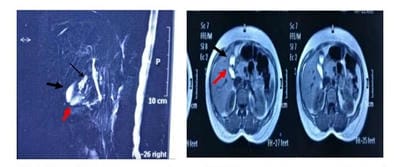
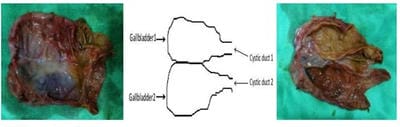
A duplicated gallbladder is a rare congenital anomaly with an incidence of 1:4000 live births. They can remain asymptomatic and identified incidentally or present as acute cholecystitis, empyema, torsion, cholecystoenteric fistula, Gall bladder lump, or carcinoma. Here the current case is about discussing a case of a 25-year-old female who presented with symptomatic gallstone disease with a duplicated gallbladder having multiple stones in both the gallbladders. MRCP performed preoperatively revealed Y type duplication (double Gall bladder with common cystic duct). Laparoscopic cholecystectomy was performed and it finally revealed H type duplication (double Gall bladder with separate cystic ducts for each Gall Bladder).

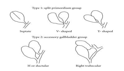
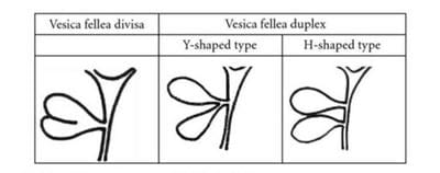
Keywords: Duplicated Gall Bladder, H type gall bladder, Boyden’s classification, Harlaftis classification

Corresponding Author How to Cite this Article To Browse
Shivanshu Misra, Consultant Gastro, Laparoscopic Surgeon, Shivani hospital and IVF, Kanpur, Uttar Pradesh, India.
Email:
Mishra A, Ansari AM, Misra S. Laparoscopic management of rare “ H type” duplication of gall bladder. Surgical Rev Int J Surg Trauma Orthoped. 2020;6(6):353-356.
Available From
https://surgical.medresearch.in/index.php/ijoso/article/view/213

Manuscript Received Review Round 1 Review Round 2 Review Round 3 Accepted
2020-12-05 2020-12-15 2020-12-21 2020-12-26
Conflict of Interest Funding Ethical Approval Plagiarism X-checker Note
No Nil Yes 9%

© 2020 by Anurag Mishra, Md Abu Masud Ansari, Shivanshu Misra and Published by Siddharth Health Research and Social Welfare Society. This is an Open Access article licensed under a Creative Commons Attribution 4.0 International License https://creativecommons.org/licenses/by/4.0/ unported [CC BY 4.0].

Introduction

Gallbladder duplication is a rare congenital anomaly with an incidence of 1 in 4000 live births [1]. The first reported human case of an accessory gallbladder was noted in the sacrificial victim of emperor Augustus in 31BC [2], but the first documented case of double accessory GBs in living humans was reported by Sherron in 1911 [3]. There is no sex predilection and they are mostly found in adults incidentally during cholecystectomy or autopsy.

There are two classification systems for duplicated gallbladders: Boyden’s anatomical classification [1] and Harlaftis classification system [4].

The Harlaftis classification (Fig 3) is the most widely used and classifies the duplicated GBs anomalies into 3 types:

  1. type I or split primordial group has only one cystic duct draining into the common bile duct. These Gallbladders are usually next to each other and share a common GB fossa. Its sub-classification includes septate, V-shaped, and Y duplicated GBs.
  2. type II anomalies, the most common form have 2 separate GBs drain into a common bile duct through independent cystic ducts (H-type), or one of the cystic ducts drains into the right or left hepatic duct (trabecular type).
  3. Type III accessory GBs include all other types not included in type I and II, including triple GBs.

Case Presentation

25 years female without comorbidities presented with complaints of recurrent colicky pain in the right upper quadrant of the abdomen for 1 year associated with nausea and vomiting. No history of fever jaundice or Pruritus. Ultrasound abdomen was suggestive of the double cavity of the gallbladder with multiple stones of size 8-9 mm with normal CBD.

A provisional diagnosis of acute calculus cholecystitis was made and MRCP was planned given abnormal ultrasound features. MRCP revealed duplicated gallbladders with multiple calculi(5-12mm) with majority calculi seen in the anteriorly placed gallbladder. The cystic duct was not well traceable but appears to be common to both gallbladder lumens (Figure 1).

surgical_213_01.jpg

Fig-1: MRCP (Thin black arrow: cystic duct; thick black and red arrows; gallbladders).

Management and course

The patient underwent laparoscopic cholecystectomy. At Surgery, both gallbladders were found in a common Gallbladder fossa with minimal adhesions to the omentum. It was H-type according to the Harlaftis classification. After meticulous dissection, both cystic ducts and their blood vessels were divided and clipped, and the 2 GBs were successfully resected (Figure 2a,2b). Gallbladders were distended with multiple calculi. On opening, no polyps or growth were seen (Figure 2c). Both intraoperative and postoperative periods were uneventful and the patient was discharged on postoperative day two. The final histopathology report was suggestive of chronic cholecystitis.

surgical_213_02.jpg

Fig-2: Resected specimen; 2A: Duplicated gallbladders, 2B; Schematic representation of specimen. 2C; Opened gallbladder’s lumen.

Discussion

surgical_213_03.jpg

Fig-3: Harlaftis classification system for duplicated gallbladders.


Boyden's classification (Fig 4) of gallbladder duplication is as follows:

  1. Vesica fellea divisa (bilobed or bifid gallbladder, double gallbladder with a common neck),
  2. Vesica fellea duplex (double gallbladder with two cystic ducts),

Y-shaped type (the two cystic ducts uniting before entering the common bile duct),

H-shaped type (ductular type, the two cystic ducts entering separately into the biliary tree).

surgical_213_04.jpg

Fig-4: Boyden’s classification of gallbladder duplication.

The differential diagnosis for duplicated GBs includes gallbladder diverticula, gallbladder fold, Phrygian cap, choledochal cyst, pericholecystic fluid, focal adenomyomatosis. In the past, duplicated Gallbladders were mostly found incidentally during cholecystectomy or autopsy, but these days they can be detected preoperatively by various imaging modalities. Ultrasound, as an extension of clinical examination, is usually the first imaging modality for suspected biliary diseases.

Ultrasound may diagnose gallbladder duplication but the anatomy of the ductal system cannot be defined very clearly [5,6]. MR cholangiogram becomes a handy investigation in a suspected case of duplicated GB. It is a noninvasive imaging technique and can very well identify duplicated GBs and associated ductal anomalies [7,8].

Clinical problems associated with duplicated gallbladder have similar nature to those encountered in the single gallbladder. Even the incidence and risk of the disease remain similar to a single gallbladder.

They can be diagnosed incidentally or may present as acute or chronic cholecystitis [9,10], cholelithiasis [9,10], empyema, torsion [11], cholecystocolic fistula [12], GB lump, and carcinoma. There are no specific symptoms attributable to a double gallbladder.

Once identified, symptomatic patients must undergo surgical removal of both gallbladders simultaneously because if the only gallbladder with pathology is removed first, another gallbladder may present later with similar pathology or symptoms at variable time intervals [13-16]. Cholecystectomy can be successfully performed either laparoscopically [9,10,14] or by the open method [3] but the laparoscopic approach should be the initial choice and cholangiography is recommended to aid in identifying and resecting the duplicated GBs.

Since there is no increased incidence of gallbladder disease in duplicated GBs, prophylactic cholecystectomy in asymptomatic patients with gallbladder duplication is not recommended due to limited data at present.

Conclusion

Presenting as a rare congenital anomaly, the duplicated gallbladder must be properly diagnosed and followed up by cholecystectomy preferably via the laparoscopic method, if symptomatic. Sometimes the final diagnosis can be made directly after surgery. As in our case, the MRCP report was suggestive of Y-type GBs but the final diagnosis was H-type GBs.

Reference

  1. Boyden EA. The accessory gallbladder- an embryological and comparative study of aberrant biliary vesicles occurring in man and the domestic mammals. Am J Anat. 1926;38(2)177–231.
  2. Udelsman R, Sugarbaker PH. Congenital duplication of the gallbladder associated with an anomalous right hepatic artery. Am J Surg. 1985;149(6)812-815.
  3. Sherren J. IX- A Double Gall-bladder Removed by Operation. Ann Surg. 1911;54(2)204-205.
  4. Harlaftis N, Gray SW, Skandalakis JE. Multiple gallbladders. Surg Gynecol Obstet. 1977;145(6)928–934.

  1. Goiney RC, Schoenecker SA, Cyr DR, Shuman WP, Peters MJ, Cooperberg PL. Sonography of gallbladder duplication and differential considerations. AJR Am J Roentgenol. 1985;145(2)241-243.
  2. Diaz MJ, Fowler W, Hnatow BJ. Congenital gallbladder duplication- preoperative diagnosis by ultrasonography. Gastrointest Radiol. 1991;16(3)198-200.
  3. Botsford A, McKay K, Hartery A, Hapgood C. MRCP imaging of duplicate gallbladder- a case report and review of the literature. Surg Radiol Anat. 2015;37(5)425-429.
  4. Hekimoglu K, Bayrak A, Ulu F, Coskun M. Combined use of ultrasonography, MDCT and MRCP for the diagnosis of gallbladder duplication- case report. J Dig Dis. 2010;11(2)115-118.
  5. Desolneux G, Mucci S, Lebigot J, Arnaud JP, Hamy A. Duplication of the gallbladder- A case reports. Gastroenterol Res Pract. 2009;483473.
  6. Zhou DK, Huang Y, Kong Y, Ye Z, Ying LX, Wang WL. Complete laparoscopic cholecystectomy for a duplicated gallbladder- A case report. Medicine (Baltimore). 2020;99(1)e18363.
  1. Recht W. Torsion of a double gallbladder- a report of a case and a review of the literature. Br J Surg. 1952;39 (156)342-4.
  2. Ritchie AW, Crucioli V. Double gallbladder with cholecystocolic fistula- a case report. Br J Surg. 1980;67(2)145-146.
  3. Gigot JF, Van Beers B, Goncette L, Etienne J, Collard A, Jadoul P, et al. Laparoscopic treatment of gallbladder duplication. Surg Endosc. 1997;11(5)479-482.
  4. Cueto García J, Weber A, Serrano Berry F, Tanur Tatz B. Double gallbladder treated successfully by laparoscopy. J Laparoendosc Surg. 1993;3(2)153-155.
  5. Silvis R, van Wieringen AJ, van der Werken CH. Reoperation for a symptomatic double gallbladder. Surg Endosc. 1996;10(3)336-337.
  6. Reinisch A, Brandt L, Fuchs KH. Gallbladder duplication- laparoscopic cholecystectomy 17 years after open cholecystectomy. Zentralblatt fur Chirurgie. 2009 Dec;134(6)576-579.