E-ISSN:2455-5436
P-ISSN:2456-9518
RNI:MPENG/2017/70870

Research Article

Fractures

Surgical Review: International Journal of Surgery Trauma and Orthopedics

2020 Volume 6 Number 6 November-December
Publisherwww.medresearch.in

Study of treatment for distal end radial fractures by open reduction and internal fixation with volar locking compression plates

Amritbhai Patel V.1, A. Pushkarna V.2*, S. Padhiyar H.3
DOI: https://doi.org/10.17511/ijoso.2020.i06.02

1 Vivek Amritbhai Patel, Associate Professor, Department of Orthopedics, Gujarat Adani Institute of Medical Science, Kutch, Gujarat, India.

2* Vishal A. Pushkarna, Assistant Professor, Department of Orthopedics, Gujarat Adani Institute of Medical Science, Kutch, Gujarat, India.

3 Hardik S. Padhiyar, Post Graduate Student, Department of Orthopedics, Gujarat Adani Institute of Medical Science, Kutch, Gujarat, India.

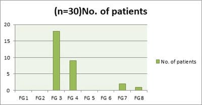
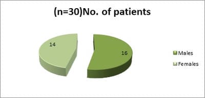
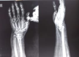
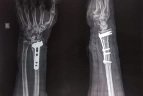
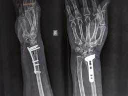
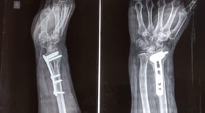
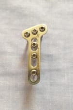
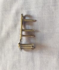
Introduction: For dorsally displaced lower end radius fractures percutaneous pinning fixation and in some studies fragment specific fixation shows their advantages. For volarly displaced fractures volarly plate fixation traditionally showed good results. The volar locking plates with their inheritance ability to provide absolute stability are attractive for their advantages. Objective: To assess the functional outcome of treating the distal end radius fractures with the use of volar locking compression plates. Materials and methods: Study of a total of 30 patients was conducted in the department of orthopedics at G K General Hospital, Bhuj, Gujarat. All 30 patients with closed fractures were included in the study. All patients were treated with open reduction and internal fixation with the use of a volar locking compression plate. The patients were followed up at one, two, three, and up to six months. Mayo wrist scoring system was used to assess final functional outcomes of treatment. Results: At the final functional assessment, as per mayo wrist scoring out of a total of 30 patients, 17 of them achieved excellent, 7 achieved good outcomes, with 5 patients exhibiting fair results and one patient had the collapse of fixation at the 3-month review. No, any patient had diminution of functional outcome. On radiological assessment, 70% of patients had callus formation and no clear fracture line was seen, 18% had callus formation but a visible fracture line was present and 12% of patients had clear visible fracture line up to final follow up. Conclusion: Open reduction and internal fixation with volar locking compression plating is a safe and effective treatment for unstable especially volarly displaced fractures of distal end radius with satisfactory functional outcome.

Keywords: Distal Radius, Fractures, Locking Plates, Volar locking compression plates

Corresponding Author How to Cite this Article To Browse
Vishal A. Pushkarna, Assistant Professor, Department of Orthopedics, Gujarat Adani Institute of Medical Science, Kutch, Gujarat, India.
Email:
Patel VA, Pushkarna VA, Padhiyar HS. Study of treatment for distal end radial fractures by open reduction and internal fixation with volar locking compression plates. Surgical Rev Int J Surg Trauma Orthoped. 2020;6(6):347-353.
Available From
https://surgical.medresearch.in/index.php/ijoso/article/view/212

Manuscript Received Review Round 1 Review Round 2 Review Round 3 Accepted
2020-11-21 2020-12-02 2020-12-12 2020-12-21
Conflict of Interest Funding Ethical Approval Plagiarism X-checker Note
No Nil Yes 5%

© 2020 by Vivek Amritbhai Patel, Vishal A. Pushkarna, Hardik S. Padhiyar and Published by Siddharth Health Research and Social Welfare Society. This is an Open Access article licensed under a Creative Commons Attribution 4.0 International License https://creativecommons.org/licenses/by/4.0/ unported [CC BY 4.0].

Introduction

Distal end radius is important in the kinematics of the radiocarpal and radioulnar joint, open reduction of articular surface and restoration of radial length, volar angulation and radial inclination are the required parts for good clinical outcome. Dorsal angulation and secondary displacement with the poor cosmetic outcome are common with conservatively treated distal end radius fractures. Because the distal radius is the foundation of the wrist joint and an indispensable part of ligamentous support, reconstruction of articular congruity and stable fixation reduces the incidence of post-traumatic osteoarthritis and allows early functional rehabilitation.

The surgical approach depends on the direction of the displacement of the distal fragment. Patients treated with dorsal platting end up with a high complication rate [1]. Although double platting has been advocated for unstable distal fractures of the radius, loss of reduction related to comminution and osteoporosis is common [2]. The volarly displaced intraarticular fractures have inheritance instability with wrist volar subluxation and loss of volar pillar. They need buttress with articular congruity for articular settlement and stability. It also needs to balance the dorsovolar ligaments of the wrist to optimize the balance and that is important for smooth wrist function.

A volar approach has been developed for fixing a dorsally angulated fracture of the distal end radius. It has several advantages, including more access to the volar surface of the distal radius, it has also the advantage of the avoidance of both dorsal dissection and it’s complications of extensor tendon injury and the interruption of blood supply to dorsal metaphyseal fragments [3]. The volar locking compression plate utilizes a threaded screw head that locks into the plate holes when the screws are tightened, providing good angular and axial stability and minimizing the possibility of screw pullout and plate loosening. This is useful in the prevention of secondary displacement of the unstable fracture in elderly patients with osteoporotic bone [3-7].

Materials and Methods

A retrospective study was conducted on 30 patients with displaced distal end radius fractures, 30 patients with distal radial fractures were treated with open reduction and internal fixation using locking compression plates, of these patients.

All had displaced fractures of distal end radius and they were fixed with volar locking plates. Out of 30 patients, 16(53.33%) were male and 14(46.67%) were female patients. In no case, any bone grafting was performed. All the fractures were treated under image intensifier television by trained orthopedic surgeons. Fractures were classified according to Frykman classification [11].18(60%) patients of Frykman grade 3,9(30%) were Frykman grade 4,2(66.67%) were Frykman grade 7 and 1(3.33%) was of Frykman grade 8.

surgical_212_01.jpg

Fig-1: Frykman classification (FG: Frykman Grade) Of the study patients.

Inclusion criteria: Failure of close reduction of displaced fractures of distal end radius with a radiographic image showing volar barton and volarly displaced fractures, >2mm of articular step-off or gap, >2mm of radial shortening.

Exclusion criteria: Fractures of the immature skeleton, open fractures, dorsal barton fractures, distal radial fractures extending to the shaft of radius, and concomitant fractures of the same limb.

surgical_212_02.jpg

Fig-2: Gender distributions of the study patients.

All procedures were performed under general or regional anesthesia. The application of the volar approach was determined according to the type of fracture and part of the fragment to be fixed first. Dissection between the flexor carpi radialis tendon and radial artery was done in the cases where the styloid was decided to be fixed first.


Dissection between the median nerve and flexor carpi radialis tendon was used in the cases with die-punch and typical volar barton fractures.

Results

Follow-up duration varied from 1 to 6 months; all 30 of 30 patients were available for the regular follow-up and final assessment. The mean age of all 30 patients (16 men and 14 women ) was 46.5 years with a minimum of 20 years to a maximum of 72 years. The mean interval between injury and surgical stabilization was 2 days ( range 1 to 4 ).28 patients were given regional anesthesia and 2 patients were given general anesthesia.19 Patients united clinicoradiologically with a good outcome. One patient had the collapse of fixation which subsequently required removal of the plate and treated with a cast and subsequently malunited. No Infection was noted in our series. No case of shoulder hand syndrome was noted in our series.

Post-operatively on functional assessment with mayo wrist score [10] (Table:1) showed 17 patients had excellent results, whereas 7 patients with good, 5 patients with fair, and 1 patient with poor results. On radiological assessment, parameters showed that the mean radial shortening was 0.2mm, mean radial inclination of 25 degrees.

Table-1: Mayo wrist Score in some patients [10].

Mayo wrist score No. of patients
Excellent 17(56.67%)
Good 7(23.33%)
Fair 5(16.67%)
Poor 1(3.33%)

surgical_212_03.jpg

Fig-3: Wrist flexion

surgical_212_04.jpg

Fig-4: Wrist Extension

surgical_212_05.jpg

Fig-5: Pre-Operative X-ray.

surgical_212_06.jpg

Fig-6: Post-Operative X-ray


surgical_212_07.jpg

Fig-7: Follow up at 1 month.

surgical_212_08.jpg

Fig-8: Follow up at 3 months.

Discussion

The surgical approach to distal radial fractures depends on the direction of displacement of the distal fragment. For dorsal comminution extraarticular and intraarticular fractures, the standard closed reduction and pinning shows good results and some study of fragment specific fixation also shows good results as compared to earlier. Nine degrees of dorsal angulation has been reported in volar plate fixation ( titanium symmetry plate ) of colle’s type distal radial fractures [3-8]. For volarly displaced fractures many studies suggest volar locking plate fixation.

The dorsal approach often requires dissection of the extensor retinaculum, and sometimes resection of the lister’s tubercle. The extensor policies longus tendon is therefore commonly exposed to mechanical attrition by the plate and screws.

Because of distal radius inheritance morphology and the majority of fall on outstretched hand happens on the extension of wrist distal radius dorsal comminution is commoner than volar comminution.

The palmer cortex since relatively flat more important in the restoration of the length. The plate also can be better contoured at this more relatively flat cortex [8]. Because of relative concave in nature, there is room for the muscle over the plate and when the plate is buried below the quadrates muscle it is avoided by the friction of the long tendons of flexor muscles.

The intraarticular fractures need absolute stability [9]. The fracture stability does not come with the plate bone interface but it is a multifactorial origin. The locking plates make the screws and plate a single construct and pass the axial and angulatory deforming forces from distal fragment to proximal fragment. Here comes the difference between locking and nonlocking plate and screws construct. In the present study, there were no complications with the flexor tendon or wrist joint impingement, nor there any cutting out of screws or implant loosening.

First, the plate is put over the volar surface held by the K wire. The orientation of the plate was checked under image intensifier television. The plate is fixed by a corticle screw to snugly fit with the volar cortex. The hole used for the corticle screw was an oval hole to slide the plate in either longitudinal direction. The distal hole is fixed with locking screws. The proximal shaft screws 2-3 in number fixed with locking screws.

In recent times many locking plates come with pre-contoured volar angulation. If they are not with precontouring manual contouring of the plates must be done to provide sufficient volar tilt. It may help fracture reduction to some extent, though the direction of the distal locking screws must be adjusted accordingly. The present study required adjustment of the plate and screws according to the normal inclination of the palmer cortex of the distal radius.

surgical_212_09.jpg


Fig-9: Locking plate with one cortical screw in oblong hole

surgical_212_11.jpg

Fig-10: Plate and Screw single construct.

surgical_212_12.jpg

Fig-11: Locking Plate and Screws

surgical_212_13.jpg

Fig-12: “ T and Oblique T ” Designs with different Lengths.

surgical_212_14.jpg

Fig-13: Fragment Specific plates and instruments.

Conclusion

Volar plate fixation is a simple and traditional approach for intraarticular fractures mainly for volar displaced fractures. Volar locking construct provides absolute stability being a single construct design of plate and locking screws has been revolutionary. It also suffices the fixation for some dorsally displaced fractures of the distal radius.

They provide early mechanical stability in these extremely unstable fractures. Early stability provides some minimal movements possible at the wrist. These will help with cartilage nutrition and fewer chances of osteoarthritis.


What does the study add to the existing knowledge?

The volar surface plates also minimize the chances of tendon attrition. However despite our early results satisfactory long-term study warrants. A volar locking plate is an effective approach for intraarticular and majorly volarly displaced fractures. They have simplified the approach for volarly displaced intraarticular lower end radius and associated fractures which have the potential for serious outcomes.

Author’s contribution

Dr. Vivek Amritbhai Patel: Concept, study design

Dr. Vishal A. Pushkarna: Manuscript preparation

Dr. Hardik S. Padhiyar: Manuscript preparation

Reference

  1. Campbell DA. Open reduction and internal fixation of intra articular and unstable fractures of the distal radius using the AO distal radius plate. J Hand Surg Br. 2000;25(6)528-534.
  2. Rikli DA, Regazzoni P. Fractures of the distal end of the radius treated by internal fixation and early function- A preliminary report of 20 cases. J Bone Joint Surg Br. 1996;78(4)588-592.
  3. Leung F, Zhu L, Ho H, Lu WW, Chow SP. Palmar plate fixation of AO type C2 fracture of distal radius using a locking compression plate- a biomechanical study in a cadaveric model. J Hand Surg Br. 2003;28(3)263-266.
  4. Wong KK, Chan KW, Kwok TK, Mak KH. Volar fixation of dorsally displaced distal radial fracture using locking compression plate. J OrthopSurg (Hong Kong). 2005;13(2)153-157.
  5. Lindau T, Arner M, Hagberg L. Intraarticular lesions in distal fractures of the radius in young adults- A descriptive arthroscopic study in 50 patients. J Hand Surg Br. 1997;22(5)638-643.
  1. Fok MW, Klausmeyer MA, Fernandez DL, Orbay JL, Bergada AL. Volar plate fixation of intra-articular distal radius fractures- a retrospective study. J Wrist Surg. 2013;2(3)247-254.
  2. Frykman G. Fracture of the distal radius including sequelae- shoulder-hand-finger syndrome, disturbance in the distal radio-ulnar joint and impairment of nerve function- A clinical and experimental study. Acta Orthop Scand. 1967;38(108)1-61.
  3. Osada D, Viegas SF, Shah MA, Morris RP, Patterson RM. Comparison of different distal radius dorsal and volar fracture fixation plates- a biomechanical study. J Hand Surg Am. 2003;28(1):94-104.
  4. Jenkinson R, Kreder HJ. Principles of Internal Fixation of Fractures, In- Papadakos PJ, Gestring ML (eds) Encyclopedia of Trauma Care. Springer, Berlin, Heidelberg. 2015.
  5. Cooney WP, Bussey R, Dobyns JH, Linscheid RL. Difficult wrist fractures. Perilunate fracture-dislocations of the wrist. Clin Orthop Relat Res.
  6. Shehovych A, Salar O, Meyer C, Ford DJ. Adult distal radius fractures classification systems- essential clinical knowledge or abstract memory testing?. Ann R Coll Surg Engl. 2016;98(8)525-531.