E-ISSN:2456-9518
P-ISSN:2455-5436
RNI:MPENG/2017/70870

Research Article

Anatomical

Surgical Review: International Journal of Surgery, Trauma and Orthopedics

2020 Volume 6 Number 4 July-August
Publisherwww.medresearch.in

Importance of critical point of Sudeck in colorectal surgery: an anatomical study in cadaver

Jain P.1, Sharma D.2, Somashekar U.3, Kumar A.4*
DOI: https://doi.org/10.17511/ijoso.2020.i04.03

1 Pramesh Jain, Consultant, Cardio-thoracic and Vascular Surgery, Jabalpur, Madhya Pradesh, India.

2 Dhananjaya Sharma, Professor and Head, Department of Surgery, NSCB Medical College, Jabalpur, Madhya Pradesh, India.

3 Uday Somashekar, Associate Professor, Department of Surgery, NSCB Medical College, Jabalpur, Madhya Pradesh, India.

4* Atul Kumar, Assistant Professor, Department of Surgery, Government Medical College, Ratlam, Madhya Pradesh, India.

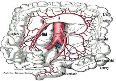
Background: To determine the incidence of the presence of a macroscopic anastomosis between the superior rectal artery and the last sigmoidal branch in a cadaveric population by means of macroscopic dissection. Also to measure the length of the inferior mesenteric artery from its origin from the abdominal aorta to Sudeck’s point and to determine the diameter of the anastomotic artery, if present. Material and Methods: Cadavers were dissected with midline vertical abdominal incision. After identifying the inferior mesenteric artery, it was ligated and divided from its origin as near as possible from the aorta. Results: Macroscopically looking for anastomosis between the superior rectal artery and the last sigmoid artery and measured its diameter. Among 40 cadavers, 34 were males and 6 were females. A macroscopic anastomosis between the superior rectal artery and the last sigmoid artery could be identified in 32 (80.0%) and was absent in 8 (20.0%). The mean length was 43.6 (±.46) mm from the origin of the inferior mesenteric artery to Sudeck’s point. The mean diameter of this anastomotic vessel was 1.9 (±0.65) mm. Conclusion: Sudeck’s point may be considered important during surgery because of the uncertainty of the presence of a macroscopic anastomosis between the superior rectal artery and the last sigmoidal artery (absent in 20.0%) and small diameter of the anastomotic vessel which fulfill the need of the caudal stump.

Keywords: Sudeck’s point, Macroscopic anastomosis, Superior rectal artery

Corresponding Author How to Cite this Article To Browse
Atul Kumar, Assistant Professor, Department of Surgery, Government Medical College, Ratlam, Madhya Pradesh, India.
Email:
Jain P, Sharma D, Somashekar U, Kumar A. Importance of critical point of Sudeck in colorectal surgery: an anatomical study in cadaver. Trauma and Orthopedics. 2020;6(4):237-243.
Available From
https://surgical.medresearch.in/index.php/ijoso/article/view/193

Manuscript Received Review Round 1 Review Round 2 Review Round 3 Accepted
2020-07-08 2020-07-21 2020-08-9 2020-08-25
Conflict of Interest Funding Ethical Approval Plagiarism X-checker Note
No Nil yes 8%

© 2020 by Jain P, Sharma D, Somashekar U, Kumar A and Published by Siddharth Health Research and Social Welfare Society. This is an Open Access article licensed under a Creative Commons Attribution 4.0 International License https://creativecommons.org/licenses/by/4.0/ unported [CC BY 4.0].

Introduction

In 1907, Sudeck [1] described a critical point at the origin of the last sigmoidal arterial branch from the inferior mesenteric artery. He concluded this point was essential to be retained during colorectal surgeries as ligations of this may lead to ischemia of the distal colon. Surgery remains the most useful method of colorectal cancer to cure the disease. To avoid early postoperative morbidity (mainly due to anastomotic leak) and achieve good long term results, low incidence of tumor recurrence and disease-free survival [2], surgeons should have in-depth knowledge of the vascular anatomy and its variation. The colonic blood supply contains various weak points known as watershed areas as Griffith’s point at the splenic flexure, Sudeck’s point at the rectosigmoid junction, and at the ileocaecal junction [3,4,5,6]. These watershed areas are more vulnerable to ischemic injury [7,8,9,10].

The classical anatomical descriptions of the blood supply to the left colon do not fully explain the occurrence, distribution, extent, and severity of many vascular disorders of the large intestine. The inferior mesenteric artery directly arises from the abdominal aorta as the last anterior aortic branch 3-4 cm above to aortic bifurcation. The inferior mesenteric artery gives rise to 3 main divisions (a) left colic artery (b) 2-3 sigmoidal artery (c) continues as the superior rectal artery. In the 1950s the increasing number of operations for aortic aneurysms and high ligations of an inferior mesenteric artery during resection for colonic carcinomas resulted in numerous instances of large bowel ischemia with gangrene, stricture, and colitis. The incidence of ischemia of colon following high ligation of the inferior mesenteric artery in surgery of cancer of colon and rectum was reported as 2.4 to 22% [11]. Ligations of the inferior mesenteric artery proximal or distal to Sudeck’s point during resection of the sigmoid colon can be crucial in post-operative blood supply to the remaining stump depending on an anastomosis between the superior rectal artery and the last sigmoid artery [12]. If the anastomosis is absent blood will have to flow up the superior rectal artery through Sudeck’s point and down the last sigmoid artery to supply the proximal part of the caudal stump when sigmoid resection is done. This is not possible if the superior rectal artery has been ligated as the part of the intestine supplied by last sigmoid branch may not fulfill the demands of the caudal stump due to insufficient blood supply in the absence of an anastomosis and

this may lead to post-operative ischemic colitis [13] and necrosis and later stricture [14]. Various authors have questioned the validity of Sudeck’s point. Some say it is only a misnomer and that it has no surgical importance [6,15]. There is also confusion regarding the presence of an anastomosis around the Sudeck’s point.

Materials and Methods

Study design and Setting: This cadaveric study was carried out in the Mortuary Department of Forensic medicine and Toxicology and Department of Surgery, NSCB Medical College and Hospital, Jabalpur, M.P. between July 2009 to October 2010.

Inclusion criteria: All available adult (above 18 years of age) cadavers in the Mortuary with the following exceptions:

  • Cadavers with any intraabdominal injury.
  • Cadavers with any gross intraabdominal pathology/ tumor.
  • Cadavers which need viscera preservation for toxicology.
  • Cadavers with previous surgery to rectosigmoid junction.

Methods: All fresh cadavers were dissected by using standard dissection methods and instruments. Cadavers were dissected with midline vertical abdominal incision from xiphoid to the pubic symphysis. After opening the anterior abdominal wall, the reflection of parietal peritoneum overlying the posterior abdominal wall was dissected to reach the inferior mesenteric artery. After identifying the inferior mesenteric artery, it was ligated and divided from its origin as near as possible from the aorta. En-bloc specimen with descending colon, sigmoid colon, and upper rectum with mesentery was dissected out along with the inferior mesenteric artery. Then the inferior mesenteric artery was dissected further to its sigmoidal branches and the superior rectal artery. Further, the last sigmoidal artery and the superior rectal artery dissected up to the gut wall. The area between the superior rectal artery and the last sigmoid artery was subsequently carefully dissected to see the presence of any anastomosis between them. The length of the inferior mesenteric artery from its origin from the abdominal aorta to the critical point of Sudeck was measured. If an anastomosis was present between the last sigmoidal artery and the superior rectal artery, its diameter was measured with the help of a


Vernier’s calipers.

Evaluation of the response to intervention:

Statistical Analysis- After getting the required information, the collected data were coded, tabulated, and analyzed P<0.05 was taken as statistically A descriptive analysis was done on all variables to obtain a frequency distribution. The mean + SD and ranges were calculated for quantitative variables.

Results

This cadaveric study was conducted in the Mortuary, NSCB Medical College, Jabalpur between July 2009

– Oct 2010. During this study, 41 fresh cadavers were dissected. One cadaver was found to have a colonic mass and was excluded from the study.

Macroscopically looking for anastomosis between the superior rectal artery and the last sigmoid artery and measured its diameter. Among 40 cadavers, 34 were males and 6 were females. A macroscopic anastomosis between the superior rectal artery and the last sigmoid artery could be identified in 32 (80.0%) and was absent in 8 (20.0%). The mean length was 43.6 (±.46) mm from the origin of the inferior mesenteric artery to Sudeck’s point. The mean diameter of this anastomotic vessel was 1.9 (±0.65) mm.


Table-1: Cadaveric study of the 40 samples.

S. No. Length of IMA*(mm) The diameter of the anastomotic vessel (mm)
1 45 2
2 52 3
3 60 2.5
4 47 3
5 49 2
6 54 2.5
7 55 1.5
8 45 2
9 50 3
10 42 2.5
11 40 Absent
12 35 1.5
13 40 2
14 35 2.5
15 37 1
16 42 1.5
17 34 2
18 38 0.5
19 42 1.5
20 40 Absent
21 44 Absent
22 40 1.5
23 40 1.5
24 41 1
25 45 2.5
26 38 Absent
27 42 2
28 44 0.5
29 40 Absent
30 46 Absent
31 42 2
32 48 1.5
33 42 1.5

34 44 2
35 45 Absent
36 42 2
37 44 1.5
38 44 2.5
39 48 Absent
40 42 1.5
Sum 1743 60
Average 43.575 1.875
Std Dev 5.462635318 0.6475
*Length of the inferior mesenteric artery from its origin of the aorta to the critical point of Sudeck

surgical_193_01.jpg

Fig-1: Graphical representation of Sudeck’s point.

Discussion

Sudeck, in 1907 describes a critical point in the superior rectal artery after observing gangrene of the upper rectum in two patients whose superior rectal artery has been ligated just distal to the last sigmoidal artery branch of an inferior mesenteric artery during conservative operation for cancer rectum pull-through procedures. He later did injection experiments by injecting a radio-opaque dye into relevant parts and subsequently taking radiographs to search for anastomosis between the last sigmoid artery and the superior rectal artery if any [1].

He found such a vessel lacking in most cases. After ligation of the superior rectal artery below Sudeck point, there are few rectal vessels filled. Later on, Hartmann corroborated the Sudeck’s results.

Later many other authors [16-18] questioned the existence of a critical point of Sudeck, its validity, and importance during surgery. They averred that the techniques of Sudeck and Drummond were erroneous and open to misinterpretation because

they did not take into account [6]:

  • The viscosity of the injectate,
  • The adequacy of arterial filling in the postmortem state,
  • A confirmation of radiographic findings by surgical dissection.

Later Drummond [19] studying sigmoid vessels by injecting dye and taking roentgenograms found the same results as Sudeck. He affirmed that the Sudeck’s point should be considered attentively by surgeons during resections of the sigmoid colon.

To rectify these errors, Griffith [6] injected barium and gelatine mixture under pressure and took radiographs followed by dissection of the en bloc specimen. He found that there was an adequate anastomosis between the last sigmoidal artery and the superior rectal artery. He, therefore, stated that the conclusions of Sudeck and Drummond were unreliable and misleading.

Many studies [20,21] found that there were many anatomical variations in the inferior mesenteric artery and sigmoidal arteries. Sutherland [22] found the number of sigmoidal arteries varies from one to seven in number and they did not form any definite or constant pattern. All these authors used an injectate nearly equaling the viscosity of blood and used high pressure for injecting radio-opaque material followed by dissection of a specimen. One important point to be considered in the injection of radioopaque substance is the incomplete filling of the anastomotic vessel many times [5,23]. There are many studies that confirm the presence of an anastomosis between the last sigmoidal artery and the superior rectal artery. Greenberg [24] found an anastomosis in 38 of 71 specimens (51.4%), Miechel et al [3] found a definite anastomosis in 39 of 75 specimens (52%). VonTonder,et al [12] 2007 identified an anastomosis in 61 of 64 cadavers


(95.3%). Michel et al [3] ligated10 random specimen of 75, distal to Sudeck’s point, and injected a dye into the aorta. He demonstrated that the dye reached the superior rectal artery in all 10 specimens and therefore concluded that an anatomical bypass does co-exist. Autopsy specimens have completely relaxed arteries subjected to higher internal pressure than normal and may give erroneous data at variance with the true condition during life. One important other factors which highlighted by Michels [3] was the presence of shunt not demonstrated by roentgenogram techniques.

Some studies [25-27] have suggested that ischemic colitis is more likely to occur in the watershed area of the colon. Longo et al [13] reported in a retrospective study, a rectosigmoid region involved in 40% of 47 patients with non-occlusive ischemia. The diagnosis was made during surgery, by endoscopy or barium enema. Yamakazi et al [28] reported cases in which ischemic colitis (developed in the watershed area of the colon) resulting from incomplete anastomosis of marginal arteries. They proposed Griffith’s point and Sudeck’s point as high-risk areas for ischemic colitis.

Yamazaki et al [14,28] reported a case of sigmoidectomy for the distal colon carcinoma who developed ischemic stricture, distal to the anastomosis. In this sigmoidectomy division of the superior rectal artery was done immediately distal to Sudeck’s point. This case highlighted the validity of Sudeck’s view. A recent study by Von Tonder et al [12] found a macroscopic anastomosis between the last sigmoidal artery and the superior rectal artery in 61 of 64 cadavers (95.3%) by mean of standard dissection methods. This vessel had a mean diameter of 1.9 mm which may not be sufficient to supply the proximal part of the distal stump in colorectal surgeries. On the other hand, such an artery may dilate acutely at the time of ligation or adapt overtime to carry increase load and demand.

Allison et al [26] studied anastomotic leak in low anterior resection (colorectal resection) with an angiographic evaluation of small arteries and their collaterals. They suggested that disruption of small arterial collateral during colorectal resection might be implicated for an anastomotic leak as these small arteries are not capable of developing further collaterals and may not be able to meet the demands of the proximal part of the caudal stump. In the present study, a macroscopic anastomosis was present in 32 of 40 cadavers (80.0%). These

findings are comparable with findings of Von Tonder et al [12] (95.3%), Griffith [6,15] (48%), Greenberg [24] (51%). Due to the uncertainty of anastomosis (absent in 20.0%), surgeons should pay attention to retaining Sudeck’s point during colorectal surgery. Another point of consideration is the small diameter of the anastomotic vessel found in the present study with a mean diameter of 1.9mm, which is similar to that found by Von Tonder et al [12] in their study.

This small-diameter may not be sufficient enough to fulfill the requirements of the proximal part of the caudal stump. This along with the extent of submucosal plexus anastomosis supplying caudal stump needs to be investigated. Griffith [15] and Michels et al [4] have described 3 plexuses in the wall of the colon. The largest of which is rich submucosal plexus. A study [26] observed small arterial and collateral in the colorectal region and found disruption of these arterioles in an anastomotic leak in low anterior resection.

Various factors affect the measured size of the anastomotic vessel. Cadaveric studies have disadvantages of slight shrinkage and deformation. Cadaveric specimens have completely relaxed arteries and the arterial lumen is collapsed so the diameter measurement is the diameter of a collapsed artery. Sometimes small vessels are difficult to dissect and may not easily demonstrable. Therefore measurement reports might defer somewhat from the in vivo diameter.

Further studies from in vivo samples as well as the relationship between vessel diameter and its capability to meet the needs of the area supplied, need to perform. Postoperative colitis ischemia necrosis and stricture can be avoided by ligation proximal to Sudeck’s point, especially where the anastomosis is absent or insufficient. The length of the inferior mesenteric artery from its origin at aorta to Sudeck’s point was found to be a mean of 43.6mm. This length is sufficient for ligation of an inferior mesenteric artery proximal to Sudeck’s point without harming any autonomic nervous plexus running on the great vessel regardless of the presence of anastomosis.

Limitation

  1. Small sample size
  2. Chances of bias
  3. Single-center trial

Conclusion

During colorectal surgery length of the inferior mesenteric artery from its origin at the abdominal aorta to Sudeck’s point is sufficiently long for ligation proximal to Sudeck’s point especially when anastomosis between the superior rectal artery and the last sigmoidal branch might be absent. The diameter of the anastomotic vessel between the last sigmoidal artery and the superior rectal artery may be insufficient to carry blood to meet the demand of the caudal stump after colorectal anastomosis. Hence it can be concluded that Sudeck’s point may be considered important during surgery because of the uncertainty of the presence of a macroscopic anastomosis between the superior rectal artery and the last sigmoidal artery (absent in 20.0%) and small diameter of the anastomotic vessel which fulfill the need of the caudal stump.

What does the study add to the existing knowledge

During surgery, an effort should be made to identify the presence of an anastomosis between the last sigmoid artery and the superior rectal artery and to retain the Sudeck’s point. It becomes more important when an anastomosis is absent or is considered insufficient to meet the demand of the caudal stump. The diameter of the anastomotic vessel between the last sigmoidal artery and the superior rectal artery may be insufficient to carry blood to meet the demand of the caudal stump after colorectal anastomosis.

Author’s contribution

Dr. Pramesh Jain: Concept, data collection and discussion

Dr. Prof. Dhananjaya Sharma: Concept and guidance

Dr. Uday Somashekar: Concept and discussion

Dr. Atul Kumar: Discussion and data collection

Reference

  1. Michels NA, Siddharth P, Kornblith PL, Parke WW. Routes of collateral circulation of the gastrointestinal tract as ascertained in a dissection of 500 bodies. Int Surg. 1968;49(1)8-28.
  1. Michels NA, Siddharth P, Kornblith PL, Parke WW. The variant blood supply to the descending colon, rectosigmoid and rectum based on 400 dissections, Its importance in regional resections- a review of medical literature. Dis Colon Rectum. 1965;49;251-278.
  2. Sakorapas GH, Zouros E, Peros G. Applied vascular anatomy of the colon and rectum- Clinical implication for surgical oncologist. Surg Onco. 2006;15(4)243-255.
  3. Sudeck P. U¨ber die gefa¨ssversorgung des mastdarmes in hinsicht auf die operative gangr¨an. Mu¨nchen Med Wchnschr. 1907;54;13-14.
  4. Griffiths JD. Surgical anatomy of the blood supply of the distal colon. Ann R Coll Surg Engl 1956;19(4)241-256.
  5. Bower TC. Ischemic colitis. Surg Clin North Am. 1993;73;1037-1053.
  6. Landreneau RJ, Fry WJ. The right colon as a target organ of nonocclusive mesenteric ischemia. Arch Surg. 1990;125(5)591-594.
  7. Farman J. Ischemic colitis. Abdom Imaging. 1995;20;85-86.
  8. Shoji BT, Becker JM. Colorectal disease in the elderly patient. Surg Clin North Am. 1994;74;293-316.
  9. Meyers MA. Griffith’s point- Critical anastomosis at the splenic flexure. AJR Am J Roentgenol. 1976;126(1)77-91.
  10. Tonder JJV, Boon JM, Becker JH, Schoor ANV. Anatomical consideration on Sudeck’s critical point and its relevance to colorectal surgery. Clin Anat. 2007;20(4)424-427.
  11. Bacon HE, Smith CH. The arterial supply of the distal colon pertinent to abdominoperineal proctosigmoi- dectomy, with preservation of the sphincter mechanism. Ann Surg. 1948; 127(1)28-33.

  1. Longo WE, Ballantyne GH, Gusberg RJ. Ischemic colitis- patterns and prognosis. Dis Colon Rectum. 1992; 35(8)726-730.
  2. Griffiths JD. Extramural and intramural blood-supply of colon. Br Med J. 1961;1(5222)323-326.
  3. Yamazaki T, Shirai Y, Sakai Y, Hatakeyama K. Ischemic stricture of the rectosigmoid colon caused by division of the superior rectal artery below Sudeck’s point during sigmoidectomy- report of a case. Surg Today. 1997;27(3)254-256.
  4. Hartmann H. Some considerations upon high amputation of the rectum. Ann Surg. 1909;50(6)1091-1094.
  5. Rubesch R. Uber die vermeidung der darmgangran bie rectumoperationen. Beitr Z Klin Chir Tubing. 1910;67;480-493.
  6. Drummond H. The arterial supply of the rectum and pelvic colon. Br J Surg. 1914;1(4)677.
  7. Steward JA, Rankin FW. Blood supply of the large intestine- it’s surgical considerations. Arch Surg. 1933;26(5)843-891.
  8. Dixon CF. Anterior resection for malignant lesions of the upper part of the rectum and lower part of the sigmoid. Ann Surg. 1948;128(3)424-429.
  9. Pope CE, Judd ES. The arterial blood supply of the sigmoid, rectosigmoid and rectum. Surg Clin North Am. 1929; 9;957.
  1. Greenberg MW. Blood supply of the rectosigmoid and rectum. Ann Surg. 1950;131(1)100-108.
  2. Patrício J, Bernades A , Nuno D, Falcão F, Silveira L. Surgical anatomy of the arterial blood-supply of the human rectum. Surg Radiol Anat. 1988;10(1)71-75.
  3. Yamazaki T, Shirai Y, Tada T, Sasaki M, Sakai Y, Hatakeyama K. Ischemic colitis arising in watershed areas of the colonic blood supply- a report of two cases. Surg Today. 1997;27(5)460-462.
  4. Guttormson NL, Bubrick MP. Mortality from ischemic colitis. Dis Colon Rectum. 1989;32(6)469–472.
  5. Zuloaga J, Arias-Diaz J, Fernandez-Diaz S, Balibrea JM, Balibrea JL. Late ischemic stricture following anterior rectal resection. Rev Esp Enferm Dig. 2008;100(1)49-52.
  6. Goligher JC. The blood-supply to the sigmoid colon and rectum with reference to the technique of rectal resection with restoration of continuity. Br J Surg. 1949;37(146)157-162.
  7. Sunderland S. Blood supply of the distal colon. ANZ Jour Surg. 1942;11(4)253-263.
  8. Allison AS, Bloor C, Faux W, Arumugam P, Widdison A, Lloyd-Davies E, et al. The angiographic anatomy of the small arteries and their collaterals in colorectal resections- some insights into anastomotic perfusion. Ann Surg. 2010;251(6)1092-1097.