E-ISSN:2455-5436
P-ISSN:2456-9518
RNI:MPENG/2017/70870

Research Article

Morbidity

Surgical Review: International Journal of Surgery, Trauma and Orthopedics

2020 Volume 6 Number 3 May-June
Publisherwww.medresearch.in

Study of clinical presentation and management of intestinal obstruction and its evaluation with respect to morbidity and mortality

Nutan K.1*, Charokar K.2, Bharang K.3
DOI: https://doi.org/10.17511/ijoso.2020.i03.05

1* Kumari Nutan, PG Resident, Peoples College of Medical Sciences and Research Centre, Bhopal, Madhya Pradesh, India.

2 Kailash Charokar, Associate Professor, Department of Surgery, Peoples College of Medical Sciences and Research Centre, Bhopal, Madhya Pradesh, India. https://orcid.org/0000-0002-0540-6726

3 Krishna Bharang, Associate Professor, Department of Surgery, Peoples College of Medical Sciences and Research Centre, Bhopal, Madhya Pradesh, India.

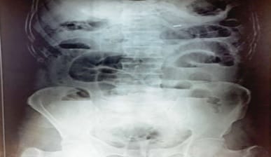
Introduction: Intestinal obstruction is one of the common surgical emergencies. Often the patients presenting with intestinal obstruction may be extremely ill and require prompt evaluation and definitive treatment. Aim: The aim of this study was to evaluate the clinical pattern and surgical management outcome of Intestinal Obstruction.Material and Methods: This prospective descriptive study was conducted at Peoples College of Medical Sciences and Research Centre and Hospital during the period from December 2017 to April 2019. The patients presenting with clinical features of acute intestinal obstruction were included in the study after their informed consent. Patients below the age of 14 years were excluded. A predesigned and validated proforma was used to record data. Results: A total of 50 patients were included in the study.The mean age of patients was 35.85±10.99 years. The pattern of intestinal obstruction observed in patients was small bowel obstruction, followed by a large bowel obstruction, and the remaining with features of both. The common causes of intestinal obstruction were adhesion and bands, followed by tuberculosis. Adhesiolysis and release of bands were performed in 41%, resection, and anastomosis in 49%. Wound infection was the most common complication observed. A mortality rate of 4% was observed. Conclusion: Small bowel obstruction was the common type of intestinal obstruction. Prompt diagnosis and early surgical treatment after resuscitation is key to favourable treatment outcomes.

Keywords: Intestinal obstruction, Adhesions, Stricture, Emergency laparotomy, Resection-anastomosis

Corresponding Author How to Cite this Article To Browse
Kumari Nutan, PG Resident, Peoples College of Medical Sciences and Research Centre, Bhopal, Madhya Pradesh, India.
Email:
Kumari N, Charokar K, Bharang K. Study of clinical presentation and management of intestinal obstruction and its evaluation with respect to morbidity and mortality. Surgical Review Int J Surg Trauma Orthoped. 2020;6(3):166-172.
Available From
https://surgical.medresearch.in/index.php/ijoso/article/view/188

Manuscript Received Review Round 1 Review Round 2 Review Round 3 Accepted
10-06-2020 20-06-2020 25-06-2020 29-06-2020
Conflict of Interest Funding Ethical Approval Plagiarism X-checker Note
No Nil Yes 17%

© 2020 by Kumari Nutan, Kailash Charokar, Krishna Bharang and Published by Siddharth Health Research and Social Welfare Society. This is an Open Access article licensed under a Creative Commons Attribution 4.0 International License https://creativecommons.org/licenses/by/4.0/ unported [CC BY 4.0].

Introduction

Intestinal obstruction is a common surgical emergency. It can occur at any age and accounts for a significant number of emergency surgical admissions. Patients presenting with intestinal obstruction may be often extremely ill and require prompt assessment and resuscitation, with intensive monitoring of their vital signs and clinical progress. The disease carries significant morbidity and mortality.

Intestinal obstruction is usually a mechanical obstruction of the bowel, which may be either partial or complete, leading to proximal distension of the bowel with stasis of fluid and gaseous contents. The intestine responds to obstruction by increasingly forcefully prolonged contractions, and in the lumen of the intestine proximal to obstruction, the secretions accumulate with gases leading to progressive distension of the bowel. The intestinal contents are not absorbed and there is a net secretion of extracellular fluid into the intestinal lumen, leading to significant loss of body fluids.

Enteric bacteria proliferate in the stagnant fluid, with increasing translocation of bacteria and toxins through the bowel wall into the peritoneal cavity with exudates. In cases of closed-loop obstruction, as seen in obstructed hernia, volvulus, or when the base of loop is snared by adhesions, strangulation develops leading to grave systemic complications due to bowel ischemia, infarction, and gangrene. Mechanical intestinal obstruction accounts for 5-15% cases of severe abdominal pain of sudden onset requiring hospitalization[1,2].

Adynamic bowel obstruction (colonic pseudo-obstruction) is usually seen in hospitalized or immobile patients who have non-intestinal conditions (chest infection, hypothyroidism, orthopedics operations, retroperitoneal hematoma or inflammation, and diabetes mellitus). Metabolic problems like hypokalaemia, uremia, and tricyclic antidepressants predispose to this condition. It is clinically indistinguishable from mechanical colonic obstruction.

Clinical features of intestinal obstruction are varying degrees of intestinal colic, vomiting, constipation, and abdominal distension, depending on the etiology, level of obstruction in the gastrointestinal tract, and the duration of the pathological process. Clinically, intestinal obstructions are classified as high or proximal small bowel, distal small bowel,

and large bowel obstruction.

Intestinal obstruction is a symptom complex of a disease with diverse aetiologies of wide geographical variations worldwide[3]. Considering the etiological causes and controlling the risk factors for bowel obstruction is important in decreasing morbidity and mortality. The present study was undertaken with a view to describe the clinicopathological spectrum of intestinal obstruction, its management, and the treatment outcomes. The study aims to share our experience, directed towards meeting the diagnostic and treatment challenges posed by complex clinical situations arising in these patients.

Methods

Settings: This study was conducted in the Department of Surgery at the People’s College of Medical Sciences and Research Centre, Bhopal.

Duration and type: Between December 2017 to April 2019. This is was a prospective observational study.

Sampling methods:Non-probability criteria sampling. A total of 50 patients were included in the study after the informed written consent.

Inclusion criteria: Patients above 14 years of age, diagnosed with intestinal obstruction were included in the study.

Exclusion criteria: The cases of acute abdomen having perforation peritonitis, pancreatitis, enterocolitis, adynamic ileus, and ascites were excluded. All those who did not consent were excluded from the study.

Data Collection Procedure: Data was recorded and collected in predesigned validated proforma by the principal researcher.

Surgical procedure: The treatment strategy was executed were conservative management to operative emergency laparotomy. Patients with normal hemodynamic status, mild abdominal signs, and with few air-fluid levels were subjected to conservative treatment. The patients were evaluated on 8 hourly bases to monitor the clinical progression. Based on the progression of symptoms and signs the treatment plan was tailored. A serial X-ray abdomen if clinically indicated to monitor the progression or regression of the air fluids. Those patients who responded favorably within 24 to 48 hours were managed conservatively and were


subjected to second-line investigations to find the final cause of obstruction. Patients with the frank presentation of acute intestinal obstruction were optimized and operated in Emergency for Exploratory Laparotomy. Operative treatment: The type of operative procedure performed was selected based on surgical principles to match the operative findings of the case.

Adhesiolysis with the release of bands, resection-anastomosis, release of obstruction with Hernia repair, and temporary intestinal diversion was performed. The site of obstruction, extent, degree of distension, type of peritoneal fluid, presence of ischemic changes, or any other significant findings recorded in the operative findings.Postoperatively patients were managed on nasogastric suction, intravenous fluids, adequate electrolytes supplements, appropriate antibiotics, analgesics, and parenteral nutrition wherever indicated. Complications if any were detected early and managed accordingly.

Ethical considerations and permission: The approval of the Institutional research advisory committee and the institutional ethical Committee was obtained at People’s College of Medical Sciences and Research Centre, Bhopal for conducting this research in the institute.

Statistical Analysis: Simple descriptive statistics with mean, mode, median, range, and percentage were used for analysis. Data was compiled using MS Excel and analyzed using SPSS software version 20. A P-value of less than 0.5 was considered significant and a p-value of less than 0.01 was considered highly significant.

Results

In the present study, the majority of patients belonged to 31 to 40 years of age (32%) followed by 21 to 30 (30%). The mean age of patients was 35.85±10.99 years. The majority of patients with intestinal obstruction were male (82%).

Table-1:Socio-demographic profile of the study group (N=50).

Patients parameters Number of Patients Percentage (%)
Age (in years)
< or equal to 20 3 6
21-30 15 30
31-40 16 32
41-50 12 24
>50 4 8
Total 50 1
Gender
Male 41 82
Female 9 18

Abdomen pain was presenting feature in all the patients, about 88% had symptoms of distension, 84% had vomiting and 80% had constipation. Tenderness in the abdomen was present in 98% of the patients, guarding in 28% and rigidity in 18%.

The pattern of intestinal obstruction observed in patients was small bowel obstruction (SBO) 60%, large bowel obstruction (LBO) 20%, and the remaining 20% features of both.

The scout films of the patients revealed multiple air-fluid levels in the small bowel (60%), large bowel (20%), and both small and large bowel (20%). The number of the air-fluid level was 4 in the majority of patients (52%).The ultrasonography revealed dilated bowel loops in 72% of the patients, and in 14% interbowel fluid was visualized sonologically.

Additionally, in 10% of the cases, intraperitoneal mas was visualized. The majority of patients 37(74%) were treated surgically (Table 4) and the remaining 13 (26%) were treated conservatively.

Table-2: Etiology of intestinal obstruction (N=37).

Age groups Cause Frequency (%)
< or equal to 20 Intussusception 1 2.7
21-30 Adhesions and Bands 18 48.6
31-50 Stricture 4 10.8
Tuberculosis 5 13.5
Obstructed hernia 1 2.7
>50 Volvulus 3 8.1
Carcinoma colon 3 8.1
Foreign body 2 5.4

Table-3: Surgical procedures performed (N=37).

Operative procedure performed Frequency (%)
Adhesiolysis and release of bands 15 41
Resection and anastomosis 18 49
Foreign body removal with primary repair 2 5
Resection with stoma 2 5

The mean length of hospital stay was 10.6 ± 4.7 days (Table 8.). 10% of the patients had less than 3 days of hospital stay, 18% had between 4-7 days, 48% had between 8-14 days, and 24% had more than 14 days. Those patients who developed postoperative complications stayed longer in the hospital.


Table-4: Post-operative complications (N = 37).

Complications Number of cases Percentage (%)
Wound infection(SSI) 6 16.6
Systemic Sepsis 4 10
Pneumonia 3 8
Wound Dehiscence 1 3
No complications 21 57
*Death 2 4

A total of 14 postoperative complications were recorded in the study group in Table 9. Surgical site infection was the most common complication accounting for 16.6% of cases. In the present study, there were two deaths.

surgery_188_01.jpg

Fig-1: Multiple small bowel air-fluid levels.

surgery_188_02.jpg

Fig-2: Compound volvulus.

surgery_188_03.jpg

Fig-3: Compound volvulus.

Discussion

In the present study, the majority of patients belonged to 31 to 40 years of age (32%) followed by 21 to 30 (30%). The mean age of patients was 35.85±10.99 years. The majority of patients with intestinal obstruction were male (82%). Bankole AO et al (2017) in their study included patients with a relatively higher mean age of 45.6 ± 14.8 years as compared to the present study. The male preponderance in the present study was similar to the reference study [4]. Moghadam AG et al (2017), in their retrospective study also observed male preponderance (65%) as compared to females(35%) [5].Findings of the present study were also supported by Ahmad MS et al (2015) in which the author reported a preponderance of males in 68.75%[6].

Symptoms of intestinal obstruction usually depend upon the level of obstruction. However predominant symptom in the present study was pain observed in all the patients followed by abdominal distension (88%), vomiting (84%) and constipation (80%). These findings were similar to study by Bankole AO et al (2017) in which the author documented pain in 99% patients followed by absolute constipation in 97.1% and abdominal distension in 87.6% patients [4]. Our findings were also similar to the findings of Murat K et al (2012) [7] and Kahn TS et al (2013) [8] in which pain was observed in 100% and 98.6% patients respectively.

In the present study, the most common sign associated with intestinal obstruction was abdominal tenderness (98%) followed by distension (96%), guarding (28%), and rigidity (18%). However, the most common sign was abdominal distension reported by Bankole AO et al (2017) [4] and Kuremu RT et al (2006) [9]. Bankole AO et al (2017) [4] reported cardiovascular disease (hypertension) as the most common co-morbidity. Murat K et al (2012) also reported cardiovascular disease as the most common co-morbidity in their patients [7]. However, the most common comorbidity in the present study was diabetes followed by hypertension and TB.

In the present study, the X-ray abdomen was suggestive of air-fluid levels in all the patients, however, the number of the air-fluid level was 4 in the majority of patients (52%). These findings were similar to study by Priscilla SB et al (2017) [10] and Malik AM et al (2010) [3] in which most common radiological feature was multiple air-fluid levels


followed by dilated bowel loops. However, the diagnostic accuracy and specificity of abdominal radiography are low, but they are the predominant investigations in low resource settings and greatly contribute to the initial diagnostic and therapeutic decision making. The most common findings are dilated bowel loops and the presence of more than two air-fluid levels [11,12]. Another investigation for the diagnosis of intestinal obstruction includes abdomen sonography. USG findings revealed dilated bowel loops with or without inter-bowel fluid.

The most common type of obstruction in the present study was SBO (60%) followed by LBO in 20% of patients, and the remaining 20% had features of combined presentation. These findings were supported by Moghadam AG et al (2017), in which the most common type of bowel obstruction was small bowel (66.7%) [5]. Vincenzo Neri et al (2016) also observed acute mechanical small bowel obstruction as the most frequent (71.2%) type of bowel obstruction[15]. The most common causes of intestinal obstruction in the present study were adhesion and bands(48.6%), followed by tuberculosis(13.5%), stricture (10.8%), volvulus(8.1%), foreign body(5.4%) and carcinoma colon(8.1%) respectively.

This reflects the change in the spectrum of causes of intestinal obstruction and the fact that adhesions are an emerging surgical problem as the most common cause of intestinal obstruction even in developing countries. These causes were similar to the study by Bankole AO et al (2017), in which the causes of mechanical intestinal obstruction(MIO) were adhesions 48.5%, colorectal tumors 25.7%, external hernias 15.2%, volvulus 5.7%, intussusception 1.9%, granulomatous infection 1.9% and internal hernia 0.9% [4]. In the study of Nasiruddin S et al (2019) done on 50 cases of acute intestinal obstruction, 36% of the cases were due to post-operative adhesions who have undergone previous surgeries [13]. Gogineni JC et al (2020) observed the most common cause of intestinal obstruction requiring surgical intervention was obstruction due to nontuberculous strictures, closely followed by adhesions [14].

The present study findings were also supported by Vincenzo Neri et al (2016) in which they reported adhesion - relate obstructions as the most common cause of obstructions [15]. Lawal OO et al (2005) also reported adhesions as the most common cause accounting for 44.4% of cases [16]. Our findings were contrasting to findings of Okeny PK et al

(2011) [17] and Ohene-Yeboah et al (2006) [18], in which the authors documented hernia as the most common cause of MIO. The observed difference in the present study and reference studies could be explained by the fact that these studies were conducted more than 10 years ago, and etiology have greatly changed from hernia to adhesions in recent years.

Management of intestinal obstruction is directed at correcting physiologic derangements caused by the obstruction, bowel rest, and removing the source of obstruction [3]. The majority of patients in the present study required surgery (74%). Of the 37 patients operated in the present study, adhesiolysis with the release of bands and resection- anastomosis was the commonly performed surgical procedure in 41% and 49% patients respectively. These findings were in line with the study by Bankole AO et al (2017) in which 80% of patients had an operative intervention and 20% had conservative management [7].

Vincenzo Neri et al (2016) performed surgery in 100% cases of acute intestinal obstruction. The choice of surgery was based on intraoperative pathological findings. They also performed adhesiolysis (34.6%) followed by small bowel resection (11.5%) based upon underlying etiology [15]. The present study findings were also supported by the study of Moghadam AG et al (2017), where the most common type of surgery was adhesiolysis (33.3%) followed by Resection and ostomy (16.6%) [5].

In the present study, wound infection was the most common complication observed in 16.6% of patients followed by sepsis in 10% and Pneumonia in 8% respectively. These findings were similar to the study by Moghadam AG et al (2017), the most common postoperative complication was wound infection which constituted 18 (30%) [5]. The most common postoperative complication was surgical site infection, affecting 39.3% of operated cases in a study by Bankole AO et al (2017) [4]. Kahn TS et al (2013) also reported wound-related sepsis as the most common complication[8].

The major predictors of treatment outcomes of intestinal obstruction were: duration of illness before surgical intervention, intra-operative findings, completion of Intra-operative procedures (bowel resection and anastomosis, and length of hospital stay [19]. In the present study, death was observed in 2 cases where the associated complica-


tions were sepsis along with bilateral Pneumonia. The test of significance observed a highly significant statistical association between outcome and complications (p<0.01). These findings were supported by Bankole AO et al (2017), [4] The most common cause of death was related to sepsis, which accounted for 66.7% of the mortality, similarly, Kahn TS et al (2013) also reported sepsis as the most common cause of death [8].

Perioperative mortality was significantly higher in the elderly group, due to the major incidence of cardiovascular and respiratory fatal events directly related to pre-existing comorbidities [19]. In the present study, the mortality rate was observed in 4%. The mortality rate recorded in the present study was much lower as compared to 14.3% that was reported by Bankole AO et al (2017). [4] The mortality rate reported by Moghadam AG et al (2017) was 9.2% [5]. The mortality rate in the present study was higher as compared to 2.5% reported in the study by Soressa U et al (2016) [20].

Lengths of stay in the hospital were dependent upon the severity of disease and treatment modality required for intestinal obstruction. Mean Length of stay in hospital in the present study was 10.66±4.76 days. Length of stay was 8 to 14 days for 48% of patients followed by more than 14 days for 24% of patients. The length of stay documented in the present study was relatively short as compared to the study by Moghadam AG et al (2017), in which the overall median length of hospital stay was 26 days (range 1 to 72days). The longer hospital stays in the reference study could be due to a higher incidence of complications i.e. patients who had postoperative complications stayed longer in the hospital [5].

Limitations

Small sample size. Non-probability criteria sampling has limitations for the generalizability of the study findings.

Conclusion

Small bowel obstruction is the common type of intestinal obstruction with a male preponderance. Clinically stable patients should be treated initially on conservative management with continuous clinical monitoring for the resolution of obstruction. They should be subjected to diagnostic evaluation to diagnose the final cause of obstruction. Clinically

unstable patients with acute intestinal obstruction and those who do not resolve on conservative therapy within 48 hours should be treated with emergency surgery. The decrease in the incidence of obstructed hernia and an increase in the adhesive obstruction is a change in the pattern of intestinal obstruction. Abdominal tuberculosis is emerging as another common cause of acute bowel obstruction.

What does the study add to the existing knowledge?

The current study adds to the previous studies and concludes that prompt diagnosis and early surgical treatment after resuscitation is key to favorable treatment outcomes.

Author’s contributions

Dr.Kumari Nutan, Dr.Kailash Charokar, and Dr.Krishna Bharang designed the present study. Dr.Kumari Nutan reviewed literature, collected all the data, performed the statistical analysis, and drafted the manuscript. All the authors have reviewed the manuscript and edited it. All the authors have read and approved the final manuscript.

Reference

  1. Gore RM, Silvers RI, Thakrar KH, Wenzke DR, Mehta UK, Newmark GM, et al. Bowel Obstruction. Radiol Clin North Am. 2015;53(6)1225-1240.
  2. Fitzgerald J, Edward F. Small Bowel Obstruction. Emerg Surg. 2010;74-79.
  3. Malik AM, Shah M, Pathan R, Sufi K. Pattern of Acute Intestinal Obstruction- Is There a Change in the Underlying Etiology?. Saudi J Gastroenterol. 2010;16(4)272-274.
  4. Bankole AO, Osinowo AO, Adesanya AA. Predictive factors of management outcome in adult patients with mechanical intestinal obstruction. Niger Postgrad Med J. 2017;24(4)217-23

  1. Moghadam AG, Harooni SA. Pattern of dynamic intestinal obstruction in adults. IAIM. 2017;4(10)230-235.
  2. Ahmad MS, Shadab M, Omar S, Mallik AZ, Ansari MA. Causes, evaluation, and surgical management of mechanical small bowel obstruction. Int Surg J. 2015;2(4)492-495.
  3. Murat K, Onder A, Polat S, Aliosmanoglu I, Arikanoglu Z, Taskesen F, et al. Mechanical bowel obstruction and related risk factors on morbidity and mortality. J Curr Surg. 2012;2;55-61.
  4. Kahn TS, Wani ML, Wani SN, Kenu BA, Misgar AS, Fazili A, et al. Clinicopathological profile and management of acute mechanical small bowel obstruction- A prospective study. Arch Clin Exp Surg. 2013;2(3)154-160.
  5. Kuremu RT, Jumbi G. Adhesive intestinal obstruction. East Afr Med J. 2006;83(6)333-336.
  6. Priscilla SB, Edwin IA, Kumar K, Gobinath M, Arvindraj VM, Anandan H. A Clinical Study on Acute Intestinal Obstruction. Int J Sci Study. 2017;5(2)107-110.
  7. Thompson WM, Kilani RK, Smith BB, Thomas J, Jaffe TA, Delong DM, et al. Accuracy of abdominal radiography in acute small-bowel obstruction- does reviewer experience matter?. AJR Am J Roentgenol. 2007;188(3)W233-W238.
  8. Maconi G, Porro GB. Ultrasound of the gastrointestinal tract. Intestinal obstruction. 2007;27–34.
  9. Nasiruddin S, Patil S, Pinate AR. A clinical study of aetiology of acute intestinal obstruction. Int Surg J. 2019;6(3)783-787.
  1. Gogineni JC, Tonape T, Athavale VS, Kumar S, Kutty SA, Sri Likhita K. A clinical study on the surgical management of intestinal obstruction. Int Surg J. 2020;7(1)150-6.
  2. doi: https://doi. org/10. 18203/2349-2902. isj20195961.
  3. Neri V. Management of Intestinal Obstruction. Actual Problems of Emergency Abdominal Surgery. 2006;13-36.
  4. Laval OO, Olayinka OS, Bankole JO. Spectrum of causes of intestinal obstruction in adult Nigerian patients. S Afr J Surg. 2005;43(2)34-36.
  5. Okeny PK, Hwang TG, Ogwang DM. Acute bowel obstruction in a rural hospital in Northern Uganda. East Cent Afr J Surg. 2011;16(1)65-70.
  6. Ohene-Yeboah M, Adippah E, Gyasi-Sarpong K. Acute intestinal obstruction in adults in Kumasi, Ghana. Ghana Med J. 2006;40(2)50-54.
  7. Oldani A, Gentile V, Magaton C, Calabrò M, Maroso F, Ravizzini L, et al. Emergency surgery for bowel obstruction in extremely aged patients. Minerva Chir. 2020;75(1)11-14.
  8. Soressa U, Mamo A, Hiko D, Fentahun N. Prevalence, causes and management outcome of intestinal obstruction in Adama Hospital, Ethiopia. BMC Surg. 2016;16(1)38.