E-ISSN:2455-5436
P-ISSN:2456-9518
RNI:MPENG/2017/70870

Research Article

Fracture

Surgical Review: International Journal of Surgery, Trauma and Orthopedics

2020 Volume 6 Number 2 March-April
Publisherwww.medresearch.in

A study of functional outcome of bimalleolar fracture treated with fully threaded 4 mm cancellous cannulated screw in medial malleolus fracture

P Vala G.1, Patel N.2*, Vora J.3
DOI: https://doi.org/10.17511/ijoso.2020.i02.02

1 Gaurav P Vala, Assistant Professor, CU Shah Medical College and Hospital, Surendranagar, Gujarat, India.

2* Neel Patel, Resident, CU Shah Medical College and Hospital, Surendranagar, Gujarat, India. https://orcid.org/0000-0002-2570-7155

3 Jinesh Vora, Associate Professor, CU Shah Medical College and Hospital, Surendranagar, Gujarat, India.

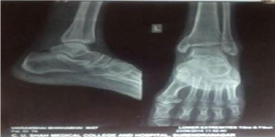
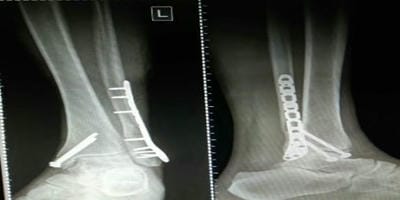
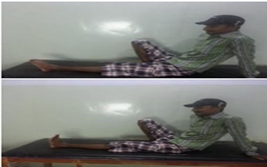
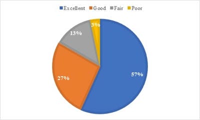
Aim: To analyse the functional outcome of Medial malleoli after Internal Fixation of medial malleolar fracture with fully threaded 4 mm cancellous cannulated screws. Introduction: Ankle fractures are one of the commonest fractures encountered by an orthopaedic surgeon with the majority being treated with open reduction and tension band wiring but it has a midterm and long term complications like hardware prominence, low-grade infection and hardware removal. Purpose of this study on medial malleolus fracture is to evaluate functional outcome after open reduction and fully threaded 4 mm cancellous cannulated screw fixation which has a very low complication rate. Materials and Method: This is a prospective study done in c u shah medical college and hospital Surendranagar. In the present study 30 cases of bimalleolar ankle fracture.Common Mechanism of Injury was Road traffic accidents and fall from height. the diagnosis was confirmed by Anteroposterior, lateral and Mortise radiograph. Classifications used are Lauge-Hanse Classification and DenisWeber Classification. Fully threaded 4 mm Cannulated cancellous screw was used for medial malleolus fixation. Post-op protocol: Postoperatively limb were immobilized in a plaster splint for 6 weeks and limb were elevated. Results: According to Baird and Jackson scoring system out of 30 cases, 56.67% were excellent and 26.67% were good, 13.34% were fair and 3.32% were poor. Conclusion: Open reduction and tension band wiring fixation of medial malleolus has biomechanically proved stable fixation but it has a midterm and long term complications like hardware prominence, low-grade infection and hardware removal.

Keywords: Medial malleolus, 4mm fully threaded cancellous cannulated screw

Corresponding Author How to Cite this Article To Browse
Neel Patel, Resident, CU Shah Medical College and Hospital, Surendranagar, Gujarat, India.
Email:
Vala GP, Patel N, Vora J. A study of functional outcome of bimalleolar fracture treated with fully threaded 4 mm cancellous cannulated screw in medial malleolus fracture. Surgical Review Int J Surg Trauma Orthoped. 2020;6(2):72-78.
Available From
https://surgical.medresearch.in/index.php/ijoso/article/view/159

Manuscript Received Review Round 1 Review Round 2 Review Round 3 Accepted
10-03-2020 20-03-2020 25-03-2020 30-03-2020
Conflict of Interest Funding Ethical Approval Plagiarism X-checker Note
No Nil Yes 17%

© 2020 by Gaurav P Vala, Neel Patel, Jinesh Vora and Published by Siddharth Health Research and Social Welfare Society. This is an Open Access article licensed under a Creative Commons Attribution 4.0 International License https://creativecommons.org/licenses/by/4.0/ unported [CC BY 4.0].

Introduction

The ankle joint is usually highly susceptible to injuries. This is because it is relatively mobile and bear much of the stresses associated with weight-bearing. The ankle joint supports moreextremity is The Ankle joint which bears upto five times the body weight per unit area than any other joint in the body [1].

Lauge-Hansen recognized four patterns based on pure injury sequences and taken into account at the time of Ankle injury, deforming force direction and position of the foot.

All Bimalleolar fracture are treated operatively for achieving a good functional outcome. Traditionally all medial malleolar fracture is treated with open reduction and tension band wiring which is biomechanically proved stable fixation in which tension surface of fracture bone converts the distracting tensile force into compressive force [2] but it has a midterm and long term complications like implant prominence, low-grade infection and hardware removal, loosening of K wire[3].

Rovinsky in his study shows that tension band is more technically more advanced over cancellous cannulated screw fixation for small fragment fracture [4]

The present study had used other methods of medial malleolus fixation which is open reduction and fully threaded 4 mm cancellous cannulated screw fixation in which good fixation was achieved with pointed forceps. The present study had used fully threaded 4 mm cancellous cannulated screws instead of 16 threaded cancellous cannulated screws because it has better purchase of distal fragment. The current study had used this method

Methodology

This study was done to evaluate the functional outcome of the ankle joint after medial malleolus fixation by fully threaded 4 mm cancellous cannulated screws. This is a prospective study (from May 2018 to October 2019) of 30 patients treated at c u shah medical and hospital.

It includes the patients presenting themselves in casualty and OPD of Department of orthopaedics and newly diagnosed as bimalleolar fractures.

When the patients were seen for the first time after injury, a thorough history was taken concerning the

time of injury, mechanism of injury, any significant past or personal history. Patients were examined giving special importance to whether the fracture was open or closed, presence of gross swelling, fracture blisters and presence of other associated injuries.

Routine investigations were done as were necessary. The diagnoses were confirmed by anteroposterior, lateral and mortise radiographs. Stabilization was done with a below-knee slab.

Inclusion criteria

  1. Age above 16 years.
  2. Bimalleolar fracture
  3. Gustello Anderson type A and B

Exclusion criteria

  1. Pilon fracture.
  2. Trimalleolar fracture.
  3. Ankle fracture-dislocation
  4. Gustello Anderson type C

Before patients were taken up for surgery, they were put on foot elevation and anti-inflammatory drugs for a few days to reduce ankle swelling.

Surgical methods: Spinal anaesthesia was given to the patient. Inflate a tourniquet in all cases. First lateral malleolus fixed with plating and then medial malleolus fracture fixed with open reduction and fully threaded 4.00 mm cancellous cannulated screws.

Approach to the fibula: Direct lateral approach over fibula was taken with dissection plane between peroneus Tertius anteriorly and peroneus longus and brevis posteriorly. Soft tissue and periosteum were cleared around the fracture edges. Fracture reduction is done by reversing the force that caused the fracture. Primary fixation was done by using k wires. Later on,the final reduction achieved by fibula anatomical plate.

Approach to medial malleolus: Anteromedial approach was taken for medial malleolus fracture fixation. Soft tissue and periosteum were cleared. The primary reduction achieved by pointed reduction forceps and visualised under IITV. Insert appropriate size of k wire perpendicular to the fracture as possible to the fracture plane. After that final fixation achieved by fully threaded 4 mm cancellous cannulated screws with washers.

Postoperative: Limb were immobilized in a below-


knee plaster cast for 6 weeks. Antibiotics and analgesics are given for 5 days. The dressing was done regularly and sutures were removed on average 15 days (decided according to the wound inspection).

Follow up: Weight-bearing is restricted for 6 weeks. At 6 weeks the plaster was removed. Clinical examination was done regarding the movement of the ankle.

At 6 weeks x-ray of the ankle was taken in Anteroposterior and lateral views and looked for signs of fracture union and then were advised partial weight-bearing once the fracture showed signs of the union.

Patients were advised to keep the limb at elevation to night times and perform active movements of ankle joints. It was then gradually increased to full weight-bearing.

Regular follow up was done at 6 weeks, 3 months and thereafter at monthly intervals till 6 months after the operation until the fracture united (Figure 1).

Follow up X-rays were taken to assess fracture union and the condition of the implant (Figure 2).

surgery_159_01.jpg

Fig 1(a): Pre-OP.

surgery_159_02.jpg

Fig 1(b):6 monthsfollow up.

surgery_159_03.jpg

Fig-2: Follow up X-rays were taken to assess fracture union and the condition of implant.

Assessment of results: Baird and Jackson scoring[5]system was used to evaluate the patients at the end of the 6th month. Finally, the correlation between the results of the score and clinical data were used to evaluate the functional outcome of ankle joint after internal fixation of Bimalleolar fracture.

Baird and Jackson Scoring System

1) Pain Score a. No Pain 15

  1. Mild pain with strenuous activity 12
  2. Mild pain with activities of daily living 8
  3. Pain with weight-bearing 4
  4. Pain at rest 0

2) Stability of ankle

  1. No clinical Instability 15
  2. Instability with sports activities 5
  3. Instability with activities of daily living ability to Walk 0

3) Able to walk

  1. Able to walk desired distances without limp or pain 15
  2. Able to walk desired distances with mild limp or pain 12
  3. Moderately restricted inability to walk 8
  4. Able to walk short distances only 4
  5. Unable to walk 0

4) Able to run

  1. Able to run desired distances without Pain 10
  2. Able to run desired distances with slight pain 8
  3. A moderate restriction inability to run with mild pain 6

  1. Able to run short distances only 3
  2. Unable to run 0

5) Ability to work

  1. Able to perform usual occupation without restrictions 10
  2. Able to perform usual occupation with restrictions in some strenuous activities 8
  3. Able to perform usual occupation with substantial restriction 6
  4. Partially disabled; selected jobs only 3
  5. Unable to work 0

6) The motion of the ankle

  1. Within 10 of uninjured ankle 10
  2. Within 15 of uninjured ankle 7
  3. Within 20 of uninjured ankle 4
  4. <50 of the uninjured ankle, or dorsiflexion <5 0

7) Radiographic result

  1. Anatomical with intact mortise (normal medial clear space, Normal 2mm superior joint space, no talar tilt) 25
  2. Same as above with mild reactive changes at the joint margins 15
  3. Measurable narrowing of the superior joint space, superior joint space 2mm, or talar tilt >2mm 10
  4. Moderate narrowing of the superior joint space, with superior space between 2 and 1mm. 5
  5. Severe narrowing of the superior joint space with superior joint space <1 mm widening of the medial clear space, severe reactive changes 0

A Score According to the Baird and Jackson Scoring System

Excellent:96-100

Good:91-95

Fair:81-90Poor:0-80

Maximum Possible Score- 100

Results

This was a prospective study includes 30 cases of bimalleolar ankle fractures who were treated surgically at c u shah medical college and hospital, Surendranagar from May 2018 to October 2019. Injury was more common in males 19 63.34%)

and females 11 (36.66%).age distribution for age 16-30 in 11 patients (36.66) 31-40 in 5 patients (16.66) 41-50 in 8 patients(26.66) 51-60 in 4 patients (13.34) 61 and above in 2 patients (6.66) The mean age was 43.83.

The left side was more commonly involved -16 patients (53.34%) and right sides are 14 patients (46.67%). The most common mode of injury is Road Traffic Accidents -17 patients (56.67%), followed by slip Injury contributed to 8 patients (30%) and fall from height were 4 patients (13.34%).

The most common injury pattern seen in this study was supination external type injury 12 patients (40.10%), followed by supination adduction type were 1 patient (3.33%), pronation external type were 16 patients (53.34%) and pronation abduction type was 1 patient (3.33%) Danis Weber type A was found to be most common 20 patients (66.67%), Type B was found in 7 patients (23.34%) and type C was found in 3 patients (10%)

According toBaired and Jackson scoringsystem 17 patients (56.67%) had an excellent result, 8 patients (26.67%) had a good result, 4 patients (13.34%) had a fair result and 1patient (3.32%) had a poor result.

Gender, Age of the patients, Mechanism of Injury (According to Lauge-Hansen classification) and Types of Fracture (According to Danis-Weber classification) was not found to be statistically significantly associated with the functional outcome

surgery_159_04.jpg

Fig-3: Patient classification according to Baired and Jackson scoring system.

Discussion

The present studywas compared with Rakesh Singh et al(6) which was done on 22 patients bimalleolar fracture between January 2016 to January 2017 in which all of the patients' medial malleolus treated


by tension band wiring and another study was Dr Maruthi CV et al (7) which was done on 14 patients medial malleolar fracture between January 2010 to October 2014 in which all patients treated by tension band wiring.

This study was consists of 30 cases of Bimalleolar ankle fractures treated at CU Shah Medical College and hospital Surendranagar.

The bimalleolar fracture had a Male predominance with 63.34% and Male: female ratio of 19:11, which is comparable with the study by Rakesh Singh et al (6) and dr. Maruthi cv et al(7).

Table-1: Sex distribution in various studies.

Study No of Patients Male: Female % of males
Rakesh Singh [5] 22 17:5 77.2
Maruthi CV [6] 13 9:4 69.23
Present study 30 19:11 63.34

In this study, Bimalleolar fractures were common in the fourth decade of life.

Mean age of patients was 43.83. compare to Rakesh Singh et al study [6]

Table 2: Mean age distribution in various studies.

Study Number of Patients Mean age
RakeshSingh [6] 22 37.3
Present study 30 43.83
Mohapatra A,Raj K [7] 84 43.8
Roberts SR [8] 25 40

The most common mode of injury is Road Traffic Accidents-17 patients (56.67%) which were in accordance with the study by Rakesh Singh et al [6] and MaruthiCV et al[7].

Table-3: Mode of Injury in various studies.

Study Number of Patients The common mode of Injury
RakeshSingh [5] 22 (9) Road Traffic Accidents
Maruthi cv [6] 14 (11) Road Traffic Accidents
Present study 30 (17) Road Traffic Accidents

Out of 30 patients, 16 patients (53.34%) were pronation external pattern, 12 patients (40%) were supination external pattern, 1 patients (3.33%) were supination adduction pattern, 1 patients (3.33) were pronation abduction compare to Rakesh Singh et al [6] study

Table-4:Common type of injury.

Study Number of patients Most commontype percentage
Rakesh Singh [5] 22 SER 54.6
Present study 30 PER 53.34
ParataneniPrathap [9] 30 SER 46.6
Roberts RS [8] 25 SER 53.34

Average time required for the radiological union was 18.5 weeks which was compared to study by Rakesh Singhet al [6] and MaruthiCV et al [7].

Table-5: Radiological union in various studies.

Study Follow-up period Mean for Radiological Union
RakeshSingh[5] 6 Months 14.6 Weeks
MaruthiCV [6] 6 Months 18 Weeks
Present study 6 Months 18.5 Weeks

According to Baird and Jackson score at the end of 6 months, Out of 30 patients in the current study, 17 (56.67%) patients had excellent, 81 (26.67%) patients had good, 6 (05%) patients had fair and 1 (3.34%) patient had poor results compare with the study done by Rakesh Singh et al [6] and Maruthi CV et al [7].

Table-6: Comparative results in various studies.

Study Excellent Good Fair Poor
Rakesh Singh [5] 12(54.6%) 7(31.8%) 2 (9%) 1(4.5%)
Maruthi CV [6] 11%(78.57%) 3(21.43%)    
Present study 17(56.6 7%) 8(26.67%) 04(13.34%) 01(3.32%)

In the present study, any loss of reduction and implant failure was not observed. In a study done by Mac D and SZABO RM [3] show loss of reduction with the use of tension band wiring as a result of k wire become loosened and migrated proximally.

The present study did not have any case of non-union which were similar to the study result of S.K.Nurul [10] who achieved a 100% union rate in both groups.this study show more time required for the union than treated with tension band wiring.

In the current study, Gender, Age of the patients, Mechanism of Injury (According to Lauge-Hansen classification) and Types of Fracture (According to Daanis-Weber classification) was not found to be statistically significantly associated with the functional outcome

Conclusion

It was observed that Bimalleolar fracture treated with ORIF gain Excellent to Good Ankle function after 6 months by using Baird and Jackson scoring system. Open reduction plus tension band wiring in medial malleolus fracture have more stable fixation Tension band wiring has a midterm and long term complications like implant prominence, low-grade


infection, implant removal. 16 threaded 4 mm cancellous cannulated screw has more Compression at fracture site but low purchase in the distal fragment

What does the study add to the existing knowledge?

This study showeda fully threaded 4 mm canncellous cannulated screw construct to prevent shear and to decrease strain at the fracture.Fully threaded 4 mm cancellous cannulated screw has

better purchase in the distal fragment but low compression at fracture site so pointing reduction forceps were used for better reduction. The current study shows medial malleolus fixation with open reduction and fully threaded 4mm cancellous cannulated screw fixation has a comparable functional outcome with very less midterm and longterm complications like hardware prominence, low-grade infection and hardware removal.

Author’s contribution

Dr. Gaurav P.Vala: Concept, study design

Dr. Neel Patel: Statistical analysis

Dr Jinesh Vora: Manuscript preparation

Reference

  1. I Davidovitch Egol AK. Ankle Fractures- In; Bucholz WR, Charles, Heckman JD, Paultornetta, Editors; Rockwood and Greens Fractures in Adults. Lippincott Williams and Wilkins Publishers. 7th Ed;2009;19752017.
  2. AO. Manual of Principles of fracture fixation. AO Trauma. 1;249-254.
  3. Mack D, Szabo RM. Complications of tension-band wiring. J Bone Joint Surg. 2005;67(9)1936-1941.
  4. Rovinsky D, Haskell A, Liu Q, Paiement GD, Robinovitch S. Evaluation of a new method of small fragment fixation in a medial malleolus fracture model. J Orthop Trauma. 2000;14(6)420-425.
  1. Singh R, Pandey A, Sharma G. Functional results of bimalleolar ankle fractures treated by plating and Tension band wiring technique- A prospective study. Int J Orthop. 2019;5(3)379-382.
  2. Maruthi CV, Shivanna. Closed intramedullary interlocking nailing for fracture shaft of femur- Prospective study. Int J Orthop. 2017;3(1)602-604.
  3. Mohapatra A, Raj K. Functional outcome after surgical treatment of ankle fracture using Baird Jackson score. Int J Res Orthop. 2018;4(4)638-641.
  4. Roberts RS. Surgical treatment of displaced ankle fractures. Clin Orthop Relat Res. 1983;172;164-170.
  5. Parvataneni Prathap DA, Kondlapudi A, Hariprasad S. Functional outcome in surgical management of Bimalleolar fractures in adults. Int J Orthop. 2016;2(4)72-76.
  6. Alam N, Parviz S. Comparative study of malleolar fractures by tension-band and malleolar screw. BOS J. 2007;12(1)13-19.