E-ISSN:2455-5436
P-ISSN:2456-9518
RNI:MPENG/2017/70870

Research Article

ERCP

Surgical Review: International Journal of Surgery, Trauma and Orthopedics

2020 Volume 6 Number 2 March-April
Publisherwww.medresearch.in

A rare complication of ERCP: colocutaneous fistula

Mishra N.1, Goyal S.2*
DOI: https://doi.org/10.17511/ijoso.2020.i02.11

1 Navneet Mishra, Associate Professor, Department of General Surgery, L.N. Medical College and J.K. Hospital, Bhopal, Madhya Pradesh, India.

2* Sakshi Goyal, PG Resident, Department of General Surgery, L.N. Medical College and J.K. Hospital, Bhopal, Madhya Pradesh, India.

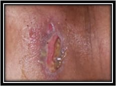
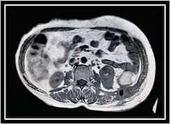
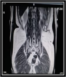
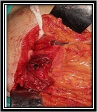
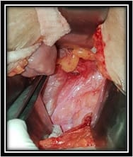
Endoscopic Retrograde Cholangiopancreatography (ERCP) is the most widely used diagnostic and therapeutic modality for hepatobiliary and pancreatic diseasesbut is not free from complications. Though rare, the common complications are migration of biliary stent, perforation, pancreatitis, hemorrhage, cholangitis, intra-abdominal sepsis, obstruction, etc. Here, we report a rare complication of ERCP presented as colocutaneous fistula. Colocutaneous fistulas are abnormal communications between the colon and the abdominal skin. They can occur spontaneously in patients with inflammatory bowel disease (most common) as well as after an injury or a surgical procedure. About one-third of fistulas close spontaneously with medical treatment and surgery being reserved for failures after optimal medical treatment. This case of colocutaneous fistula following ERCP presented to us was successfully treated as a single staged procedure without any complication. To the best of our knowledge, this is the first case reported of colocutaneous fistula following ERCP.

Keywords: ERCP, Colocutaneousfistula, Single staged procedure

Corresponding Author How to Cite this Article To Browse
Sakshi Goyal, PG Resident, Department of General Surgery, L.N. Medical College and J.K. Hospital, Bhopal, Madhya Pradesh, India.
Email:
Mishra N, Goyal S. A rare complication of ERCP: colocutaneous fistula. Surgical Review Int J Surg Trauma Orthoped. 2020;6(2):134-138.
Available From
https://surgical.medresearch.in/index.php/ijoso/article/view/154

Manuscript Received Review Round 1 Review Round 2 Review Round 3 Accepted
11-02-2020 22-02-2020 27-02-2020 05-03-2020
Conflict of Interest Funding Ethical Approval Plagiarism X-checker Note
No Nil Yes 16%

© 2020 by Navneet Mishra, Sakshi Goyal and Published by Siddharth Health Research and Social Welfare Society. This is an Open Access article licensed under a Creative Commons Attribution 4.0 International License https://creativecommons.org/licenses/by/4.0/ unported [CC BY 4.0].

Introduction

Endoscopicretrograde cholangiopancreatography (ERCP) is the most widely used diagnostic and therapeutic modality of various pancreatic and biliary diseases. Overall, complications occur in 5%-10% of cases following ERCP with or without sphincterotomy[1].

The incidences of post-ERCP pancreatitis, hemorrhage, cholangitis, and perforation are 3.5%-3.8%, 0.9%-1.3%, 1.0%-5.0%, and 0.1%-1.1%, respectively. The overall mortality rate after ERCP is 0.3% and can be reduced by early recognition and prompt treatment of these complications[2,3].

Recently, a patient in our care experienced colocutaneous fistula following ERCP.Colocutaneous fistulas are abnormal communications between the colon and the abdominal skin. They can occur spontaneously in patients with IBD (most common), malignancy, appendicitis, diverticulitis, radiation, tuberculosis/actinomycosis, and ischemia or after an injury or a surgical procedure.

Surgical procedures performed in patients with neoplastic diseases, inflammatory bowel diseases, or adhesions often result in postoperative fistula formation[4]. Enterocutaneous fistula represents one of the most protracted and difficult problems in colorectal surgery with substantial morbidity and mortality rate. There has been only one previously published report on a secondary duodenal fistula after ERCP-related duodenal perforation[5].

To the best of our knowledge, this is the first case reported of colocutaneous fistula following ERCP. The fistula was successfully treated surgically without any complication.

Case Report

A 55-year-old woman was admitted to our surgery department with the complaints of foul-smelling pus discharging sinus in the left lower back associated withthe occasional passage of gas from the sinus and on and off fever with chills and rigors.

She had a history of cholelithiasis with choledocholithiasis for which cholecystectomy and ERCP with biliary stenting were done in August 2019.

The entire procedure was uneventful. Immediately followed this, within a week she developed vague

dull aching abdominal pain and within a month of post -ERCP she developed the above-mentioned complaints. Both CBD and PD stents were removed on 11 Dec.

'19 in our hospital. On examination, there was a single oval-shaped sinus 2x1cm in the left lower back with purulent discharge (10-15ml/day) with erythematous and indurated surrounding tissue and slightly local temperature raised. She was clinically diagnosed with subcutaneous abscess.

surgery_154_01.jpg

Fig-1: Sinus (2 x 1 cm)with an indurated margin in the left lower back.

Investigations- All blood investigations were within normal limits. USG whole abdomen was s/o abscess. MRI findings revealed fluid and air-filled sinus tract of 4-5cm in left lumbar region communicating skin, subcutaneous tissue, abdominal wall to retroperitoneal space. MRI sinogram revealed an abscess in a subcutaneous and intermuscular plane extending till posteriorly pararenal space. X-ray thoracolumbar spine reveals no pathology.

surgery_154_02.jpg

Fig-2


surgery_154_03.jpg

Fig-3

Figure 2 and 3. MRI showing fluid and air-filled sinus tract in the left lumbar region communicating skin, subcutaneous tissue, abdominal wall to retroperitoneal space

Intraoperative findings:The patient was planned for fistulous tract excision. Intraoperatively origin of the fistulous tract was localized with the help of methylene blue and it was found that its origin was from the splenic flexure of colon communicating with left lumbar skin via retroperitoneal muscles. So, it was then, and there converted to exploratory laparotomy. Firstly left colon was mobilized along with the splenic flexure.

Fistulous tract was then resected along with a small portion of splenic flexure followed by an end to end colo-colic anastomosis and two abdominal drains were placed in the left paracolic and left retroperitoneal defect.

Flatus tube was passed through the anal opening across anastomotic site till transverse colon. Wound closure of both the left lumbar and left paramedian was done.

The postoperative period was uneventful. Flatus tube was removed on POD 5 and the patient was discharged on POD7.

surgery_154_04.jpg

Fig-4: Fistulous tract originat the splenic flexure.

surgery_154_05.jpg

Fig-5: Extension till retroperitoneal space.

Microscopic findings

1) A section of dye stained fistulous tract of about 3cm showed tract lined by granulomatous tissue consisting of a multinucleated giant cell, histiocytes, and chronic inflammatory infiltration.


2) A resected segment of colon splenic flexure measuring about 8 x 3 x 0.5 cm showing no significant pathology. A thickened area was identified 3cm from the resected margin which showed submucosal edema with congested serosa with acute inflammatory cell infiltration suggestive of acute serositis.

Discussion

Colocutaneous fistulas are abnormal communications between the colon and the abdominal skin. Their etiologies may vary and thus the treatment is based on the underlying cause.

Enterocutaneous (EC) fistulas have been described as surgical tragedies. Most EC fistulas occur following abdominal surgeries and about 15-25% of spontaneous EC fistulas are the result of underlying diseases such as Crohn's disease, radiation enteritis, or diverticular disease [4,6].

Colocutaneous fistula presenting as a complication of ERCP is rare; perhaps seen for the first time. Presentation in affected patients could be either delayed or acute (i.e., in the immediate postoperative period). Substantial morbidity is associated with the presence of colocutaneous fistulas.

Morbidity depends on the underlying cause where medical treatment and stabilization precede attempts at surgical intervention [7].

dfsaA common acronym used to describe EC fistula care protocol is "SNAP," which stands for management of skin and sepsis, nutrition, the definition of fistula anatomy, and proposing aprocedure to address the fistula[8,9]. Surgical treatment is reserved for patients whose fistula cannot be resolved with nonsurgical therapy.

Favorable and unfavorableprognostic factors for spontaneous fistula closurehave been described in terms of patient prognosis. 1) surgical etiology fistula 2) length >2cm, end fistula 3) low output <200ml/hr 4) no sepsis and balanced electrolytes are favourable factors whereas 1) ileal, jejunal , nonsurgical etiology 2) length <2cm, multiple fistulas 3) high output >500 ml/hr 4) sepsis and electrolyte disturbances are unfavourable [4,10-15].

The aims of surgeryare to do the refunctionalization of the entire bowel;resection of the fistula with end-to-end anastomosis of the bowelandsecure abdominal wall closure.To facilitate early feeding

and decompression of the proximal bowel, selected cases require diverting ileostomy or jejunostomy.

Our patient had a chronic fistula that occurred following ERCP;this fistula was treated as the single staged procedure where the fistulous tract was resected completely followed by resection of the affected portion of bowel and end to end colocolic anastomosis with a flatus tubebypassing the anastomotic site.

Conclusion

Colocutaneous fistulas exhibit various etiologies and courses of presentation and arestill remains a surgical challenge. Appropriate management depends on the time of diagnosis. We had performed a single staged procedure where the patient showed good postoperative recoveryand avoided most of the morbidities that accompany colocutaneous fistulas. Hence, colocutaneous fistula can be added as another differential diagnosis of the complications of ERCP.

Reference

  1. Freeman ML. Adverse outcomes of ERCP avoidance and management. Gastrointest Endosc Clin N Am. 2003;13(4)775-798.
  2. Vezakis A, Fragulidis G, Nastos C, Yiallourou A, Polydorou A, Voros D. Closure of a persistent sphincterotomy-relatedduodenal perforation by placement of a covered self-expandablemetallic biliary stent. World J Gastroenterol. 2011;17(40)4539-4541.
  3. Erdmann D, Drye C, Heller L, Wong MS, Levin SL. Abdominal walldefect and enterocutaneous fistula treatment with the vacuum assistedclosure (VAC) system. Plast Reconstr Surg. 2001;108(7)2066-2068.
  4. Berry SM, Fischer JE. Classification and pathophysiology of enterocutaneous fistulas. Surg Clin North Am. 1996;76(5)1009-1018.

  1. Anderson MA, Fisher L, Jain R, Evans JA, Appalaneni V, Ben-Menachem T, et al. Complications of ERCP. Gastrointest Endosc. 2012;75(3)467-473.
  2. Draus JM Jr, Huss SA, Harty NJ, Cheadle WG, Larson GM. Enterocutaneous fistula- are treatments improving?. Surg. 2006;140(6)570-576.
  3. deWeerd L, Kjaeve J, Aghajani E, Elvenes OP. The sandwich design- a newmethod to close a high output enterocutaneous fistula and an associated abdominal wall defect. Ann Plast Surg. 2007;58(5)580-583.
  4. Christensen M, Matzen P, Schulze S, Rosenberg J. Complicationsof ERCP- a prospective study. Gastrointest Endosc. 2004;60(5)721-731.
  5. Kaushal M, Carlson GL. Management of enterocutaneous fistulas. Clin Colon Rectal Surg. 2004;17(2)79-88.
  1. Samad S, Anele C, Akhtar M, Doughan S. Implementing a proforma for multidisciplinary management of an enterocutaneous fistula- a case study. Ostomy Wound Manage. 2015;61(6)46-52.
  2. Schecter WP. Management of enterocutaneous fistulas. Surg Clin North Am. 2011;91(3)481–491.
  3. Lloyd DA, Gabe SM, Windsor AC. Nutrition and management of enterocutaneous fistula. Br J Surg. 2006;93(9)1045-1055.
  4. Martinez JL, Luque-de-Leon E,Mier J, Blanco-Benavides R, Robledo F. Systematic management of postoperative enterocutaneous fistulas- factors related to outcomes. World J Surg. 2008;32(3)436–443.
  5. Campos AC, Andrade DF, Campos GM, Matias JE, Coelho JC. A multivariate model to determine prognostic factors in gastrointestinal fistulas. J Am Coll Surg. 1999;188(5)483-490.
  6. Evenson AR, Fischer JE. Current management of enterocutaneous fistula. J Gastrointest Surg. 2006;10(3)455-464.