E-ISSN:2455-5436
P-ISSN:2456-9518
RNI:MPENG/2017/70870

Research Article

Osteosynthesis

Surgical Review - International Journal of Surgery, Trauma and Orthopedics

2020 Volume 6 Number 1 January-February
Publisherwww.medresearch.in

Evaluation of results of minimally invasive plate osteosynthesis for humeral shaft fractures. A study involving 40 patients

Sharma A.1, Sharma G.2*, Bidoliya V.3, Nagina K.4
DOI: https://doi.org/10.17511/ijoso.2020.i01.05

1 Ashwini Sharma, Associate Professor, Department of Orthopaedics, Chirayu Medical College hospital, Bhopal, Madhya Pradesh, India.

2* Gourav Sharma, Assistant Professor, Department of Orthopaedics, Chirayu Medical College hospital, Bhopal, Madhya Pradesh, India.

3 Vinaydeep Bidoliya, Senior Resident, Department of Orthopaedics, Chirayu Medical College hospital, Bhopal, Madhya Pradesh, India.

4 Kirtiraj Nagina, Senior Resident, Department of Orthopaedics, Chirayu Medical College hospital, Bhopal, Madhya Pradesh, India.

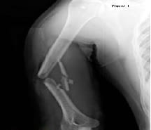
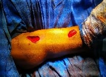
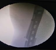
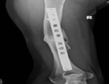
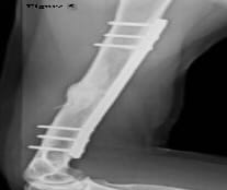
Introduction: Fracture of humeral shaft account for roughly 3% of all fractures. Previously, non-operative treatment has been accepted modality of treatment. Three main operative techniques are in vogue for treating displaced humeral shaft fractures namely intramedullary nailing, conventional plating osteosynthesis (CPO) and minimally invasive plate osteosynthesis (MIPO). Material and Methods: 40 fractures of humerus shaft were treated with MIPO technique, in a prospective study between December 2015 and September 2017 at our institute. The cases were followed up for a minimum period of 2 years. Results: The average age was 41 years (23-71 years). Twenty-three (57.5%) were males and 17 (42.5 %) females. Twenty-nine cases (72.5%) had injury in their dominant arm. The mean surgical time was 45.5 minutes and the mean radiation exposure was for 85.3 seconds. Conclusion: MIPO is a better choice for treating humeral shaft fractures than CPO, though there is no significant difference between MIPO and CPO in terms of operative time, fracture union rate, and fracture union time.

Keywords: CPO- Conventional plate osteosynthesis, MIPO-minimally invasive plate osteosynthesis, Humerus shaft fracture

Corresponding Author How to Cite this Article To Browse
Gourav Sharma, Assistant Professor, Department of Orthopaedics, Chirayu Medical College hospital, Bhopal, Madhya Pradesh, India.
Email:
Sharma A, Sharma G, Bidoliya V, Nagina K. Evaluation of results of minimally invasive plate osteosynthesis for humeral shaft fractures. A study involving 40 patients. Surgical Review Int J Surg Trauma Orthoped. 2020;6(1):27-34.
Available From
https://surgical.medresearch.in/index.php/ijoso/article/view/151

Manuscript Received Review Round 1 Review Round 2 Review Round 3 Accepted
26-10-2019 06-11-2019 14-11-2019 18-11-2019
Conflict of Interest Funding Ethical Approval Plagiarism X-checker Note
No Nil Yes 15%

© 2020 by Ashwini Sharma, Gourav Sharma, Vinaydeep Bidoliya, Kirtiraj Nagina and Published by Siddharth Health Research and Social Welfare Society. This is an Open Access article licensed under a Creative Commons Attribution 4.0 International License https://creativecommons.org/licenses/by/4.0/ unported [CC BY 4.0].

Introduction

Fractures of humeral shaft account for roughly 3 % of all fractures [1-3]. Previously, non-operative treatment has been accepted modality of treatment. Non operative treatment includes POP-u- cast and hanging cast. Sarmiento [4] popularized functional bracing of humeral shaft fractures to mitigate stiffness caused by cast treatment.

However, a high rate of nonunion up to 10-40 % was reported in humeral shaft fracture patients with nonoperative treatment. Nowadays treatment of humeral shaft fractures continues to generate controversy in orthopaedic community. Biber et al [5] opined that there is still no gold standard for the treatment of humeral shaft fractures and there is currently insufficient evidence for a clear superiority of either of the methods.

Three main operative techniques are in vogue for treating displaced humeral shaft fractures namely intramedullary nailing, conventional plating osteosynthesis (CPO) and minimaslly invasive plate osteosynthesis (MIPO). Intramedullary nail and plate are the conventionally used surgical methods.

Intramedullary nailing of humerus has its set of problems as long learning curve, shoulder stiffness, iatrogenic communition. Currently, open reduction and plate fixation remains to be the golden standard for humeral shaft fractures [6,7]. CPO has also got its own disadvantages such as surgical site infection, radial nerve palsy and extensive soft tissue dissection.

Recently, minimally invasive plate osteosynthesis (MIPO) techniques with encouraging results in humeral shaft fracture patients have been reported [8-11]. MIPO not only carries advantage of smaller incisions but also eliminates disadvantage of shoulder stiffness as caused by IMN at the site of insertion. It seems to imply that MIPO is superior to conventional plate osteosynthesis (CPO).

Material and Methods

Study setting: This study was performed in Chirayu Medical College and Hospital, a tertiary level facility under department of Orthopaedics.

Duration of study: Study was conducted from December 2015 to September 2017.

Sampling methods: all consecutive patients being admitted with humeral shaft fractures were

considered for this study and subjected to inclusion and exclusion criteria.

Sample size: 40 patients fulfilled the inclusion criteria. Minimum follow-up period was 2 years.

Inclusion criteria: displaced diaphyseal fracture of humerus between 21 and 75 years and who consented to participate in the study. The operative procedure was performed within 10 days of the injury. The fractures were classified as per the AO-ASIF trauma classification [12].

Exclusion criteria: coexisting medical disorders (such as a malignant tumor and hyperparathyroidism), vascular insufficiency of the upper limb, polytrauma patients with an injury severity score [13] of >16 points and psychiatric patients.

Ethical considerations: This study was approved by the Institutional Ethical Committee.

Preoperative planning: A routine preoperative clinical evaluation of the affected arm was carried out noting the swelling, abrasions, contusion, puckering of skin and distal neurovascular deficit, including the status of the radial nerve. Standardized anteroposterior (AP) and lateral (Lat) radiographs of the humerus, with the patient supine, arm abducted to 30° at the shoulder, elbow extended, and forearm supinated, were taken (Fig 1). These radiographs were also used to template the appropriate length of implant and planning the number and position of screws and their order of insertion. These fractures were fixed with 4.5-mm narrow locking compression plate (LCP).

surgery_151_01.jpg

Fig-1: Standardized anteroposterior (AP) and lateral (Lat) radiographs of the humerus.


Data collection and analysis: was done using WPS office suite

surgery_151_02.jpg

Fig-2: Distal incision was made along the lateral border of the biceps.

surgery_151_03.jpg

Fig-3: Site of incision was confirmed under the image intensifier

Surgical procedure: Operation table was rotated at 90 degrees so that head end becomes foot end and vice versa. The procedure was done in the supine position under general anesthesia or interscalene block, with the arm by the side of the body and the forearm in full supination. No side table or arm support was used. The image intensifier was positioned on the opposite side of the operating table as the arm to be operated. Two small incisions were used each 3 cm in length. Proximal incision between the biceps and the medial border of deltoid, in the lower half of deltoid utilizing the deltoid-pectoral approach was made. Dissection was done down to humerus. Distal incision was made along the lateral border of the biceps,

approximately 3 cm proximal to the flexion crease (Figure 2). The site of incision was confirmed under the image intensifier and 10-hole or 12-hole LCP was used and site of incision was altered, if required (Figure 3). The biceps was retracted medially to expose the musculocutaneous nerve, which overlies the brachialis muscle. The brachialis muscle was split in two halves and the musculocutaneous nerve retracted medially, and the radial nerve was protected by the lateral half of the brachialis muscle.

An extra-periosteal tunnel was created by passing long periosteum elevator, used as a tunneling instrument, deep to the brachialis muscle from proximal to distal incision. Care was taken to pass the tunneling instruments anteriorly or anteromedially to avoid the chances of injury to the radial nerve. After creating the tunnel, LCP of appropriate length was passed through the tunnel. The plate position and reduction was visualized on the image intensifier.

Traction was applied to restore length avoiding distraction. Direct pressure from lateral side was applied on proximal fragment to nullify pull of deltoid. Rotation was checked cortical step sign and medullary canal diameter. C-arm was used to drill the distal hole in humerus with forearm in supination and elbow in 90 degree flexion. Ensuring that the position of the plate on the distal fragment was central, it was fixed with a locking screw and, similarly, the proximal fragment was also fixed. After confirmation of the reduction alignment, the fixation was completed with a minimum of two screws in both fragments.

The rotational deformity was minimized using the 'cortical step sign' and the 'diameter difference sign' described by Krettek [14]. The operative time (defined as the time, from the skin incision to wound closure) and duration of radiation exposure (in seconds) was recorded though the doses were not calculated.

Postoperatively, arm was immobilized in an arm pouch sling. The standard protocol of mobilization exercises were started from day 2, as far as the patient's pain permitted. The time to union, the need for secondary procedure, and complications were noted.

Results

The average age was 41 years (23-71 years). Twenty-three (57.5%) were males and 17 (42.5 %)


females. Twenty-nine cases (72.5%) had injury in their dominant arm. The current study had seven cases of C1 and A2 type; six cases of B3, B1, and A1 type; four cases of B2 type; three cases of C2 type; and one case of A3 type of fractures.

(Table 1) Road traffic accident was the most common mode of injury, being reported by 31 (77.5 %) cases; the rest sustained injury following fall on an outstretched hand (four cases) and direct trauma (two cases).

Table-1: Distribution of cases based on several factors

  Male Female
Sex 23 17
AO class C1 7
  A2 7
  B3 6
  B1 6
  A1 6
  B2 4
  C2 3
  A3 1
Mean surgical time 45.5 min  
Mean radiation time 85.3 seconds  
Mean follow up 33 months  
Average union 14.9 weeks  

The mean surgical time was 45.5 minutes (range: 30-60 minutes) and the mean radiation exposure was for 85.3 seconds (range: 70- 120 seconds). The mean follow-up of our cases was 33 months (range: 24-43 months). Union was observed at a mean period of 14.9 weeks (range: 12-23 weeks) (Figure 4, 5).

surgery_151_04.jpg

Fig-4: Mean follow-up of the cases was 33 months.

surgery_151_05.jpg

Fig-5: Union observed mean period of 14.9 weeks.

In three cases, where there was scanty callus at 12 weeks, the current study infiltrated bone marrow taken from the patient's iliac crest at the fracture site, and these patients showed good union at 18-24 weeks. The current study accepted up to 5° of varus /valgus angulation intraoperatively and on following these patients up, the remaining seven cases, four had 3° of varus, two had 3° valgus, and one case had 5° varus angulation at the end of 2 years; however, this did not affect their functional outcome. The current study did not have any case of radial nerve palsy or musculocutaneous nerve injury. Shoulder function was assessed using UCLA shoulder score [15]. All patients had excellent score (more than 27). Elbow function was assessed using MPES [16]. All patients had good to excellent elbow function at final follow-up. The current study did not have one case of secondary procedure and no case hardware removal.

Discussion

Although conservative treatment has been used for humeral shaft fractures yet due to relatively high incidence of nonunion and high velocity trauma, operative treatment has gained popularity. There are many reports in the literature of good results with nailing technique; problems with insertion site morbidity and union rates have dampened the original enthusiasm for this mode of treatment [17,18].

Shoulder pain has been reported after antegrade intramedullary nailing in 16-37% of patients in recent studies. Bhandari et al found that reoperation and shoulder impingement were significantly more


common after intramedullary nailing than after plate fixation [19]. Conventional plate osteosynthesis remains the gold standard of fixation for humeral shaft fractures. It provides enough stability to allow early upper extremity weight bearing in polytrauma patients and produces minimal shoulder or elbow morbidity. A prospective randomized comparison of CPO and intramedullary nail fixation of humeral shaft fractures found no significant difference in the function of the shoulder and elbow , but shoulder impingement occurred more often with intramedullary nailing and a second surgical procedure was required in more patients with intramedullary than a plate.

A meta-analysis of the literature that included 391 patients concluded that reoperation and shoulder impingement were significantly more common after intramedullary nailing than after MIPO but radial nerve palsy was more common in CPO group as compared to MIPO [20]. vande Wall B et al in their study of 76 patients concluded absolute stability for simple humeral shaft fractures leads to a significantly shorter time to radiological union compared to relative stability [21].

Extensive soft tissue dissection and radial nerve palsy associated with CPO led orthopedic surgeons to minimally invasive plate osteosynthesis (MIPO) of humeral shaft fractures. Minimal invasive plating (MIPO) techniques for humeral shaft fractures appear to have fewer complications and higher union rates compared to open reduction and internal fixation (ORIF). Apivatthakakul et al in their cadaveric study observed that mean distance of nerve from plate was 3.2 mm [22].

However the current study didn’t encounter any radial nerve palsy in the present study. The danger zone for the musculocutaneous nerve lies, on average 18.37%-42.67% of the humeral length from the lateral epicondyle [23] Shetty et al in their study of 32 patients reported two cases of the musculocutaneous nerve neuropraxia, just above the elbow [24]. The present study did not encounter any such injury in the current cases.

Livani [10] et al reported 15 patients with MIPO done through two small incisions proximal and distal to the fracture; all fractures united within 12 weeks except one with brachial plexus palsy. As MIPO has been popular in other long bones its use for humerus fractures has been suggested, however risk to radial nerve injury has been a concern. All of our patients underwent surgery within 10 days of

injury. Majority of the patients in the present series returned to their preinjury activity levels by around 18 weeks. The current study have used delto-pectoral and Henry approach for proximal and distal incisions. Tunneling was initially difficult, but technique was mastered with increasing number of cases. Rotation and angulation was less than 10 degree. All patients united within 12-18 weeks with average union time of 14.7 weeks. The duration for bone union is better than that Zhiquan [8] et al for their series of MIPO but worse than that reported by Shetty [24] et al and comparable with CPO series [25,26].

Union was delayed in a case of comminuted fracture. Possible explanation for delayed union was comminution at fracture site and hampered vascularity due to severe soft tissue injury. Bone marrow injection expedited union in this case and union was achieved within 4 weeks. An added advantage with MIPO is that it is devoid of the entry-point problems of intramedullary nailing such as rotator cuff impingement.

The mean surgical time was 45.5 minutes (range: 30-60 minutes) and the mean radiation exposure was for 85.3 seconds (range: 70- 120 seconds). The mean follow-up of our cases was 33 months (range: 24- 43 months). Union was observed at a mean period of 14.9 weeks (range: 12-23 weeks).

In none of the cases shortening was more than 2 cm. In present series out of 40 patients five patients suffered some complication. However, there was no incidence of surgical site infection, nonunion, plate breakage, complex regional pain syndrome, and neurovascular compromise and compartment syndrome in any of our patients. In the present study, there were three patients with symptomatic delayed union but pain disappeared after union of fractures. Two cases of shoulder stiffness were managed with physiotherapy and one patient regained full movement.

However, Ismail et al in their small study of ten patients used the less invasive osteosynthesis technique via a modified anterolateral approach [27]. They concluded that MIPO minimises the soft tissue damage, allows for improved and more rapid bone healing, less infection, less postoperative pain, and promotes early recovery. It is important to note, however, that this technique is technically demanding. It requires the orthopaedic surgical expertise since the surgical exposure and fracture reduction is limited.


Xue et al in their study on mid shaft humeral fractures the conventional method of open reduction and internal fixation of fractures of the mid-distal humeral shaft, the stripping of the soft tissues and periosteum around the fracture site is unavoidable [28].

This may compromise the poor blood supply to the distal fracture fragments, thereby increasing the risk for non-union. The findings of the present study showed that MIPO caused less damage to the accessory nutrient arteries and their blood flow, unlike the case with ORIF, where they were frequently damaged and often necessitated ligation. And it has been confirmed that these accessory arteries is crucial to fracture healing and ligation of them will lead to adverse outcome.

Put together, these findings indicate that MIPO might be superior to ORIF in preserving the blood supply of the mid-distal portion of the humeral shaft. And because the fracture pattern is unpredictable, it is preferable to use a minimal invasive approach to preserve the remaining blood supply and minimize the iatrogenic disruption of the perfusion.

Some drawbacks of MIPO also need to be considered. Closed reduction required for MIPO is technically difficult; therefore, the surgeon performing the procedure should have received sufficient training and the surgery is prolonged. Further, frequent intraoperative fluorescent examination may be necessary to ensure proper reduction, thereby further extending the operation time. Moreover, angulation deformity is an inherent risk of closed reduction.

Tets worth et al in their review paper on MIPO humerus opined that this technique is based on the anterior humeral shaft providing a relatively safe surface for plate application, and limited open exposures proximally and distally allow percutaneous insertion of the necessary implant [11]. They were able to search more than 40 articles regarding MIPO, and it compares favorably to other available forms of treatment with excellent functional outcomes and a lower rate of iatrogenic radial nerve injury. Larger randomized controlled trials comparing this method with other accepted techniques, including nonsurgical management, are necessary to better define the role of MIPO in the management of humeral shaft fractures.

One weakness of the present study was small sample size. A study with large number of

patients with control group of patients can give us more conclusive evidence. Another weakness was that it was single center study.

Conclusion

The cost-effectiveness of MIPO is driven by low revision rates and high uneventful healing rates. Similar union rates and time to union for MIPO and CPO were observed, which is consistent with existing literature that has failed to identify either as the clinically superior technique. However, more high-quality randomized control trials are needed to further confirm this conclusion in future.

What this study adds to existing knowledge?

The current literature recommends plaster of Paris cast or functional bracing as primary treatment of mid shaft humeral fracture with acceptable alignment, although failure rates appear high. Surgical intervention is recommended in cases fracture nonunion, inability to maintain stability with bracing, floating elbow, multiple injury, polytrauma patients. With evolutions in surgical treatment for humeral shaft fractures from open plating to intramedullary interlocking nailing to MIPO; the risk of radial nerve palsy and union rates observed are likely to change. Shorter incisions, minimal soft tissue stripping, less incidence of radial nerve palsy and less operating time as observed in the present study (comparable with CPO) will make treatment of these common fractures less complex. Many surgeons are not familiar with nuances MIPO and cannot resist proclivity for open conventional plate osteosynthesis. This article provides food for thought for surgeons who are less inclined to MIPO. Future research is needed to determine the optimal treatment strategies for fracture cases of severe bone loss.

Authors’ Contribution

Dr. Ashwini Kumar Sharma had conceptualized the study, prepared the study protocol, conducted the data collection, analysis and manuscript writing. He has verified all the drafts and approved the final draft.

Dr. Gaurav Sharma had provided key inputs on methodology during protocol preparation, supported data compilation and analysis. He has also edited all the drafts and approved the final draft of the


manuscript.

Dr. Vinaydeep Bidoliya and Dr. Kirtiraj Nagina did data collection and postoperative dressings of patients.

Reference

  1. Ekholm R, Adami J, Tidermark J, Tidermark J, Hansson K, Törnkvist H, Ponzer S. Fractures of shaft of humerus, An epidemiological study of 401 fractures. J Bone Joint Surg. 2006; 88(11)1469-1473.
  2. Strohm PC, Reising K, Hammer T, Sudkamp NP, Jaeger M, Schmal H. Humeral shaft fractures- where are we today. Acta Chir Orthop Traumatol Cechoslov. 2011;78(3)185-189.
  3. Cole PA, Wijdicks CA. The operative treatment of humeral diaphyseal fractures. Hand Clin. 2007;23(4)437-448.
  4. Sarmiento A, Kinman PB, Galvin EG, Schmitt RH, Phillips JG. Functional bracing of fractures of the shaft of the humerus. J Bone Joint Surg Am. 1977;59(5)596-601.
  5. Foster RJ, Dixon GL Jr, Bach AW, Appleyard RW, Green TM. Internal fixation of fractures and nonunion of humeral shaft, Indications and results in a multi-centre study. J Bone Joint Surg. 1985;67(6)857-864.
  6. Tetsworth K, Hohmann E, Glatt V. Minimally invasive plate osteosynthesis of humeral shaft fractures- current state of the art. JAAOS-J Am Acad Orthopaed Surg. 2018;26(18)652-661.
  7. Jawa A, McCarty P, Doomberg J, Harris M, Ring D. Extrarticular distal third diaphyseal fractures of humerus, A comparison of functional bracing and plate fixation. J Bone Joint Surg. 2006;88(11)2343-2347.
  1. Zhiquan A, Bingfang Z, Yeming W, Chi Z, Peiyan H. Minimally invasive plating osteosynthesis (MIPO) of middle and distal third humeral shaft fractures. J Orthop Trauma. 2007;21(9)628-633.
  2. Ziran BH, Belangero W, Livani B, Pesantez R. Percutaneous plating of the humerus with locked plating- technique and case report. J Traum Acute Care Surg. 2007;63(1)205-210.
  3. Livani B, Belangero W, Andrade K, Zuiani G, Pratali R. Is MIPO in humeral shaft fractures really safe?- Postoperative ultrasonographic evaluation. Int Orthopaed. 2009;33(6)1719-1723.
  4. Biber R, Bail HJ, Geßlein M. Humeral shaft fractures. Unfallchirurg. 2018;121(9)747-758.
  5. Müller ME, Nazarian S, Koch P, Schatzker J. The comprehensive classification of fractures of long bones. Springer Science & Business Media. 2012 Dec 6.
  6. Baker SP, o'Neill B, Haddon Jr W, Long WB. The injury severity score- a method for describing patients with multiple injuries and evaluating emergency care. J Trauma Acute Care Surg. 1974;14(3)187-196.
  7. Krettek C, Miclau T, Grün O, Schandelmaier P, Tscherne H. Intraoperative control of axes, rotation and length in femoral and tibial fractures-Technical note. Injury. 1998;29(3)C29-C39.
  8. Amstutz HC, Sew Hoy AL, Clarke IC. UCLA anatomic total shoulder arthroplasty. Clin Orthop Relat Res. 1981;(155)7-20.

  1. Petsatodes G, Karataglis D, Papadopoulos P, Christoforides J, Gigis J, Pournaras J. Antegrade interlocking nailing of humeral shaft fractures. J Orthop Sci. 2004;9(3)247-252.
  2. Longo UG, Franceschi F, Loppini M, Maffulli N, Denaro V. Rating systems for evaluation of the elbow. Brit Med Bullet. 2008;87(1)131-161.
  3. Apivatthakakul T, Arpornchayanon O, Bavornratanavech S. Minimally invasive plate osteosynthesis (MIPO) of the humeral shaft fracture- Is it possible?; A cadaveric study and preliminary report. Injury. 2005; 36(4)530-538.
  4. Bhandari M, Devereaux PJ, McKee MD, Schemitsch EH. Compression plating versus intramedullary nailing of humeral shaft fractures- a meta-analysis. Acta Orthop. 2006;77(2)279-284.
  5. Hu X, Xu S, Lu H, Chen B, Zhou X, He X, et al. Minmally invasive plate osteostnthesis v/s conventional fixation techniques for surgically treated humeral shaft fractures- a meta-analysis. J Orthop Surg Res. 2016;11(1)59.
  6. Vande Wall B, Theus C, Link BC, Van Veelen N, Van de Leeuwen RJH, Ganzert C, et al. Absolute or relative stability in plate fixation for simple humeral shaft fractures. Injury. 2019;50(11)1986-1991.
  7. Santori FS, Santori N. The Exp Nail for the treatment of diaphyseal humeral fractures. J Bone Joint Surg Br. 2002;84(3)280.
  8. Shetty MS, Kumar MA, Sujay KT, Kini AR, Kanthi KG. Minimally invasive plate osteosynthesis for humerus diaphyseal fractures. Ind J Orthop. 2011;45(6);18 521-526.
  1. Apivatthakul T, Patiyasikan S, Luevitoonvechkit S. Danger zone for locking screw placement in minimally invasive plate osteosynthesis (MIPO) of humeral shaft fractures- A cadaveric study. Int J Care Injured. 2010;41(2)169-172.
  2. Fernandez DellOca AA. The principle of helical implants- Unusual ideas worth considering. Injury. 2002;33(1)A1-A27.
  3. Ismail HD, Boedijono DR, Hidayat H, Simbardjo DS. Minimal Invasive Plate Osteosynthesis (MIPO) Technique Using Anterolateral Approach for Treating Closed Proximal Humerus Fracture. Malays Orthop J. 2012;6(1)18-24.
  4. Xue Z, Jiang C, Hu C, Qin H, Ding Q, An Z. Effects of different surgical techniques on mid-distal humeral shaft vascularity: open reduction and internal fixation versus minimally invasive plate osteosynthesis. BMC Musculoskeletal Disord. 2016;17(1)370.
  5. Dabezies EJ, Banta CJ 2 nd , Murphy CP, d'Ambrosia RD. Plate fixation of the humeral shaft for acute fractures, with and without radial nerve injuries. J Orthop Trauma. 1992;6(1)10-13.